肝脏影像解剖:精彩图解肝分叶分段
- 格式:pdf
- 大小:372.43 KB
- 文档页数:7

肝脏的分叶分段(解剖超声声像图)肝脏的分叶分段(解剖+超声声像图)楼主yufeionly 2012-04-19 21:32:29 大家再来看看肝脏的分叶分段: ·先来看看解剖如何分分叶:分叶、分段:图示:超声声像图图:第1、2、3、4段:第2、3、4、5、6、7、8段:大家一起来学习学习吧,应该对我们工作还是有帮助的。
嘿嘿。
喜欢的送朵花啦!沙发回复cherry8383 2012-04-19 21:39:20 哇!太知我心了最近还真在学习全面的肝八分段!!十分感谢贵友的分享啊!!得慢慢体会才行,呵呵。
板凳回复yufeionly 2012-04-19 21:41:59 回复 cherry8383 没事哈。
我也是整理了一下图片而已嘛。
大家一起学习,共同进步!!嘿嘿4楼回复cherry8383 2012-04-19 21:45:29 对于门脉左支“工”形的那个分段,我持怀疑,因为那个切面应该剑突下横切面来的,横切面下如何分上下?!它只有前后左右之分!大家说呢?请指点一二!5楼回复yufeionly 2012-04-19 21:52:51 回复 cherry8383 我觉得结合解剖来看,既然能够打出“工”形分支,那么就能够显示左外叶的上、下两段。
而且好像不是垂直的横切,我记得书上这个断面是:右肋缘下斜切第一肝门声像图。
所以我认为还是不完全是横切,而是斜切,,不然应该打不出“工”形分支实际操作中,探头也是剑突下横切,探头往上一点。
嘿嘿。
拙见哈。
大家来讨论讨论吧!6楼回复cherry8383 2012-04-19 21:53:44 如此分法大家理解吗?我也是最近才学习透的,不对之处请指正哈!7楼回复cherry8383 2012-04-19 21:58:29 回复 yufeionly 也有理,有时真的很难把握其精确度,不像CT,哈!8楼回复yufeionly 2012-04-19 22:01:45 回复 cherry8383 再来两张图:。


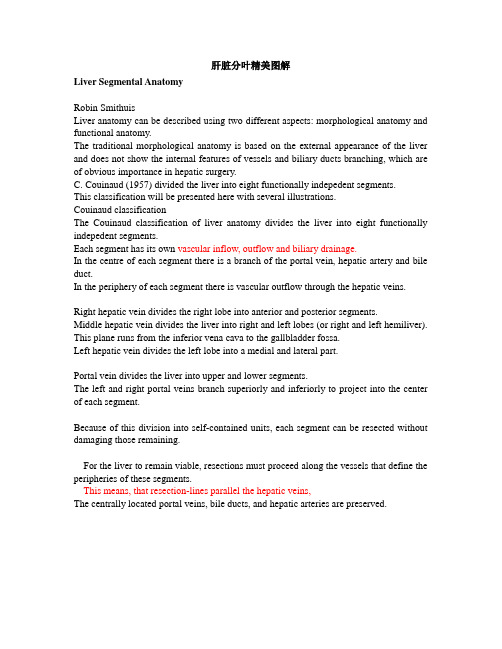
肝脏分叶精美图解Liver Segmental AnatomyRobin SmithuisLiver anatomy can be described using two different aspects: morphological anatomy and functional anatomy.The traditional morphological anatomy is based on the external appearance of the liver and does not show the internal features of vessels and biliary ducts branching, which are of obvious importance in hepatic surgery.C. Couinaud (1957) divided the liver into eight functionally indepedent segments.This classification will be presented here with several illustrations.Couinaud classificationThe Couinaud classification of liver anatomy divides the liver into eight functionally indepedent segments.Each segment has its own vascular inflow, outflow and biliary drainage.In the centre of each segment there is a branch of the portal vein, hepatic artery and bile duct.In the periphery of each segment there is vascular outflow through the hepatic veins. Right hepatic vein divides the right lobe into anterior and posterior segments.Middle hepatic vein divides the liver into right and left lobes (or right and left hemiliver). This plane runs from the inferior vena cava to the gallbladder fossa.Left hepatic vein divides the left lobe into a medial and lateral part.Portal vein divides the liver into upper and lower segments.The left and right portal veins branch superiorly and inferiorly to project into the center of each segment.Because of this division into self-contained units, each segment can be resected without damaging those remaining.For the liver to remain viable, resections must proceed along the vessels that define the peripheries of these segments.This means, that resection-lines parallel the hepatic veins,The centrally located portal veins, bile ducts, and hepatic arteries are preserved.根据Couinaud分段Segments numberingThere are eight liver segments.Segment 4 is sometimes divided into segment 4a and 4b according to Bismuth.The numbering of the segments is in a clockwise manner (figure).Segment 1 (caudate lobe) is located posteriorly. It is not visible on a frontal view.顺时钟数字标记段The illustrations above are schematic presentations of the liversegments.In reality however the proportions are different.On a normal frontal view the segments 6 and 7 are not visible because they are located more posteriorly.The right border of the liver is formed by segment 5 and 8.Although segment 4 is part of the left hemiliver, it is situated more to the right.Couinaud divided the liver into a functional left and right liver (in French 'gauche et droite foie') by a main portal scissurae containing the middle hepatic vein. This is known as Cantlie's line.Cantlie's line runs from the middle of the gallbladder fossa anteriorly to the inferior vena cava posteriorly.On this illustration it looks as if the medial part of the left lobe is separated from the lateral part by the falciform ligament. However it actually is the left hepatic vein, that separates the medial part (segment 4) from the lateral part (segments 2 and 3).The left hepatic vein is located slightly to the left of the falciform ligament.从前方看,看不到六,七段Transverse anatomyThe far left figure is a transverse image through the superior liver segments, that are divided by the hepatic veins.The right figure shows a transverse image at the level of the left portal vein.At this level the left portal vein divides the left lobe of the liver into the superior segments (2 and 4A) and the inferior segments (3 and 4.The left portal vein is at a higher level than the right portal vein.左:在左门静脉以上水平右:在左门静脉水平The image on the far left is at the level of the right portal vein. At this level the right portal vein divides the right lobe of the liver into superior segments (7 and 8) and the inferior segments (5 and 6).The level of the right portal vein is inferior to the level of the left portal vein.At the level of the splenic vein, which is below the level of the right portal vein, only the inferior segments are seen (right image).左:在右门静脉水平右:在脾静脉水平Caudate lobeThe caudate lobe or segment 1 is located posteriorly.The caudate lobe is anatomically different from other lobes in that it often has direct connections to the IVC through hepatic veins, that are separate from the main hepatic veins.The caudate lobe may be supplied by both right and left branches of the portal vein.On the left a patient with cirrhosis with extreme atrophy of the right lobe, normal volume of the left lobe andhypertrophy of the caudate lobe.Due to a different blood supply the caudate lobe is spared from the disease process and hypertrophied to compensate for the loss of normal liverparenchyma肝硬化尾叶增大注意:右叶缩小Other Classifications and V ariantsThere are many other anatomical and functional descriptions of the liver anatomy.In the classical description the external appearance of the liver is used to describe the anatomy.However there are many differences between this classical model and the fuctional models, as popularized by Couinaud and Bismuth.A more detailed discussion of the various models is given in reference 4.Classical AnatomyThe classical description of the liver anatomy is based on the external appearance.On the diaphragmatic surface, the ligamentum falciforme divides the liver into the right and left anatomic lobes, which are very different from the functional right and left lobes (or right and left hemiliver).In this classical description, the quadrate lobe belongs to the right lobe of the liver, but functionally it is part of left lobe.Bismuth's classificationThis classification is very similar to the Couinaud classification, although there are small differences. It is popular in the United States, while Couinaud's classification is more popular in Asia and Europe.According to Bismuth three hepatic veins divide the liver into four sectors, further divided into segments.These sectors are termed portal sectors as each is supplied by a portal pedicle in the centre.The separation line between sectors contain a hepatic vein.The hepatic veins and portal pedicels are intertwined, as are the fingers of two hands.The left portal scissura divides the left liver into two sectors: anterior and posterior.Left anterior sector consists of two segments: segment IV, which is the quadrate lobe and segment III, which is anterior part of anatomical left lobe.These two segments are separated by the left hepatic fissure or umbilical fissure.Left posterior sector consists of only one segment II. It is the posterior part of left lobe.V ariationsIn the Couinaud classification little attention is given to the high prevalence of anatomical variations which occur, especially in the right hemiliver.Using volumetric acquisition techniques, such as magnetic resonance imaging or spiral computed tomography scanning, detailed insight into the individual segmental anatomy can now be obtained in a non-invasive manner (2,3).The significance of this anatomical insight lies in the planning of anatomical resections, whereby the relationship between tumour and individual segmental anatomy can be depicted in a three-dimensional format.Three dimensional liver imaging is of most practical value if a resection of one or more segments or sectors is considered, especially in the right hemiliver.In these cases, 3D liver imaging can demonstrate the precise location of the scissuras to the surgeon pre-operatively.The 8 anatomical points of interest: 注意下面8个解剖标志the inferior vena cava, the right hepatic vein, the medial hepatic vein, the left hepatic vein, the deep ligament venosum, the superficial ligament venosum, the left lobe tip and the right portal veinFigure 1: Definition of liver's segments (left) and sample of liver surgery planification (right).圖12. 肝臟的解剖節段 A. 前表面; B. 后表面。
肝脏CT分段肝脏从表面划分的左叶、右叶、方叶和尾叶没有真正反映其内部管道系统的构造特征,因而不适应肝脏外科进行部分肝切除的需要。
CT医师对病灶的准确分段,对外科医师有很大的帮助。
根据格利森系统的分布,将肝脏分为左、右两个半肝,再进一步分为8段(奎纳德分类):I段:尾状叶。
Ⅱ段:相当于左外叶上段。
Ⅲ段:左外叶下段。
Ⅳ段左内叶。
Ⅴ段:右前叶下段。
Ⅵ段:右后叶下段。
Ⅶ段:右后叶上段。
Ⅷ段:右前叶上段。
肝脏的分段主要是根据肝内的管道系统而命名。
门静脉、肝动脉、肝胆管三者伴行包裹在同一Glisson鞘内,故称为Glisson系统或门脉系统。
据门脉系统分布所作的肝脏分段,称为门脉肝段。
肝静脉与门静脉呈插指状的关系,按照肝静脉引流区域所作的分段,称为静脉肝段。
由于肝内胆管是与肝内门静脉伴行,故在肝胆管外科中均采用门脉肝段的命名。
Couinaud根据门脉系统肝段按顺时针方向标以罗马数字从ⅠⅡⅢⅣⅤⅥⅧ,其中左内叶及尾状叶不再分段。
肝脏分8个段,主要被肝静脉系统和门静脉系统分割。
肝中静脉将肝分成左右两叶。
肝右静脉分肝右叶为右前、右后两部分。
肝左静脉分肝左叶为左内叶、左外叶。
门静脉系统走行于肝段内。
Ⅰ段为尾状叶,CT示在门、腔静脉之间,Ⅱ段(靠上)与Ⅲ段(靠下)构成左外叶,Ⅳ段为方叶,也是左内叶,Ⅴ段(靠下)与Ⅷ(靠上)段构成肝右前叶,Ⅵ段(靠下)与Ⅶ段(靠上)构成肝右后叶。
至于Ⅱ段与Ⅲ段、Ⅴ段与Ⅷ段、Ⅵ段与Ⅶ段分界,粗略方法以肝内门静脉分支或肝门平面为分界标志,出现以上平面所显示的是靠上方的,Ⅱ段、Ⅶ段、Ⅷ段,以下层面就是Ⅲ段、Ⅴ段、Ⅵ段。
Ⅴ段与Ⅵ段、Ⅶ段与Ⅷ段之间以肝右静脉分界。
影像学上横断面上以肝静脉为界,2,3;5,8;6,7 的分界大约门静脉左右分支平面。
1、肝脏分段的意义:可以了解病灶所处位置,尤其对于肝脏恶性肿瘤患者,从而指导治疗。
肝脏的分段和肝脏的解剖密切相关,了解了肝脏的解剖并且应用到影像学的分析中去,也就可以熟练的了解肝脏的分段了。
肝脏影像解剖:精彩图解肝分叶分段
解剖是万病之本,掌握基本解剖才能对疾病有更深的认识。
一、基础知识
Couinaud分段法将肝脏分为8段,段的编号依据顺时针进行,门静脉分支分布于肝段内,而肝静脉位于肝段间,每段功能上是独立的,有独立的血液、胆汁引流道。
每段的中心是门静脉、肝动脉和胆管,周围是血液流出的肝静脉。
尾状叶为Ⅰ段
左外上、下叶为Ⅱ、Ⅲ段
左内叶为Ⅳ段(Bismuth分类法,第4段又分为4a和4b段)
右前叶上、下为Ⅴ、Ⅷ段
右后叶上、下为Ⅵ、Ⅶ段
门静脉将肝分为上、下部分,门静脉左右支发出上、下分支分别进入每段的中心。
肝中静脉:将肝脏分为左叶和右叶。
肝左静脉:将肝左叶分为内侧段(II、III)和外侧段(IV)。
肝右静脉:将肝脏分为右前段(V、VIII)和右后段(VI、VII)。
各段按顺时针方向排列,I段(尾状叶)在后方。
上方的图为肝分段示意图,下图为实际正面观察,后方的VI段和VII段被遮挡。
Cantlie’s线是指胆囊窝前方中点与下腔静脉的连线,将肝脏分为左半肝和右半肝。
右半肝可见V和VIII段,IV段更靠近右侧,尽管它属于左半肝。
二、横断面解剖图示
(1)肝上部水平:由肝中静脉、肝右静脉、镰状韧带分段。