肝脏的分叶
- 格式:ppt
- 大小:2.66 MB
- 文档页数:74
人体解剖学知识:肝脏功能及解剖学结构分析肝脏是人体内最大的脏器,位于右上腹部,是人体的化学实验室和代谢中心。
肝脏的主要功能包括分泌胆汁、负责血糖的调节、合成蛋白质、代谢脂肪、解毒和存储维生素、矿物质等。
在人体内,肝脏是最重要和最繁忙的器官之一,下面我们来探讨一下肝脏的解剖学结构和功能。
一、肝脏解剖学结构肝脏的重量约为1.2-1.5kg,位于腹膜腔内,分为左、右、中三个叶,右叶最大,左叶次之,中叶最小,其中右叶占肝脏总重量的60%左右。
肝脏外表呈三角形,稍微隆起,下表面为平坦的底面,上表面呈不规则凸面,各裂隙浅深不一,隔以许多次级的小叶,每个小叶由同心排列的肝细胞构成,每个肝细胞的周围都有毛细血管,形成了肝窦。
肝脏的血管很丰富,动脉和门静脉的分支进入肝窦,牵涉到的静脉形成肝静脉,最后汇入下腔静脉。
这些血管在肝窦中形成毛细血管,通过毛细血管内的净化作用,毒素、废物和过多的细胞代谢产物被分解和排出。
肝脏将大部分药物和生物化学物质处理掉,并将其转化为身体能够利用的形态或排泄出体外。
二、肝脏的功能1.分泌胆汁肝脏是人体内唯一的胆汁合成脏器,每天可分泌约500毫升的胆汁,经过胆囊逐渐储存,当食物进入小肠时,胆囊收缩将胆汁送入小肠,帮助消化和吸收食物中的脂肪、油脂和脂溶性维生素。
2.负责血糖的调节人体内的血糖是由肝脏进行调节和维持的。
当血糖水平低于正常值时,肝脏会释放糖原储备,将其转化为葡萄糖,并释放入血液中,以保持正常的血糖水平。
如果血糖水平过高,肝脏会转化这些多余的葡萄糖为糖原储备,维持血糖平衡。
3.合成蛋白质肝脏负责合成人体内的许多蛋白质,如凝血因子、白蛋白和胆红素等。
这些蛋白质对于人体的正常功能至关重要。
4.代谢脂肪肝脏的代谢作用包括脂肪代谢。
当人体内的脂肪摄入过多时,肝脏会将其转化为脂肪酸和甘油,通过血液循环运输到其他组织和器官中,以供能源使用。
5.解毒和存储维生素、矿物质等肝脏是人体内最重要的解毒器官之一,能够处理和中和许多有害的毒素和废物。
正常肝脏解剖及组织结构
肝脏是人体最重要的内脏器官之一,位于腹腔的右上部。
它的重量约
为1500克,占据了腹腔的大部分空间。
肝脏具有多种重要的生理功能,
如合成胆汁、代谢蛋白质和糖类、储存维生素和矿物质、解毒和排泄等。
一、肝脏的外部结构
肝脏呈橡皮筋形,由左、右两个叶组成。
右叶较大,位于心脏的下方,左叶较小,位于心脏的左上方。
肝脏的上表面呈现光滑的圆顶形,称为圆
顶面;下表面呈现凹陷的形状,称为凹陷面。
肝脏的前缘与腹壁相连,后
缘紧邻胃、十二指肠及胰腺。
二、肝脏的内部结构
1.肝叶
肝脏的两个叶之间有间叶隔分隔。
肝脏内部的组织结构由肝小叶组成,每个肝小叶都是由数百个肝细胞群组成的。
肝细胞是肝脏最基本的生物学
单位,具有多种功能。
它们分泌胆汁,将胆汁输送到肝小管中,最终汇集
成胆管。
2.血液供应
3.胆汁系统
肝细胞产生的胆汁通过肝小管排泄到胆管中,最终经过胆总管进入十
二指肠。
胆汁的主要组成成分是胆盐、胆固醇、磷脂等,它在消化过程中
起到了重要的作用。
4.淋巴系统
肝脏还有一套独立的淋巴系统。
它将排泄物和细菌从肝脏引流到周围的淋巴结,以避免感染和其他并发症的发生。
5.神经系统
总的来说,肝脏是一个复杂的器官,具有多种生理功能,包括合成、代谢、解毒、排泄等,对维持人体的健康起着重要作用。
了解肝脏的正常解剖和组织结构对于我们理解其功能和相关疾病的发生有着重要意义。


肝脏分叶标准
肝脏的分叶标准主要有以下几种:
1. 肝脏的分叶是根据其内部的管道系统特征,特别是门静脉、肝静脉和肝胆管的分布和走向来进行的。
其中,五叶指的是尾状叶、左肝外叶、左内叶、右前叶和右后叶这五个叶。
2. 肝脏的分段则是在进行肝脏手术时具有指导意义的,主要是依据肝内管道系统的位置来进行的。
具体来说,它是以正中裂为界,将肝划分为左、右两半,叫做左、右半肝;左半肝又划分为左内叶和左外叶;右半肝划分为右前叶和右后叶。
其中,尾状叶也自成一段。
在影像诊断和外科手术中,还会采用其他的分叶标准,如根据肝脏外形分为左叶、右叶、方叶和尾状叶等。
此外,国内常用的肝段划分法还包括根据Glisson系统和肝静脉的走行,将肝分为五叶六段等。
以上信息仅供参考,如有需要,建议咨询专业医师。
肝脏解剖结构一、肝脏的大体形态肝脏是人体中最大、最重的实质脏器,在成人它的重量大约为 1200 ~ 1500 克,约为体重的五十分之一。
在婴儿时期,肝脏所占的比例比成人要大的多,约为体重之十八分之一。
肝脏分为左右两叶,右叶比左叶大。
在成人,右叶大约为左叶之六倍。
肝脏左右径约25.8cm,前后径约15.2cm,上下径约5.8cm。
自下腔静脉左缘至胆囊窝中点的正中裂将肝脏分为左半肝和右半肝。
自脉切迹至肝左静脉入下腔静脉处的左叶间裂将左半肝分为左内叶和左外叶,左段间裂将左外叶分为上下两段。
肝右叶间裂将右半肝分为右前叶和右后叶,右段间裂又将右前叶、右后叶分别分成上下两段(图1-1)。
从肝脏的脏面看,有肝方叶和肝尾状叶(图1-2)。
肝方叶前缘为肝脏的下缘,其左缘为肝圆韧带,后缘为第一肝门,右缘为胆囊窝。
肝尾状叶位于肝脏后方,其左缘为静脉韧带,右缘为下腔静脉窝,下缘为第一肝门。
Glisson鞘内包括肝动脉、门静脉和胆管,经肝脏面的第一肝门出入肝脏,此三者进化论在肝内还是肝门附近,都在一起走行。
Couinaud根据肝内门静脉干的颁布范围,将肝脏分为八段(图1-3)。
Ⅰ段为尾状叶,Ⅱ段为左外叶上段,Ⅲ段为左外叶下段,Ⅳ段为左内叶,Ⅴ段为右前叶下段,Ⅵ段为右后叶下段,Ⅶ段为右后叶上段,Ⅷ段为右前叶上段。
图1-1 肝脏的膈面结构(吴孟超五叶四段肝脏分叶法)1.左外叶上段2.左外叶下段3.右后叶上段4.右后叶下段5.右前叶6.左内叶7.左冠状韧带8.右三角韧带9.左三角韧带10.肝圆韧带11.镰状韧带12.右冠状韧带图1-2 肝脏的脏面结构(吴孟超五叶四段肝脏分叶法)1.左外叶上段2.左外叶下段3.左内叶4.右前叶5.右后叶上段6.右后叶下段7.下腔静脉8.尾状叶9.胆囊10.肝圆韧带图1-3 Couinaud肝脏分段法1.右肝静脉2.中肝静脉3.左肝静脉4.门静脉2.肝脏被腹膜皱折形成的肝周韧带固定在上腹部,包括肝圆韧带、镰状韧带、冠状韧带和左右三角韧带等。


人类肝脏的解剖和生理特点肝脏是人体最大的内脏器官之一,位于腹腔右上部,重量约为1.5千克。
本文将通过解剖和生理特点两个部分来介绍人类肝脏。
一、解剖特点1. 位置肝脏位于腹腔右上部,紧贴膈肌、胃、十二指肠和胰腺。
其形状呈椭圆形,前缘凸出,后缘微凹。
在肝脏下方有胆囊,胆管和肝动脉、肝门静脉,形成肝门三角。
2. 分叶结构肝脏可以分为左叶和右叶,两个叶之间有肝门三角穿过,左叶上有一叶突向后缘,称为尾状叶。
每个叶内部再分为多个小叶,小叶间由结缔组织相连。
小叶内部有肝细胞和空间,空间中运行着肝窦的血液,从而完成肝脏的主要功能。
二、生理特点1. 代谢功能肝脏具有代谢,包括物质代谢和能量代谢。
物质代谢主要指体内物质的合成、分解和转化,例如蛋白质、脂肪、糖类等;能量代谢主要指葡萄糖的原始材料,肝脏能够储存和释放能量,保持人体的平衡状态。
2. 解毒功能肝脏是人体重要的解毒器官,能够清除体内的有害物质。
它会将体内次氯酸盐、酒精、化学药品等有毒物质转化为无毒物质,并排出体外。
3. 分泌功能肝脏能够分泌多种重要物质,其中最重要的是胆汁。
胆汁中含有胆固醇、胆盐和胆红素,能够帮助人体消化和吸收脂肪。
4. 存储功能肝脏是储存维生素、铁、锌、铜和糖原等物质的重要器官。
它可以把体内多余的营养物质储存起来,供应人体需要。
总结:肝脏是人体重要的内脏器官,具有代谢、解毒、分泌和存储等重要功能。
它具有分叶结构、肝门三角和窦道等特点,这些特点增强了肝脏的功能和适应性。
因此,保护人类肝脏健康对整个身体健康至关重要。

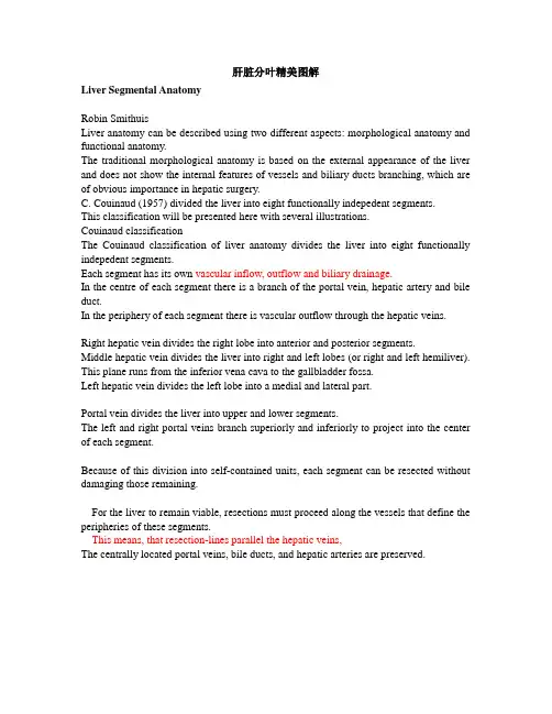
肝脏分叶精美图解Liver Segmental AnatomyRobin SmithuisLiver anatomy can be described using two different aspects: morphological anatomy and functional anatomy.The traditional morphological anatomy is based on the external appearance of the liver and does not show the internal features of vessels and biliary ducts branching, which are of obvious importance in hepatic surgery.C. Couinaud (1957) divided the liver into eight functionally indepedent segments.This classification will be presented here with several illustrations.Couinaud classificationThe Couinaud classification of liver anatomy divides the liver into eight functionally indepedent segments.Each segment has its own vascular inflow, outflow and biliary drainage.In the centre of each segment there is a branch of the portal vein, hepatic artery and bile duct.In the periphery of each segment there is vascular outflow through the hepatic veins. Right hepatic vein divides the right lobe into anterior and posterior segments.Middle hepatic vein divides the liver into right and left lobes (or right and left hemiliver). This plane runs from the inferior vena cava to the gallbladder fossa.Left hepatic vein divides the left lobe into a medial and lateral part.Portal vein divides the liver into upper and lower segments.The left and right portal veins branch superiorly and inferiorly to project into the center of each segment.Because of this division into self-contained units, each segment can be resected without damaging those remaining.For the liver to remain viable, resections must proceed along the vessels that define the peripheries of these segments.This means, that resection-lines parallel the hepatic veins,The centrally located portal veins, bile ducts, and hepatic arteries are preserved.根据Couinaud分段Segments numberingThere are eight liver segments.Segment 4 is sometimes divided into segment 4a and 4b according to Bismuth.The numbering of the segments is in a clockwise manner (figure).Segment 1 (caudate lobe) is located posteriorly. It is not visible on a frontal view.顺时钟数字标记段The illustrations above are schematic presentations of the liversegments.In reality however the proportions are different.On a normal frontal view the segments 6 and 7 are not visible because they are located more posteriorly.The right border of the liver is formed by segment 5 and 8.Although segment 4 is part of the left hemiliver, it is situated more to the right.Couinaud divided the liver into a functional left and right liver (in French 'gauche et droite foie') by a main portal scissurae containing the middle hepatic vein. This is known as Cantlie's line.Cantlie's line runs from the middle of the gallbladder fossa anteriorly to the inferior vena cava posteriorly.On this illustration it looks as if the medial part of the left lobe is separated from the lateral part by the falciform ligament. However it actually is the left hepatic vein, that separates the medial part (segment 4) from the lateral part (segments 2 and 3).The left hepatic vein is located slightly to the left of the falciform ligament.从前方看,看不到六,七段Transverse anatomyThe far left figure is a transverse image through the superior liver segments, that are divided by the hepatic veins.The right figure shows a transverse image at the level of the left portal vein.At this level the left portal vein divides the left lobe of the liver into the superior segments (2 and 4A) and the inferior segments (3 and 4.The left portal vein is at a higher level than the right portal vein.左:在左门静脉以上水平右:在左门静脉水平The image on the far left is at the level of the right portal vein. At this level the right portal vein divides the right lobe of the liver into superior segments (7 and 8) and the inferior segments (5 and 6).The level of the right portal vein is inferior to the level of the left portal vein.At the level of the splenic vein, which is below the level of the right portal vein, only the inferior segments are seen (right image).左:在右门静脉水平右:在脾静脉水平Caudate lobeThe caudate lobe or segment 1 is located posteriorly.The caudate lobe is anatomically different from other lobes in that it often has direct connections to the IVC through hepatic veins, that are separate from the main hepatic veins.The caudate lobe may be supplied by both right and left branches of the portal vein.On the left a patient with cirrhosis with extreme atrophy of the right lobe, normal volume of the left lobe andhypertrophy of the caudate lobe.Due to a different blood supply the caudate lobe is spared from the disease process and hypertrophied to compensate for the loss of normal liverparenchyma肝硬化尾叶增大注意:右叶缩小Other Classifications and V ariantsThere are many other anatomical and functional descriptions of the liver anatomy.In the classical description the external appearance of the liver is used to describe the anatomy.However there are many differences between this classical model and the fuctional models, as popularized by Couinaud and Bismuth.A more detailed discussion of the various models is given in reference 4.Classical AnatomyThe classical description of the liver anatomy is based on the external appearance.On the diaphragmatic surface, the ligamentum falciforme divides the liver into the right and left anatomic lobes, which are very different from the functional right and left lobes (or right and left hemiliver).In this classical description, the quadrate lobe belongs to the right lobe of the liver, but functionally it is part of left lobe.Bismuth's classificationThis classification is very similar to the Couinaud classification, although there are small differences. It is popular in the United States, while Couinaud's classification is more popular in Asia and Europe.According to Bismuth three hepatic veins divide the liver into four sectors, further divided into segments.These sectors are termed portal sectors as each is supplied by a portal pedicle in the centre.The separation line between sectors contain a hepatic vein.The hepatic veins and portal pedicels are intertwined, as are the fingers of two hands.The left portal scissura divides the left liver into two sectors: anterior and posterior.Left anterior sector consists of two segments: segment IV, which is the quadrate lobe and segment III, which is anterior part of anatomical left lobe.These two segments are separated by the left hepatic fissure or umbilical fissure.Left posterior sector consists of only one segment II. It is the posterior part of left lobe.V ariationsIn the Couinaud classification little attention is given to the high prevalence of anatomical variations which occur, especially in the right hemiliver.Using volumetric acquisition techniques, such as magnetic resonance imaging or spiral computed tomography scanning, detailed insight into the individual segmental anatomy can now be obtained in a non-invasive manner (2,3).The significance of this anatomical insight lies in the planning of anatomical resections, whereby the relationship between tumour and individual segmental anatomy can be depicted in a three-dimensional format.Three dimensional liver imaging is of most practical value if a resection of one or more segments or sectors is considered, especially in the right hemiliver.In these cases, 3D liver imaging can demonstrate the precise location of the scissuras to the surgeon pre-operatively.The 8 anatomical points of interest: 注意下面8个解剖标志the inferior vena cava, the right hepatic vein, the medial hepatic vein, the left hepatic vein, the deep ligament venosum, the superficial ligament venosum, the left lobe tip and the right portal veinFigure 1: Definition of liver's segments (left) and sample of liver surgery planification (right).圖12. 肝臟的解剖節段 A. 前表面; B. 后表面。