158例肺癌性淋巴管炎的CT诊断
- 格式:pdf
- 大小:286.69 KB
- 文档页数:3

肺部癌性淋巴管炎HRCT影像特征分析姜立杰;刘树荣;李慧亭;刘亚静;屈瑞博【摘要】目的探讨肺部癌性淋巴管炎高分辨扫描的影像学特征.方法分析35例病理诊断癌性淋巴管炎患者HRCT影像学资料.结果本组病例HRCT表现:支气管血管束增粗27例,小叶间隔增厚29例,小叶核心结节15例,片状磨玻璃影12例,周围线样及索条影5例,叶间裂及胸膜结节样增厚9例.结论 HRCT对癌性淋巴管炎具有相对特征性表现,并有助于和其它弥漫性间质性肺部病变的鉴别.【期刊名称】《临床肺科杂志》【年(卷),期】2013(018)009【总页数】2页(P1655,1682)【关键词】癌性淋巴管炎;间质性肺病;高分辨CT;X线计算机【作者】姜立杰;刘树荣;李慧亭;刘亚静;屈瑞博【作者单位】053000河北衡水,衡水市哈励逊国际和平医院CT室;053000河北衡水,衡水市哈励逊国际和平医院CT室;053000河北衡水,衡水市哈励逊国际和平医院CT室;053000河北衡水,衡水市哈励逊国际和平医院核磁科;053000河北衡水,衡水市哈励逊国际和平医院口腔科【正文语种】中文癌性淋巴管炎(Pulmonary lymphangitis carcinomatous,PLC)是一种恶性肿瘤细胞在肺部淋巴系统弥漫性生长为特征的肺内转移癌[1]。
本病预后较差,50%~85%的患者生存期3~6个月。
由于PLC主要累及肺间质,其影像学表现容易与其它弥漫性间质性肺病混淆,为了提高对该病CT表现的认识,现对我院经病理证实的35例PLC患者CT资料进行回顾性分析。
资料与方法2008年3月~2011年5月来我院就诊,经病理证实的PLC患者35例,均有MSCT资料,男性14例,女性21例,年龄49~72岁,平均年龄63岁,临床多表现为咳嗽、胸闷、气短,伴或不伴有发热。
其中原发病变有肺癌24例(腺癌12例,小细胞未分化癌9例,鳞癌3例),乳腺癌7例,胃癌3例,胰头癌1例。


HRCT对肺癌性淋巴管炎的诊断价值
赵波;刘永利;李英杰
【期刊名称】《医学影像学杂志》
【年(卷),期】2015(025)010
【摘要】目的探讨肺癌性淋巴管炎的HRCT特征表现,提高对该疾病诊断的准确性.方法回顾性分析经病理证实的41例癌性淋巴管炎的HRCT表现.结果本组所有病例均可见支气管血管束增粗;小叶间隔增厚33例;胸膜不规则增厚26例.结论癌性淋巴管炎具有特征性的影像学表现,HRCT对其诊断具有很高的价值.
【总页数】2页(P1894-1895)
【作者】赵波;刘永利;李英杰
【作者单位】河北省秦皇岛市第一医院CT室河北秦皇岛 066000;河北省秦皇岛市第一医院CT室河北秦皇岛 066000;河北省秦皇岛市第一医院CT室河北秦皇岛 066000
【正文语种】中文
【中图分类】R734.2;R814.42
【相关文献】
1.肺癌性淋巴管炎的高分辨率CT诊断价值 [J], 韦红星;梁晟伟
2.肺癌性淋巴管炎的18 F-FDG PET/CT及同机HRCT研究 [J], 何海涛;刘尚大;周鹏程;许康祥;朱仁
3.肺部癌性淋巴管炎HRCT影像特征分析 [J], 姜立杰;刘树荣;李慧亭;刘亚静;屈瑞博
4.肺癌性淋巴管炎的HRCT表现 [J], 孟亮廷;李洪秀
5.肺癌性淋巴管炎的HRCT诊断价值 [J], 聂鹏
因版权原因,仅展示原文概要,查看原文内容请购买。
癌性淋巴管炎的CT 表现及其对肺癌的诊断价值孟 亮 丛立智 王立成 李可峰 【摘要】 目的 探讨癌性淋巴管炎在CT 上的征象及其对肺癌的诊断价值。
方法 对6例经手术或穿刺病理证实为肺癌且有癌性淋巴管炎CT 征象进行分析。
结果 5例肺癌有典型的癌性淋巴管炎,1例肺癌和癌性淋巴管炎CT 表现均不典型。
结论 典型的癌性淋巴管炎CT 表现是诊断肺癌的重要征象。
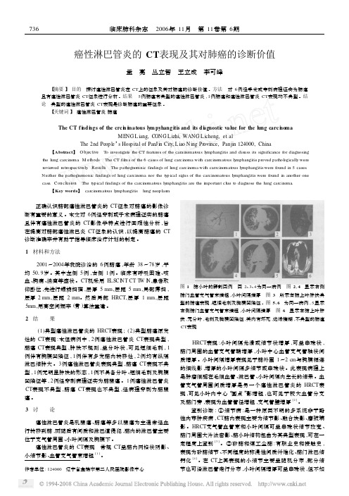
【关键词】 癌性淋巴管炎肺癌The CT f i n d i n gs of the crc i n i m a tous ly m pyhang itis and its d i a gnosti c va lue for the lung carc i n o maME NG L iang,C ONG L izhi,WANG L icheng,et alThe 2nd Peop le ’s Hos p ital of PanJ in City,L iao N ing Pr ovince,Panjin 124000,China 【Abstract 】 O bjective To investigate the CT features of the carainomat ous ly mphangitis and disuss its significance for diagnosingthe lung carcinoma .M ethods The CT fil m s of the 6cases of lung carcinoma with carcinomat ous ly mphangitis p r oved pathol ogically were revie wed retr os pectively .Resu lts The pathognomnic findings of lung carcinoma with carrcinomat ous ly mphangitis were f ound in 5cases .Neither the pathognomonic findings of lung carcinoma nor the typ ical signs of the carcinomat ous ly mphangitis were found in another one case .Conclusion The typ ical findings of the carcinomat ous ly mphangitis are the i m portant clue t o diagnose the lung carcinoma .【Key words 】 carcinomat ous ly mphangitis lung neop las m作者单位:124000 辽宁省盘锦市第二人民医院影像中心 正确认识肺部癌性淋巴管炎的CT 征象对肺癌的影像诊断有重要的意义。

肺癌性淋巴管炎16例临床分析及治疗体会目的探讨并总结肺内癌性淋巴管炎(pulmonary lymphangitic carcinomatosis,PLC)的临床特征,初步探讨大剂量糖皮质激素(甲强龙)对PLC的治疗作用。
方法分析2010~2014年我院16例肺癌性淋巴管炎的临床特征,将16例患者随机分为对照组和治疗组,对照组予常规对症治疗治疗组在对照组的基础上给予大剂量的甲强龙静脉注射(80 mg q8h),1周后判定疗效。
结果本组PLC原发肿瘤以腺癌居多,影像学表现:除可见从肺门向肺野呈放射状、条索状不均一阴影,部分条索可达胸膜并见小结节或肺野呈毛玻璃状改变;纵隔、肺门淋巴结肿大(56.25%)外,胸腔积液(75.00%)所占比例较大。
治疗组在给予吸氧等一般治疗的基础上给予大剂量激素治疗后,呼吸困难、活动能力、氧分压较对照组有所改善(P<0.05);但肺弥散功能无改善,且二氧化碳分压无明显降低(P>0.05)。
结论PLC 常发生于肺癌、乳腺癌、大肠癌、胃癌等肿瘤的肺内转移。
当患者出现上述肺间质病变,呼吸道症状经气道解痉药治疗无效,病情急进性发展时,应高度怀疑PLC,可尝试使用大剂量激素缓解临床症状。
[Abstract] Objective To evaluate and summarize the clinical features of pulmonary lymphangitic carcinomatosis (PLC)and discuss the treatment effects of large dose corticosteroids (methylprednisolone)on PLC. Methods The clinical features of 16 patients with PLC treated in our hospital from 2010 to 2014 were analyzed. The 16 patients were randomly divided into the control group and the treatment group. The control group was treated with conventional symptomatic treatment. The treatment group was given intravenous injection of large dose methylprednisolone(80 mg q8h)on the basis of the control group. The efficacy was evaluated after 1 week. Results All the PLC primary tumors was mainly adenocarcinoma and the imaging manifestations were as follows:In addition to the visible radial-pattern and cord-like shadow ranging from lung hilum to lung filed,some cords could reach pleura and the nodules were observed;or the lung filed presented frosted glass-like changes;in addition to lymphadenectasis(56.25%)of mediastinum and lung hilus,pleural effusion (75.00%)accounted for a larger proportion. In treatment group,large dose hormone was given on the basis of ordinary treatment such as oxygen uptake,after which the breathing difficulty,capacity of action and oxygen partial pressure improved than the control group(P<0.05);but pulmonary diffusion function did not improve and carbon dioxide partial pressure did not reduce significantly(P>0.05). Conclusion PLC often occurs in the lung metastasis of lung cancer,breast cancer,colorectal cancer and gastric cancer. When patients have the above interstitial lung disease,airway antispasmodic treatment is ineffective on respiratory tract symptoms,and the disease develops rapidly,PLC is highly suspicious. Large dose hormone should be tried to relieve clinical symptoms.。

158例肺癌性淋巴管炎的CT诊断【摘要】目的总结肺癌性淋巴管炎的CT表现,提高对该病的认识和诊断准确率。
方法收集经高分辨率CT检查并病理证实的肺内癌性淋巴管炎患者158例,回顾分析其CT表现。
结果全部病例均表现为不规则小叶间隔增厚,小叶间隔上常可见小结节影;支气管血管束不规则增厚,可呈串珠状或结节状阴影;小叶中心结构的增厚可造成次级肺小叶中心的蜘蛛样改变;斜裂增厚,常呈不规则结节性增厚;胸膜增厚及胸腔积液。
小叶间隔不规则增厚143例,右肺93例,左肺43例,两肺22例,胸腔积液98例,肺门、纵隔淋巴结增大28例,肺内多发小结节5例。
结论癌性淋巴管炎的CT表现具有一定的特征性表现,但需要与弥漫性线网状影的其他疾病鉴别,如结节病、间质性肺水肿等。
【Abstract】Objective To summarize the CT characteristics of pulmonary lymphangitic carcinomatosis Methods High resolution CT was performed in 158patients with histologically proved pulmonary lymphangitic carcinomatosis.Retrospective analysis of the CT.ResultsAll cases showed irregular interlobular septal thickening, the small nodules were often seen on the interlobular septum.Irregular thickening of bronchovascular bundles with beads or nodules was showed.The centrilobular structure could cause central spider like changes in secondary pulmonary lobules.Major fissure presented irregular nodular thickening with pleural effusion Interlobular septal thickening was seen in 143 cases, 93 lesions were in the right lung, 43 lesions in the leftlung, 22 cases in two lungs, pleural effusion in 98 cases, hilar and mediastinal lymph node enlargement in 28 cases, pulmonary nodules in 5 cases Conclusion Pulmonary lymphangitic carcinomatosis can show certain characteristic featureson CT,but the need to diffuse reticular shadow line identificcdion of other diseases,such as sarcoidosis,interstitial pulmonary edema.【Key words】Pulmonary lymphangitic carcinomatosis; Interlobular septum; Neoplasm; Tomography, X ray computed肺癌性淋巴管炎(PLC)也称淋巴道转移瘤、淋巴管癌病等,是肺内外肿瘤肺内转移的一种特殊类型,肺癌是引起PLC的最常见的肿瘤,其次是乳腺癌、胃癌、胰腺癌、直肠癌、肾癌、宫颈癌、食管癌、前列腺癌和未知原发部位的腺癌[1,2]。
158例肺癌性淋巴管炎的 CT诊断框架【摘要】目的:探究158例肺癌性淋巴管炎的CT诊断框架。
方法:选取2019年5月-2020年7月在本院确诊的158例肺癌性淋巴管炎患者,采用随机抽样法将上述患者分为对照组和实验组,每组患者各79例,对照组患者采取X线检查,实验组患者采取CT检查,对比两组检查方式的误诊率和患者满意度。
结果:经比较后,实验组的检查效果和患者满意度明显优于对照组,P<0.05,具有统计学意义。
结论:对于肺癌性淋巴管炎的患者实施CT检查诊断,既可以提高患者满意度,有可以提升检查效率,对临床应用具有重要意义。
【关键词】肺癌性淋巴管炎;CT;诊断框架肺癌性淋巴管炎又名为肺癌性淋巴管扩散,其发病机制是肿瘤细胞在淋巴管内发生转移引起的癌症病变,是肿瘤细胞转移的一种特殊机制。
该疾病最常见于肺部转移,其转移方式有两种,其中一种为肿瘤细胞由淋巴干道逆行与淋巴结,导致肺门淋巴结发生反流[1]。
第二种为肿瘤细胞在淋巴管内生长繁殖。
由于该疾病的表现特征与其他肺间质病变具有相似之处,会出现误诊的情况[2]。
所以,正确有效的检查方式是极为必要的。
因此,文章选取158例在院治疗患者,将上述患者采取随机抽样法分为对照组和实验组,分别采取不同的检查方式进行检查,对比两组患者的检查效果和患者满意度,具体报道如下:1、资料与方法1.1一般资料选取2019年5月-2020年7月在本院确诊的158例肺癌性淋巴管炎患者,采用随机抽样法将上述患者分为对照组和实验组,每组患者各79例,对照组患者男性60例,女性19例,年龄范围65-78岁,平均年龄(71.5±6.5)岁;实验组患者男性53例,女性26例,年龄范围67-70岁,平均年龄(68.5±1.5)岁,对比两组检查方式的检查效率和患者满意度。
两组患者一般资料经比较无差异,P>0.05,具有可比性。
1.2方法1.2.1对照组:对照组患者采取X线检查方式,具体步骤为认真核对受检者个人资料,明确适应部位,确定摄影位置,摄影前去除可影响图像的质量的金属物品和电子产品,做好X线正确防护,根据拍摄部位摆好相应体位,将中心线对准摄影部位中心,指导患者正确呼吸,最后确定图像后进行打印和传输。