血管生成
- 格式:pdf
- 大小:1.76 MB
- 文档页数:8

血管生成及其在血管疾病中的作用血管生成(vascularization)是指机体通过增生和扩张血管来满足部位代谢需求的生理过程,与许多疾病关系密切,如肿瘤、缺血性疾病、糖尿病视网膜病变、退行性病变等。
在血管生成的过程中,内皮细胞(ECs)和周围的细胞相互作用,涉及一系列信号转导途径,最终完成新的血管形成。
本文将重点讨论血管生成的机制及其在血管疾病中的作用。
一、血管生成的机制血管生成过程中,众多生长因子和细胞间粘附分子参与之间的交互作用。
主要的生长因子有:血管内皮生长因子(VEGF)、基本成纤维细胞生长因子(bFGF)、转化生长因子-β(TGF-β)等。
血管生成的过程也分为多个步骤。
1. 表达生长因子的细胞增生生长因子的表达能够使原来休眠状态的血管内皮细胞分裂增生,由内到外形成管壁中的细胞层。
VEGF被认为是调控血管生成的主要因素,它能够在受损组织中派生的内皮细胞上调节细胞增殖和存活。
2. 上皮细胞摆布在组织细胞增生后,细胞间的相互作用将使得新的内皮细胞持续扩张。
细胞间粘附分子(CAMs)参与了这个过程。
CAMs是细胞膜表面的蛋白质分子,它们通过特异性调节相邻细胞的黏着力,从而形成管腔。
3. 细胞向管腔移动细胞的移动和分化则是组成血管壁的细胞的最后一个步骤。
随着细胞的重组以及来自其他细胞类型的信号作用,内皮细胞的最终形态从伸缩形态转化为沟壑(lumenize)形态,并形成成熟的管腔。
二、血管生成在血管疾病中的作用血管生成在血管疾病中的作用主要表现为增加其疾病的发展和恶化。
1. 肿瘤血管生成肿瘤细胞生长需要营养,此过程需要透过血管发生。
肿瘤血管的生成、增生和扩散过程十分复杂,该过程中包括了对生长因子和细胞间粘附分子的调节。
肿瘤血管破坏是一种新的治疗方案,它能延长患者的存活期和消除体内的循环小突变体。
2. 缺血性疾病中的血管生成冠心病(CAD)是缺血性疾病的一种表现,其发病率也随着周期血管生成的时间增长而增加。

血管生成相关基因血管生成是指在生物体内形成新的血管,以供应氧气和营养物质。
这个过程对于维持正常生物体的生长和发育至关重要。
血管生成是一个复杂的过程,涉及许多基因的调控和相互作用。
在血管生成过程中,许多基因起到关键作用。
其中,一些基因参与血管内皮细胞的增殖、迁移和分化,调控血管的形成和分支。
另一些基因则参与血管平滑肌细胞的生长和收缩,从而调节血管的功能。
例如,血管内皮生长因子(VEGF)家族是血管生成过程中最为重要的调节因子之一。
VEGF家族成员包括VEGF-A、VEGF-B、VEGF-C和VEGF-D 等。
这些因子通过结合它们的受体,如VEGFR-1和VEGFR-2等,促进血管内皮细胞的增殖和迁移,从而促进新血管的形成。
除了VEGF家族,还有其他一些重要的基因参与血管生成过程。
例如,基本纤维生长因子(bFGF)家族和血小板衍生生长因子(PDGF)家族也起到重要的调节作用。
这些因子通过与它们的受体结合,调控血管内皮细胞和平滑肌细胞的增殖和分化,从而促进血管的形成。
此外,一些转录因子也参与血管生成的调控。
例如,转录因子HIF-1(hypoxia-inducible factor 1)在低氧条件下被激活,促进VEGF的表达,从而刺激血管生成。
另外,转录因子FOXO(forkhead box O)家族也参与血管生成过程的调控,通过调节一系列基因的表达,影响血管内皮细胞的增殖和迁移。
总的来说,血管生成过程涉及许多基因的调控和相互作用。
这些基因通过调节血管内皮细胞和平滑肌细胞的增殖、迁移和分化,参与新血管的形成。
对于进一步理解血管生成的机制和开发相关疾病的治疗方法,深入研究这些血管生成相关基因具有重要意义。

血管生成与血管新生的差异血管生成和血管新生是两个不同的过程,尽管它们都涉及到血管的形成。
在生物学中,血管生成是指在胚胎发育过程中形成全新的血管,而血管新生是指已有的血管再生,通常是由于治疗效果不佳或疾病进展而导致的。
血管生成和血管新生都是非常重要的生物学过程,对于维持整个身体的健康有很大的影响。
血管生成是胚胎发育过程中最早的生理过程之一。
在胚胎发育的早期,胚胎的细胞开始形成血管。
血管生成包括两个过程:血管内皮细胞的分化和血管形成。
血管内皮细胞是一种特殊的细胞,它们形成血管壁,从而可以将血液输送到身体的各个部位。
血管形成是指血管内皮细胞的组合形成血管。
血管生成是一种复杂的生物学过程,它涉及到多个不同的信号通路和细胞类型。
相比之下,血管新生发生在成年人身上。
当身体受到损伤或疾病时,血管新生会开始发生。
例如,如果一个组织受到损伤或失去血液供应,身体会通过血管新生重新建立血液供应。
血管新生是一种重要的恢复机制,它可以使身体快速恢复健康。
血管生成和血管新生有很多特点不同。
首先,血管生成是一个早期的过程,它开始在胚胎发育的早期出现。
相比之下,血管新生发生在成年人身上,通常是由于某种原因导致的。
其次,血管生成涉及到特定的信号途径和细胞类型,而血管新生通常是由于一般情况下血管内皮细胞被激活。
最后,血管生成是一种改变身体结构的过程,而血管新生通常只是对现有结构的修复。
尽管血管生成和血管新生有很多不同之处,但它们都是非常重要的生物学过程,对于身体的健康具有至关重要的意义。
对血管生成和血管新生的研究可以帮助我们 better understand how the body develops and repairs itself. 这些研究有望为我们治疗各种疾病提供新的方向。

血管生成生物标志物检测及其临床意义引言:血管生长是一种复杂的生物过程,涉及多种细胞、生理和生化因素的相互作用。
血管生成在许多疾病中起到关键作用,例如肿瘤的生长和转移,而且在组织再生和修复过程中也起到重要的作用。
因此,了解血管生成的机制以及发现可以用于监测和预测相关疾病的生物标志物具有重要的临床意义。
一、血管生成的机制和调控因素血管生成是指新的血管形成过程,它包括血管内皮细胞的增殖、迁移和管腔形成,以及血管周细胞(如平滑肌细胞和间充质细胞)的招募和分化。
血管生成的过程受到多种生理和病理因素的调控。
一种重要的调控机制是血管生成因子及其受体的信号通路。
血管内皮生长因子(VEGF)家族是调节血管生成最重要的因子之一。
VEGF 通过与其受体(如VEGFR-2)结合,激活下游信号通路促进内皮细胞的增殖和迁移。
除了VEGF家族外,其他因子如血小板源性生长因子(PDGF)、基础纤维生长因子(bFGF)等也参与血管生成的调控。
另一个重要的调控机制是血管生成抑制因子的作用。
如血管抑制蛋白1(Angiostatin-1)和血管抑制蛋白2(Angiostatin-2)通过干扰VEGF的信号通路,抑制血管内皮细胞的增殖和迁移,从而抑制血管生成过程。
二、血管生成生物标志物的分类和检测方法血管生成生物标志物是指可以用于检测和评估血管生成过程的生物标志物。
根据其来源和类型的不同,血管生成生物标志物可以分为细胞因子、激素、蛋白质、基因等多种类型。
最常用的血管生成生物标志物是细胞因子,如VEGF、PDGF、bFGF等。
这些细胞因子在血液和组织中的水平可以通过酶联免疫吸附实验(ELISA)或其他免疫检测方法进行测定。
这些方法具有高灵敏度和特异性,可以用于评估血管生成的活性以及疾病的进展和预后。
除了细胞因子,一些激素如雌激素和甲状腺素在血管生成中也发挥重要作用。
对这些激素的水平进行检测可以了解其对血管生成的调节作用。
蛋白质是细胞生物学中的重要参与者,在血管生成过程中也发挥着重要的作用。
1 血管生成相关机制1.1 新血管的形成与结构一个细胞需要生存,则必须围绕血管,即靠近血管约100~200μm。
这个距离是氧气弥散距离的极限。
如果没有血管供应,单个肿瘤的极限体积大小介于0.2~3mm之间,依肿瘤细胞来源不同而大小有所差异。
处于这个极限大小下,肿瘤细胞的增生与死亡达到平衡。
如果肿瘤想扩大自身体积,则必需求助于新生血管。
血管生成过程实际上就是沿着血管排列的血管内皮细胞增殖过程。
血管内皮细胞是人体内寿命最长的细胞之一。
正常情况下,它们每七年才分裂增殖一次。
如果照这种速度产生新生血管,那么血管的更新过程则非常缓慢,所以该过程一定得加快。
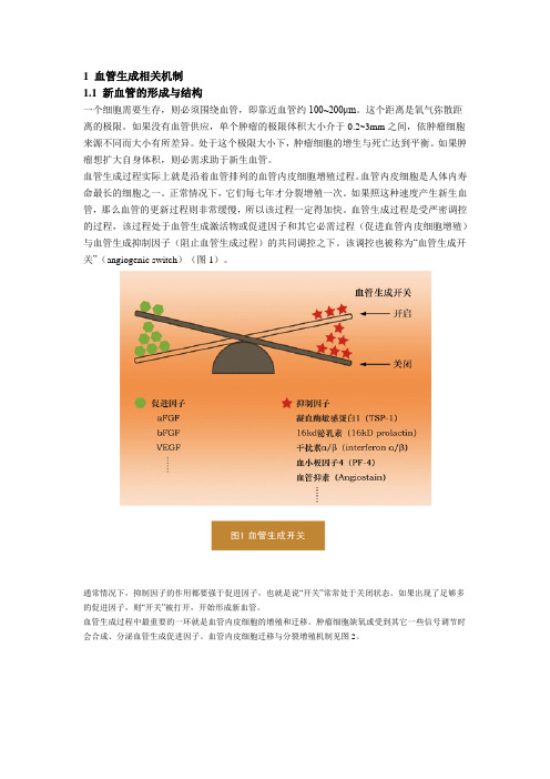
血管生成过程是受严密调控的过程,该过程处于血管生成激活物或促进因子和其它必需过程(促进血管内皮细胞增殖)与血管生成抑制因子(阻止血管生成过程)的共同调控之下。
该调控也被称为“血管生成开关”(angiogenic switch)(图1)。
通常情况下,抑制因子的作用都要强于促进因子,也就是说“开关”常常处于关闭状态。
如果出现了足够多的促进因子,则“开关”被打开,开始形成新血管。
血管生成过程中最重要的一环就是血管内皮细胞的增殖和迁移。
肿瘤细胞缺氧或受到其它一些信号调节时会合成、分泌血管生成促进因子。
血管内皮细胞迁移与分裂增殖机制见图2。
肿瘤细胞分泌的内皮细胞生长因子等物质与血管内皮细胞上的受体分子结合,刺激其释放蛋白水解酶(proteolytic enzyme)。
该蛋白水解酶可以降解血管周围的基质。
这样,为血管内皮细胞的迁移和进一步分裂做好了准备。
血管内皮细胞经过不断的分裂增殖以及向前迁移,逐渐形成管状结构,最终形成新生血管。
由于肿瘤组织中生成新生血管的过程没有受到严密调控,因而肿瘤组织中的新生血管与正常组织中的新生血管在结构上差异明显。
肿瘤组织中的新生血管非常不规则,有很多分支和旁路。
血管不完全由血管内皮细胞构成,有些地方的管壁竟然由肿瘤细胞覆盖而成。
血管的通透性非常高,因为没有正常的基底膜围绕在血管周围,血管内皮细胞间的连接非常少。
血管生成实验步骤及方法
一、技术简介
血管生成(Angiogenesis)是指源于已存在的毛细血管和毛细血管后微静脉新的毛细血管性血管的生长。
无论原发性肿瘤还是继发性肿瘤,一旦生长直径超过1-2 mm,都会有血管生成。
这是由于肿瘤细胞自身可分泌多种生长因子,诱导血管生成。
小管形成实验是测量在体外血管生成一种快速的,可量化的方法。
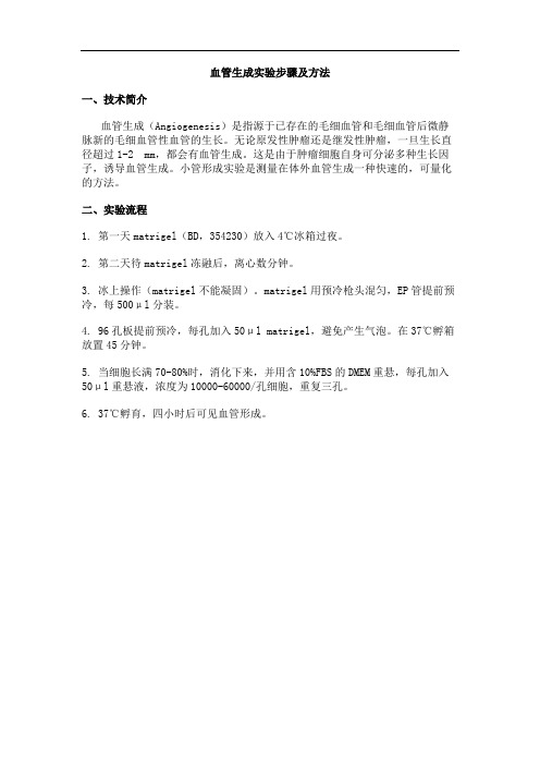
二、实验流程
1. 第一天matrigel(BD,354230)放入4℃冰箱过夜。
2. 第二天待matrigel冻融后,离心数分钟。
3. 冰上操作(matrigel不能凝固)。
matrigel用预冷枪头混匀,EP管提前预冷,每500μl分装。
4. 96孔板提前预冷,每孔加入50μl matrigel,避免产生气泡。
在37℃孵箱放置45分钟。
5. 当细胞长满70-80%时,消化下来,并用含10%FBS的DMEM重悬,每孔加入50μl重悬液,浓度为10000-60000/孔细胞,重复三孔。
6. 37℃孵育,四小时后可见血管形成。
血管生成与复杂疾病的关系随着医学技术的不断进步,越来越多的研究表明血管生成与复杂疾病之间存在着密切的关系。
血管生成是细胞和组织形成新血管的过程,它不仅涉及正常的生理过程,还与许多疾病的发生和发展密切相关。
本文将探讨血管生成与复杂疾病的关系。
一、血管生成在生理和疾病中的作用血管生成是一个复杂的生理过程,它在身体健康和稳定状态下发挥着非常重要的作用。
正常情况下,血管生成为组织和器官提供充足的氧、营养物质和免疫细胞,同时帮助排出代谢产物和毒素。
在生理过程中,血管生成的发生和调节可以通过多种信号通路进行,如血管内皮生长因子(VEGF)和转化生长因子-β(TGF-β)等。
然而,当机体处于一些病理状态下,血管生成的过程会被协同或受到干扰,从而进一步影响生理和疾病过程。
例如,在肿瘤生长和转移中,血管生成过程被肿瘤细胞激活并参与肿瘤血供的形成。
在慢性炎症和自身免疫反应中,血管生成被细胞因子和介质介导,包括炎症介质和化学因子等。
总的来说,血管生成在疾病中作为定量和预测等方面的指标被广泛研究。
二、血管生成与糖尿病、动脉粥样硬化等复杂疾病的关系随着公共健康和医疗技术的提高,复杂疾病的预防和治疗已成为医学界的重要研究方向之一。
血管生成与复杂疾病之间的关系更加引人注目。
以下是一些常见复杂疾病和血管生成的关系。
糖尿病是一种由胰岛素过少或胰岛素抵抗导致的疾病。
近年来的研究表明,糖尿病对血管生成的影响非常大。
糖尿病的发生和发展可以增加全身炎症和氧化应激,从而大大抑制下皮革细胞和内皮细胞的增殖和血管生成。
这不仅会影响组织和器官的正常生理功能,还可能导致严重的并发症,如糖尿病足和糖尿病视网膜病变等。
动脉粥样硬化是一种常见的心血管疾病,它由血管内皮细胞增生和斑块形成引起。
有研究表明,血管生成对动脉粥样硬化的发生和发展发挥了重要作用。
在动脉粥样硬化的病变大约三分之一的区域内,血管生成活跃。
临床上,血管生成的程度可以被用作动脉粥样硬化的评估指标,同时也可以预测心血管事件的风险。
第1篇一、实验目的1. 了解血管生成的基本原理和过程。
2. 掌握体内成血管实验的方法和步骤。
3. 分析实验结果,探讨血管生成过程中的关键因素。
二、实验原理血管生成是指血管新生的过程,包括血管形成和血管再生两个阶段。
血管生成在生理和病理过程中都具有重要意义,如组织修复、肿瘤生长等。
本实验采用体内成血管实验,通过构建一个模拟血管生成的模型,观察血管生成的过程,分析影响血管生成的因素。
三、实验材料1. 实验动物:C57BL/6小鼠,雌雄各5只,体重20-25g。
2. 实验试剂:血管内皮生长因子(VEGF)、血小板衍生生长因子(PDGF)、肝素钠、透明质酸酶等。
3. 实验仪器:显微镜、手术器械、组织切片机、凝胶成像系统等。
四、实验方法1. 术前准备:实验动物术前禁食12小时,自由饮水。
实验前1小时给予肝素钠0.5ml静脉注射,以防止血栓形成。
2. 手术操作:(1)将小鼠麻醉,消毒腹部皮肤。
(2)沿腹部正中线切开皮肤,暴露腹壁。
(3)将腹壁两侧的肌肉组织分离,暴露腹主动脉。
(4)在腹主动脉下端用细线结扎,形成一段无血管区域。
(5)将VEGF、PDGF、肝素钠和透明质酸酶混合均匀,注入无血管区域。
(6)缝合腹壁,无菌敷料包扎。
3. 观察与记录:(1)术后第3天、第7天、第14天,观察小鼠腹部皮肤颜色、质地及血管生成情况。
(2)取小鼠腹主动脉、肝、脾、肾等组织,进行组织切片。
(3)用显微镜观察组织切片,记录血管生成情况。
五、实验结果1. 术后第3天,观察小鼠腹部皮肤颜色正常,质地柔软,无异常。
2. 术后第7天,观察小鼠腹部皮肤颜色开始变红,质地变硬,血管生成明显。
3. 术后第14天,观察小鼠腹部皮肤颜色红润,质地柔软,血管生成更为明显。
4. 组织切片观察结果显示,实验组小鼠腹主动脉、肝、脾、肾等组织均出现血管生成现象,与对照组相比,血管密度明显增加。
六、实验讨论1. 体内成血管实验结果表明,VEGF、PDGF、肝素钠和透明质酸酶等试剂能够促进血管生成。
血管生成与血管疾病的研究进展血管是组成人体循环系统的重要结构之一,负责输送营养物质、氧气和代谢产物,保障身体各器官的正常功能。
在人类发育和生长过程中,不仅需要维持正常的血管功能,还需要不断产生新的血管,满足身体各部位的不同需求。
血管生成是一种复杂而精密的生物学过程,涉及多种细胞类型、信号通路和基因调控。
因此,对血管生成的研究一直是生物医学领域的热点之一。
血管生成的主要机制包括血管内皮细胞(EC)增殖、分化及空隙扩张、血管平滑肌细胞(SMC)增殖和迁移、上皮细胞差异化和提供支持性物质(如基质蛋白和生长因子等)。
理解这些生物学过程与机制对于治疗多种血管疾病(如心肌梗死、冠心病、癌症、眼睛疾病等)至关重要。
在过去几十年中,血管生成研究开拓了许多新领域,特别是在心血管系统、肿瘤和眼睛等领域。
值得一提的是,生长因子(GF)在这一领域中扮演了核心角色,这些因子可与EC和其它细胞类型结合,并激活特定的信号通路,进而对血管生成的不同阶段产生积极或消极的调节作用。
GF的研究依托于细胞的展种体系、动物模型及人类临床实验,在基础及应用角度产生了许多具有突破性的成果。
目前,人们广泛关注的是血管生成在肿瘤和心脑血管疾病中的作用。
在肿瘤领域中,性状多样的GF和肿瘤诱导因子(TIS)在肿瘤血供新生中起重要作用。
一些药物及干预手段,如血管内皮生长因子抑制剂和血管内皮生长因子受体阻断剂,已经通过了临床试验并获得了批准使用。
在心脑血管疾病方面,研究者们尝试通过生物材料、基因治疗和细胞治疗等方法去促进新血管的生成,改善低氧缺血的症状和治疗梗死等严重疾病。
另一方面,血管疾病也是血管生物学领域中一个备受关注的问题,包括心血管疾病、眼睛疾病等。
研究者们已经将重心从治疗向预防转移,利用基因库、高通量筛选、体内及体外模型等技术寻找与各类疾病相关的新分子标记和化合物。
通过研究血管生成机制,发展替代性疗法和生物标记物,有望为疾病预防和治疗提供新思路。
血管生成的分子机制和致病途径血管生成,指在生物体内形成和发育血管的过程,是维持器官组织正常发育、生长和修复的基础。
血管生成的分子机制和致病途径受到学术界广泛关注。
本文将就此进行阐述。
一、血管生成分子机制血管生成主要由内皮细胞(ECs)和间充质细胞(SMCs)共同完成,其中ECs促进肿瘤等形成并维持组织血量,SMCs则为血管提供支持。
两者间的交互作用极其复杂,血管生成的过程也受到许多因素的影响,如生长因子、细胞因子和细胞外基质。
1. 生长因子生长因子分为成纤维细胞生长因子(FGFs)、血小板衍生生长因子(PDGFs)、肿瘤坏死因子(TNF)、表皮生长因子(EGFs)等多种类型,这些因子对血管生成发挥着重要的作用。
(1)VEGFVEGF是维持ECs生存、促进血管造影及突破的主要因素,同时也是血管生成研究中最常见和研究最多的分子。
VEGF作用靶点主要为ECs,通过结合VEGFR-2促进ECs增殖并抑制ECs凋亡,最终促进血管内皮细胞相互作用和管腔形成。
(2)PDGFPDGF可能促进新生血管散在生长,在肿瘤等组织中起到重要作用。
PDGF受体β(PDGFRβ)和Pericytes相互作用调控了血管的生长、分化和维持。
PDGF可增强Pericytes的能力,使其成为血管管壁上的支持细胞,这对血管的稳定性和功能的维持至关重要。
(3)bFGFbFGF是一种钙离子相关的低分子质量生长因子,主要作用为调控ECs的生理功能并参与血管发生及快速增生。
bFGF可通过直接作用于细胞膜受体而刺激ECs 增殖、迁移、生存和血管内皮通透性的调节,从而促进血管生长。
(4)TGF-βTGF-β是细胞因子家族中的一员,可促进Pericytes分化形成,并维持其在血管管壁上的功能。
在ECs上,TGF-β可抑制乙型硝酸化酶表达,从而限制NO和cyclic GMP的产生,进而限制血管扩张。
TGF-β还能促进血管内皮细胞和纤维母细胞增殖,从而促进血管生长。
Cell MetabolismShort ArticleHypoxic Regulation of Glutamine Metabolism through HIF1and SIAH2Supports Lipid Synthesis that Is Necessary for Tumor GrowthRamon C.Sun1and Nicholas C.Denko1,*1Department of Radiation Oncology,James Cancer Hospital and Comprehensive Cancer Center,Ohio State University Wexner School of Medicine,Columbus,OH43210,USA*Correspondence:nicholas.denko@/10.1016/j.cmet.2013.11.022SUMMARYRecent reports have identified a phenomenon by which hypoxia shifts glutamine metabolism from oxidation to reductive carboxylation.We now identify the mechanism by which HIF-1activation results in a dramatic reduction in the activity of the key mito-chondrial enzyme complex a ketoglutarate dehydro-genase(a KGDH).HIF-1activation promotes SIAH2 targeted ubiquitination and proteolysis of the 48kDa splice variant of the E1subunit of the a KGDH complex(OGDH2).Knockdown of SIAH2or mutation of the ubiquitinated lysine residue on OGDH2 (336KA)reverses the hypoxic drop in a KGDH activity, stimulates glutamine oxidation,and reduces gluta-mine-dependent lipid synthesis.336KA OGDH2-expressing cells require exogenous lipids or citrate for growth in hypoxia in vitro and fail to grow as model tumors in immunodeficient mice.Reversal of hypoxic mitochondrial function may provide a target for the development of next-generation anticancer agents targeting tumor metabolism. INTRODUCTIONGlutamine is the most abundant amino acid in blood and is second only to glucose as a carbon source for energy production and anabolic processes.Recently,glutamine was identified as being essential for fuelling mitochondrial metabolism in rapidly dividing cancer cells transformed with either c-MYC(Gao et al.,2009;Wise et al.,2008)or Kirsten rat sarcoma viral oncogene homolog(Son et al.,2013).Inhibiting glutamine meta-bolism has been proposed as an anticancer therapy(Wise and Thompson,2010).Mitochondrial glutamine metabolism can follow either oxida-tive or reductive pathways(Holleran et al.,1995).Mitochondrial utilization of glutamine begins with a two-step conversion of glutamine to a ketoglutarate(a KG),typically by glutaminase and glutamate dehydrogenase.a KG can be either oxidized by a KG dehydrogenase(a KGDH)to succinate(standard tricarbox-ylic acid[TCA]cycle reaction)or reductively carboxylated by iso-citrate dehydrogenase(reverse TCA cycle)to isocitrate and then citrate.The reductive cycle has been shown to be favored in cells where HIF-1a is stabilized(Metallo et al.,2012b;Wise et al., 2011)or in cells with compromised electron transport capacity (Mullen et al.,2012).Glutamine-derived citrate can be trans-ported to the cytoplasm in order to generate acetyl CoA for anabolic processes such as fatty acid synthesis(Gameiro et al.,2013b;Metallo et al.,2012b).a KGDH consists of E1(oxogluterate dehydrogenase,OGDH), E2(dihydrolipoamide S-succinyltransferase,DLST),and E3(di-hydrolipoamide dehydrogenase,DLD)components that collec-tively convert a KG to succinyl-CoA and nicotinamide adenine dinucleotide(Patel and Harris,1995).The three-enzyme(E1-E2-E3)dehydrogenase complex structure is conserved in the pyruvate dehydrogenase(PDH)and branched chain a-ketoacid dehydrogenase complexes.PDH serves as the major entry point for glucose-derived pyruvate into the mitochondrial TCA cycle (Patel and Korotchkina,2001).Recent work has found that HIF1activation inhibits PDH,which blocks glucose oxidation (Kim et al.,2006;Papandreou et al.,2006).In humans, there are three major splice variants of the OGDH gene,V1 (114kDa),V2(48kDa),and V3(114kDa),although the functional differences of these enzymes remain unreported. Environmental hypoxia has a major effect on gene expression, largely by the induction of the HIF a family of transcription factors through protein stabilization(Epstein et al.,2001).However, protein destabilization by hypoxia-regulated ubiquitination (Vin˜as-Castells et al.,2010)or SUMOylation(Comerford et al., 2003)has also been reported.The E3ubiquitin ligase SIAH2 destabilizes several critical proteins in hypoxia(Qi et al.,2008). In this work,we connect the crucial HIF-1and SIAH2regulatory systems that control the metabolism of glutamine.Furthermore, we show that this circuit is necessary for the growth of cells in hypoxia and as model tumors.RESULTSHIF1Stabilization Is Necessary and Sufficient to Reduce Mitochondrial Glutamine Oxidation and a KGDH Activity Hypoxia inhibits mitochondrial glucose oxidation(Denko,2008; Kim et al.,2006),so we asked whether it could also inhibit glutamine oxidation.We found reduced mitochondrial oxygen consumption(OCR)in the head and neck tumor cell line SAS when they were treated with the HIF-stabilizing PHD inhibitor DMOG.This decrease was detected in complete basal, glucose-only,or glutamine-only media(Figure1A).We alsofound that VHL-negative renal clear cell cancer (RCC4)cells that have constitutive HIF1activation show reduced glucose and glutamine oxidation.Reintroduction of VHL into these cells restores the hypoxic regulation of HIF-1,increases normoxic mitochondrial OCR and restores its reduction in response to DMOG (Figure 1B).These experiments determined that HIF1a stabilization was sufficient to inhibit glutamine oxidation.Next,we asked whether HIF1a was necessary with the use of small hairpin RNA (shRNA)knockdown.Fig-ure 1C shows that HIF1a is essential,given that its knockdown in SAS cells blocked DMOG-dependent reduction in glutamine oxidation.Glutamine oxidation can be inhibited at any of the steps involved in its uptake or conversion to a KG.However,the addi-tion of a KG failed to restore mitochondrial OCR in DMOG (Figure S1A available online),suggesting that the block is at a KGDH itself.Therefore,we measured a KGDH enzyme activity in SAS cells and found approximately 60%reduction upon HIF stabilization,similar to the reduction in PDH activity (Figures 1D and 1E).RCC4cells have low-level a KGDH activity that is insensitive to hypoxia,and VHL reintroduction increases nor-moxic a KGDH activity and restores its hypoxic response (Fig-ure 1E).Similar HIF-responsive reductions in a KGDH and PDHactivity were seen in RKO colorectal cancer and MiaPaca2pancreatic cancer cells (Figures S1B and S1C).However,in the SAS cells with shRNA to HIF1a ,there was greatly reduced decrease in PDH (Figure 1F)and no decrease in a KGDH activity (Figure 1F).There was very modest hypoxic regulation of gluta-mate dehydrogenase (Figure S1D),NADP-dependent isocitrate dehydrogenase (Figure S1E),or carboxylase (Figure S1F),sug-gesting that these activities were not rate limiting.OGDH2Is Marked for Proteolysis in Hypoxia by SIAH2-Dependent UbiquitinationHIF-1control of a KGDH suggested that there is a HIF-1-respon-sive gene whose product interacts with a KGDH in order to inhibit it.However,gene expression studies have not identified such a gene.Therefore,we examined the a KGDH protein subunits.There was a modest hypoxia-dependent decrease in the level of E2(DSLT)but no change in E3(DLD)or in the larger splice variants of OGDH (114kDa proteins 1and 3).However,we found that hypoxia caused the complete loss of the small protein derived from splice variant OGDH2(Figure 2A).Treatment of hypoxic SAS cells with the proteasome inhibitor MG132restored OGDH2protein levels (Figure 2A).Similarly,hypoxia specifically reduced OGDH2in RKO and MiaPaca2cells,and RCC4cellsFigure 1.Hypoxia Downregulates Glutamine Oxidation(A)Mitochondrial oxygen consumption (OCR)in control and DMOG-treated SAS cells (500m M,16hr).OCR was measured in basal DMEM without serum and containing only 5mM glucose,only 2mM glutamine,or both as indicated.(B)OCR in VHL-deficient RCC4(constitutively active HIF1)and RCC4cells with VHL reintroduced treated as in (A).(C)OCR in empty vector SAS or shHIF1a SAS treated as in (A).(D)Pyruvate dehydrogenase (PDH)enzyme activity in SAS,RCC4,and RCC4-VHL cells in control conditions or after 16hr of 0.5%oxygen or 500m M DMOG.(E)a KGDH enzyme activity in SAS,RCC4,and RCC4-VHL cells treated as in (D).(F)PDH activity in shHIF1a SAS cells treated as in (D).(G)a KGDH activity in shHIF1a SAS cells treated as in (D).Data represent three independent replicates ±SD.See also Figure S1.Cell MetabolismHypoxia Downregulates a KGDH Activityhave very low levels of OGDH2that could be increased with MG132(Figure S2A).a KGDH E2has been reported to be marked for proteolysis by the E3ubiquitin ligase SIAH2upon mitochondrial disruption (Ha-belhah et al.,2004);therefore,we examined whether SIAH2could be responsible for the hypoxic destruction of OGDH2.We found that the SIAH2inhibitor Vitamin K3(Shah et al.,2009)could also restore OGDH2protein (Figure 2A).Further-more,treatment of hypoxic cells with both MG132and the deu-biquitinating enzyme inhibitor nsc632839resulted in the appear-ance of variant of OGDH27kDa larger,suggesting the addition of ubiquitin (Figure 2B).Larger,polyubiquitinated species could not be detected because of nonspecific antibody reactivity at 60kDa.We also found that MG132or VitK3could restore hypox-ic a KGDH activity (Figure 2B).These treatments had no effect on OGDH2protein levels in normoxic conditions (Figure S2B).Figure 2.SIAH2E3Ligase Is Responsible for OGDH2Protein Degradation after HIF1Stabilization(A)Western blot of SAS protein extracts from control,0.5%O 2conditions,and 0.5%O 2with 10m M MG132,50m M VitK3,or 10m M DUBI and MG132as indicated.(B)a KGDH activity in SAS cells treated with either 500m M DMOG or DMOG in combination with 10m M MG132or 50m M VitK3.(C)Western blot analysis of the indicted proteins in SAS,RCC4,and RCC4-VHL cells with and without shRNA-SIAH2as indicated cultured in normoxia or 16hr 0.5%oxygen.(D)a KGDH activity in control and shRNA-SIAH2SAS,RCC4,and RCC4-VHL cells ±16hr 500m M DMOG.(E)Mitochondrial OCR in control and shRNA-SIAH2SAS,RCC4,and RCC4-VHL cells ±16hr 500m M DMOG measured in basal,serum-free media containing only glutamine.Data from (B),(D),and (E)are two independent experiments in triplicate ±SD.See also Figure S2.In order to genetically connect SIAH2ubiquitination to HIF-dependent decrease of OGDH2protein,we reduced SIAH2protein with stable shRNA in SAS,RCC4,and RCC4VHL cells (Figure 2C,top row).This treatment decreased both the baseline and hypoxia-inducible SIAH2protein and blocked HIF-depen-dent reduction of OGDH2and the SIAH2target protein PHD3(Nakayama et al.,2004).This genetic manipulation also blocked the HIF-dependent decrease in a KGDH activity and mito-chondrial OCR in the SAS cells (Figures 2D and 2E).We also found that stable knockdown of SIAH2in VHL-deficient RCC4cells resulted in increased nor-moxic OGDH2protein levels,a KGDH activity,and mitochondrial OCR,which were all insensitive to hypoxia or DMOG(Figures 2C–2E).Finally,SIAH2knockdown in the RCC4VHL cells resulted in no change in normoxic OGDH2protein or activity but blocked the reduction of these measures in DMOG (Figures 2C–2E).HIF-Dependent Change in Glutamine Metabolism Enhances the Growth of Tumors In VivoTo determine the functional consequence of hypoxic SIAH2dependent proteolysis of OGDH2,we needed to specifically block it while allowing SIAH2to mark other proteins (Nakayama et al.,2009).Therefore,we identified the specific ubiquitinated lysine residue on OGDH2using mass spectrometry so we could mutate it and block the SIAH2-dependent destruction.We immunoprecipitated FLAG-OGDH2from hypoxic cells treated with MG132and DUBI and analyzed the precipitate at the Ohio State University Comprehensive Cancer Center proteomic coreCell MetabolismHypoxia Downregulates a KGDH Activityfacility.We identified 24mitochondrial proteins as copurifying with OGDH2,including the E2subunit of a KGDH (DSLT)and the E1b subunit of PDH (Table S1).One peptide of OGDH2was ubiquitinated at residue lysine 336(Figure S2C).We converted lysine 336to alanine by site-directed mutagenesis and stably introduced the wild-type (WT)or mutant proteins into SAS cells.The modified protein showed significant resis-tance to hypoxic degradation (Figure 3B,second and third rows)and did show high-molecular-weight polyubiquitinated species when treated with hypoxia,MG132,and DUBI (Fig-ure S3A).The cells expressing the 336KA OGDH2protein were also resistant to HIF-dependent reduction of a KGDH activity (Figure 3C)and OCR (Figure 3D).336KA OGDH2showed similar function when introduced into RKO and MiaPaca2cells (Figures S3B–S3I).We could then determine the importance of reductive gluta-mine metabolism for the growth of model tumors.Cells express-ing either WT or hypoxia-resistant OGDH2were injected into immunodeficient mice for growth as tumors.Overexpression of the WT protein had a modest inhibitory effect on tumor growth (Figure 3E),possibly because high-level expression resultedinFigure 3.Hypoxia-Resistant a KGDH Activ-ity Is Not Compatible with Tumor Growth(A)Different splice variants of OGDH.OGDH 1and OGHD3are 99%identical except at amino acid positions 153–169(represented by the inverted triangle).OGDH2is identical to OGDH1until amino acid 403.(B)Western blot of lysates from SAS cells stably expressing empty vector,FLAG-WT OGDH2,and FLAG-336KA OGDH2exposed to normoxia or 0.5%oxygen for 16hr.Lysates were probed for proteins as indicated.Note that both anti-FLAG and anti-OGDH2show OGDH2336KA that is resistant to hypoxic degradation.(C)a KGDH activity in SAS cell lines described in (B)treated with either control,16hr 500m M DMOG,or 0.5%oxygen.(D)Mitochondrial OCR in SAS cell lines described in (B)treated with either control,500m M DMOG,or 0.5%oxygen and measured in glutamine-only media.(E)Tumor volume of SAS cells expressing empty vector,OGDH-WT,and OGDH-336KA grown in nude mice (n =8–10per group).Statistically significant growth differences exist between all three groups.(F)Western blot analysis of lysates of tumors harvested after growth as described in (E).Note the decrease in markers of proliferation as tumors grow more slowly.Data from (C)and (D)are mean ±SD and tumor volumes are mean ±SE.See also Figure S3and Table S1.incomplete degradation (Figures 3B and 3F).Expression of the hypoxia-resis-tant 336KA protein had a profound inhib-itory effect (Figure 3E).Although both OGDH2-modified cell line tumors grew slower than the parental tumors,the cellsexpressing 336KA OGDH2grew significantly slower than the cells expressing WT OGDH2.Similar tumor growth inhibition of the two proteins was seen in RKO and MiaPaca2cells (Figures S3B–S3I).Analysis of lysates from these tumors showed that WT FLAG-OGDH2was expressed at intermediate levels in vivo,whereas 336KA FLAG-OGDH2was expressed at much higher levels (Figures 3F,S3E,and S3I).Furthermore,analysis of the tumor lysates for markers of proliferation (Ki67and cyclin D)indi-cated that there was reduced proliferation in the336KA OGDH2tumors.There was no increase in the apoptotic marker,cleaved caspase 3.These results suggest that reductive carboxylation of glutamine is necessary for the proliferation of tumor cells in vivo.Cellular Growth in Hypoxia Requires Glutamine-Derived Lipid ProductionThe tumors expressing 336KA OGDH2appeared to have reduced proliferation,so we examined the effect of forced gluta-mine oxidation on the proliferation of tumor cells in vitro.Reduc-tive carboxylation of glutamine in hypoxia has been proposed to be a mechanism for generating citrate for lipid synthesis when glucose entry into the TCA cycle is reduced (Le et al.,2012;Cell MetabolismHypoxia Downregulates a KGDH ActivityFigure 4.OGDH 336KA Inhibits Cell Growth under Hypoxia by Reducing Glutamine-Derived Lipid Production(A)Relative hypoxic proliferation of SAS,RCC4,and RCC4VHL cells grown for 72hr in the indicated media presented as a percentage of normoxic growth.Hypoxic growth requires either glutamine (2mM)or glutamine derivatives a KG (2mM)or citrate (2mM).(B)Hexane-soluble lipids derived from a 1hr pulse of 0.5m Ci 14C-glutamine in SAS cells expressing either empty vector or OGDH2-336KA grown for 16hr in normoxia or hypoxia.(C)Hexane-soluble lipids derived from a 1hr pulse of 0.5m Ci 14C-glucose in SAS cells as described in (B).(D)Relative hypoxic proliferation of SAS cells expressing empty vector or 336KA-OGDH2after 72hr in basal glutamine-containing media and 10%charcoal-stripped serum supplemented with either 2mM citrate or 2mM a KG.Note that WT cells have maximal hypoxic proliferation with glutamine,but 336KA-OGDH2-expressing cells require citrate.(legend continued on next page)Cell MetabolismHypoxia Downregulates a KGDH ActivityMetallo et al.,2012a;Wise et al.,2011).Removal of glutamine from the media causes significant growth inhibition in hypoxia,which can be rescued with the addition of a KG (Wise et al.,2011)or citrate (Figure 4A).Therefore,we directly measured the incorporation of either glucose or glutamine into newly synthesized fatty acids with 14C-labeled precursors.Cells expressing either empty vector or 336KA OGDH2were treated with hypoxia and pulsed with radio-labeled glucose or gluta-mine.Glutamine uptake was slightly reduced by hypoxia in all lines,whereas glucose uptake was increased (Figures S4A–S4D).Fatty acid production was measured by determining hexane-extractable counts from the pulsed cells.Both lines showed hypoxic decrease in glucose incorporation into lipids (Figure 4C).However,there was a specific reduction in glutamine incorporation into lipids in the 336KA-OGDH2-expressing cells.Glutamine-derived lipids were increased by hypoxia in empty vector cells but were further reduced in 336KA cells (Figure 4B).Consistent results were observed in RCC4and RCC4VHL cells (Figures S4E and S4F).The 336KA OGDH2cells allowed us to determine whether the inability to generate lipids from glutamine would inhibit hypoxic proliferation in vitro.We found that the 336KA OGDH2-expressing cells grew as fast as the empty vector cells in normoxia but had a reduced growth rate in hypoxia,even in complete media with glutamine and 10%charcoal stripped serum (to remove the serum lipids)(Figure 4D).This 336KA-dependent growth defect was due to an inability to produce mitochondrial metabolites,given that hypoxic growth of these cells could be rescued by addition of exogenous citrate but not exogenous a KG (which is simply oxidized instead of being used to make citrate)(Figure 4D).To establish that the growth defect was due to a lack of citrate to produce lipids,we added the desaturated lysophospholipid (C18:1)that is rapidly taken up by cells (Kamphorst et al.,2013).This lipid could rescue the glutamine-dependent growth of WT OGDH2cells and the cit-rate-dependent growth of the 336KA OGDH2-expressing cells.The saturated lysophospholipid (C24:0)that is poorly taken up by cells could not rescue either cell lines (Figures 4E and S4G).DISCUSSIONMicroenvironmental hypoxia is a significant stress that produces compensatory metabolic changes in tumor cells.The cell con-serves oxygen by reducing its use for mitochondrial energy production.However,reduction of TCA cycle reactions leads to a reduced supply of mitochondrial intermediates.Under hyp-oxic pressure,cells use alternative fuels such as glutamine to generate citrate and support proliferation (Figure 4F).Cells can use this compensation until the oxygen level becomes too low,at which point they die (Papandreou et al.,2005).Recent publications have described how low levels of intracel-lular citrate and high levels of a KG are associated with HIF-dependent reductive carboxylation of glutamine (Fendt et al.,2013;Gameiro et al.,2013a ).These elegant tracer studies areentirely consistent with our results and complement our current findings.Their conclusions focused on the changes in the me-tabolites during hypoxia but did not identify how these changes occurred.We now propose that hypoxia causes a decrease in glucose-derived citrate because of decreased PDH activity and also increases a KG levels because of decreased a KGDH activity.These changes in substrate concentrations drive the reverse reaction at isocitrate dehydrogenase.The biochemical regulation of the mammalian a KGDH complex is understudied.The E1component of the related PDH contains two proteins,the a protein with a thiamine pyro-phosphate binding domain (TBD)and the b protein with a trans-ketolase-like domain (TK).In PDH,the E1subunit functions as a heterotetramer of a 2b 2.The full-length a KGDH E1(variants 1or 3)OGDH protein contains both a TBD and TK domain and func-tions as a homodimer.The smaller OGDH variant 2contains just the TBD.However,PDHE1b copurified with OGDH2(Table S1).This suggests that OGDH2can function in the a KGDH complex by adding transketolase activity with the use of the PDH version found in the E1b enzyme.Careful biochemical purification and analysis will be needed to determine whether this is indeed the case.The mechanism by which OGDH2is specifically degraded in hypoxia is interesting,given that lysine 336is conserved on the full-length OGDH1and OGDH3.There may be variable accessibility to the lysine based on the 3D structure of the whole versus half protein or by function of its interactions with PDH E1b .It is also not yet clear how an intramitochondrial protein could be degraded by the cytoplasmic proteasome,although recent work has shown that such a mechanism exists (Azzu and Brand,2010).Fatty acid desaturation and cholesterol synthesis require molecular oxygen as an enzymatic substrate,indicating why these processes should be regulated under hypoxia.Recent work has shown that hypoxic cells preferentially take up unsatu-rated lysophospholipids from the growth media,and the level of activity of the sterol desaturase is also downregulated by hypox-ia (Kamphorst et al.,2013).Here,we show that de novo lipogen-esis is also required for the growth of cells in hypoxia in vitro and in the hypoxia that exists within the tumor microenvironment.Glutamine appears to play a crucial role in hypoxic lipogenesis because glucose flux into the mitochondria is limited by reduced PDH activity.However,it is not apparent why blocking the hypoxic downregulation of PDH (and stimulate glucose flux into the mitochondria)can also block the growth of model tumors (Hitosugi et al.,2011;McFate et al.,2008).EXPERIMENTAL PROCEDURESCellsMiaPaca2pancreatic cancer and RKO colorectal cancer cells were from ATCC.SAS head and neck cancer,RCC4renal cancer,and RCC4-VHL cells were gifts from Q.Le and A.Giaccia of Stanford University.All cells were cultured in Dulbecco’s modified Eagle’s medium (DMEM)with 10%fetal bovine serum (FBS),25mM glucose,and 4mM L-glutamine;unless stated(E)Relative hypoxic proliferation of SAS cells described in (D)in basal media with 10%charcoal-stripped serum.Supplementation with absorbable lysophos-pholipid rescues the glutamine dependence of the WT OGDH2cells and the citrate dependence of the 336KA-OGDH2-expressing cells.(F)A model illustrating how hypoxic degradation of OGDH2shifts the fate of a KG from energy production to the production of lipids.Data from panels (A)–(E)represents mean ±SD.See also Figure S4.Cell MetabolismHypoxia Downregulates a KGDH Activityotherwise,metabolic experiments were conducted in5mM glucose.Hypoxia (0.5%oxygen)was generated in a HypOxygen H35workstation.Cells were regularly tested for mycoplasma.For lipid-dependent experiments,DMEM was supplemented with10%charcoal-stripped FBS(Invitrogen).For tumor growth,53106cells were injected subcutaneously into theflank of nude mice,the growth of tumors was monitored with calipers,and volume was calculated by w23l30.52.Dehydrogenase AssayFor a KGDH activity assay,104treated cells were permeabilized with0.5% Triton X-100and incubated1mM MgCl2,0.1mM CaCl2,0.05mM EDTA, 0.2%Triton X-100,0.3mM ThDP,rotenone,3mM a KG,3mM NAD+,1mM CoA,0.75mM nitroblue tetrazolium,and0.05mM phenazine methosulfate in50mM Tris(pH7.6)at37 C for40min.Then,cells were solubilized in 10%SDS and0.01N HCl overnight.Optical density at570was determined (Molecular Devices).Slight modifications were made for PDH,isocitrate dehy-drogenase(IDH),and GDH activities(see the Supplemental Information).Oxygen Consumption Rate1–23104cells were plated in96-well Seahorse Assay plates.The next morn-ing,culture media was replaced with bicarbonate-free DMEM with or without glucose or glutamine,and oxygen consumption rate was measured.Mass SpectrometryCarboxy-tagged FLAG-OGDH2expression plasmid was purchased from Origene.FLAG-OGDHV2cells were cultured for16hr in0.5%oxygen and Mg132and DUBI for3hr.Precipitated FLAG-OGDH2was analyzed on a Thermo Scientific LTQ Orbitrap mass spectrometer equipped with a micro-spray source(Michrom Bioresources),data collected were searched by Mascot Daemon(Matrix Science).Modifications identified were methionine oxidation(variable),deamidation(variable),ubiquitination(variable),and car-bamidomethyl cysteine(fixed).Lipid Synthesis53104cells were plated in12-well plates in0.5%oxygen or0.5mM DMOG in DMEM(5mM glucose and1mM glutamine),and,16hr later,0.5m Ci14C-L-glutamine(0.5m Ci/mM)or glucose(0.1m Ci/mM)was added for1hr.Cells were rinsed with PBS,and lipids were extracted with500ul of hexane:isopro-panol(3:1)for30min.Total extractable counts are reported.StatisticsStatistical comparisons were made with a two-tailed Student’s t test, and values are indicated with*p<0.05,>0.01;**p<0.01,>0.001;***p< 0.001.SUPPLEMENTAL INFORMATIONSupplemental Information contains Supplemental Experimental Procedures, fourfigures,and one table and can be found with this article online at http:// /10.1016/j.cmet.2013.11.022.ACKNOWLEDGMENTSThis work was supported by the NCI(NCD).The authors would like to thank the proteomics core at OSU CCC.They would also like to thank Drs.Ioanna Pa-pandreou,Amato Giaccia,Ze’ev Ronai,Naduparambil Jacob,Deliang Guo, and members of the b for their helpful discussions.Received:July12,2013Revised:September7,2013Accepted:November1,2013Published:February4,2014REFERENCESAzzu,V.,and Brand,M.D.(2010).Degradation of an intramitochondrial protein by the cytosolic proteasome.J.Cell Sci.123,578–erford,K.M.,Leonard,M.O.,Karhausen,J.,Carey,R.,Colgan,S.P.,and Taylor,C.T.(2003).Small ubiquitin-related modifier-1modification mediates resolution of CREB-dependent responses to hypoxia.Proc.Natl.Acad.Sci. USA100,986–991.Denko,N.C.(2008).Hypoxia,HIF1and glucose metabolism in the solid tumour.Nat.Rev.Cancer8,705–713.Epstein,A.C.,Gleadle,J.M.,McNeill,L.A.,Hewitson,K.S.,O’Rourke,J.,Mole, D.R.,Mukherji,M.,Metzen, E.,Wilson,M.I.,Dhanda, A.,et al.(2001).C.elegans EGL-9and mammalian homologs define a family of dioxygenases that regulate HIF by prolyl hydroxylation.Cell107,43–54.Fendt,S.M.,Bell, E.L.,Keibler,M.A.,Olenchock, B.A.,Mayers,J.R., Wasylenko,T.M.,Vokes,N.I.,Guarente,L.,Vander Heiden,M.G.,and Stephanopoulos,G.(2013).Reductive glutamine metabolism is a function of the a-ketoglutarate to citrate ratio in cells.Nat Commun4,2236.Gameiro,P.A.,Laviolette,L.A.,Kelleher,J.K.,Iliopoulos,O.,and Stephanopoulos,G.(2013a).Cofactor balance by nicotinamide nucleotide transhydrogenase(NNT)coordinates reductive carboxylation and glucose catabolism in the tricarboxylic acid(TCA)cycle.J.Biol.Chem.288,12967–12977.Gameiro,P.A.,Yang,J.,Metelo,A.M.,Pe´rez-Carro,R.,Baker,R.,Wang,Z., Arreola,A.,Rathmell,W.K.,Olumi,A.,Lo´pez-Larrubia,P.,et al.(2013b). In vivo HIF-mediated reductive carboxylation is regulated by citrate levels and sensitizes VHL-deficient cells to glutamine deprivation.Cell Metab.17, 372–385.Gao,P.,Tchernyshyov,I.,Chang,T.C.,Lee,Y.S.,Kita,K.,Ochi,T.,Zeller,K.I., De Marzo,A.M.,Van Eyk,J.E.,Mendell,J.T.,and Dang,C.V.(2009).c-Myc suppression of miR-23a/b enhances mitochondrial glutaminase expression and glutamine metabolism.Nature458,762–765.Habelhah,H.,Laine,A.,Erdjument-Bromage,H.,Tempst,P.,Gershwin,M.E., Bowtell,D.D.,and Ronai,Z.(2004).Regulation of2-oxoglutarate(alpha-keto-glutarate)dehydrogenase stability by the RINGfinger ubiquitin ligase Siah. J.Biol.Chem.279,53782–53788.Hitosugi,T.,Fan,J.,Chung,T.W.,Lythgoe,K.,Wang,X.,Xie,J.,Ge,Q.,Gu, T.L.,Polakiewicz,R.D.,Roesel,J.L.,et al.(2011).Tyrosine phosphorylation of mitochondrial pyruvate dehydrogenase kinase1is important for cancer metabolism.Mol.Cell44,864–877.Holleran,A.L.,Briscoe,D.A.,Fiskum,G.,and Kelleher,J.K.(1995).Glutamine metabolism in AS-30D hepatoma cells.Evidence for its conversion into lipids via reductive carboxylation.Mol.Cell.Biochem.152,95–101.Kamphorst,J.J.,Cross,J.R.,Fan,J.,de Stanchina,E.,Mathew,R.,White, E.P.,Thompson,C.B.,and Rabinowitz,J.D.(2013).Hypoxic and Ras-trans-formed cells support growth by scavenging unsaturated fatty acids from A110,8882–8887.Kim,J.W.,Tchernyshyov,I.,Semenza,G.L.,and Dang,C.V.(2006).HIF-1-mediated expression of pyruvate dehydrogenase kinase:a metabolic switch required for cellular adaptation to hypoxia.Cell Metab.3,177–185.Le,A.,Lane,A.N.,Hamaker,M.,Bose,S.,Gouw,A.,Barbi,J.,Tsukamoto,T., Rojas,C.J.,Slusher,B.S.,Zhang,H.X.,et al.(2012).Glucose-independent glutamine metabolism via TCA cycling for proliferation and survival in B cells. Cell Metab.15,110–121.McFate,T.,Mohyeldin,A.,Lu,H.,Thakar,J.,Henriques,J.,Halim,N.D.,Wu, H.,Schell,M.J.,Tsang,T.M.,Teahan,O.,et al.(2008).Pyruvate dehydroge-nase complex activity controls metabolic and malignant phenotype in cancer cells.J.Biol.Chem.283,22700–22708.Metallo,C.M.,Gameiro,P.A.,Bell,E.L.,Mattaini,K.R.,Yang,J.,Hiller,K., Jewell, C.M.,Johnson,Z.R.,Irvine, D.J.,Guarente,L.,et al.(2012a). Reductive glutamine metabolism by IDH1mediates lipogenesis under hypox-ia.Nature481,380–384.Metallo,C.M.,Gameiro,P.A.,Bell,E.L.,Mattaini,K.R.,Yang,J.J.,Hiller,K., Jewell, C.M.,Johnson,Z.R.,Irvine, D.J.,Guarente,L.,et al.(2012b). Reductive glutamine metabolism by IDH1mediates lipogenesis under hypox-ia.Nature481,380–384.Mullen,A.R.,Wheaton,W.W.,Jin,E.S.,Chen,P.H.,Sullivan,L.B.,Cheng,T., Yang,Y.,Linehan,W.M.,Chandel,N.S.,and DeBerardinis,R.J.(2012).Cell MetabolismHypoxia Downregulates a KGDH Activity。