脑生殖细胞瘤
- 格式:ppt
- 大小:5.75 MB
- 文档页数:24
生殖细胞瘤的护理生殖细胞瘤是发源于胚生殖细胞的肿瘤,是颅内较为常见的一种恶性肿瘤,多数起源于中线附近,如松果体区、三脑室、鞍上池,生殖细胞肿瘤发病率占颅内肿瘤的0.5%-2%,而生殖细胞瘤占生殖细胞肿瘤的65%[1]。
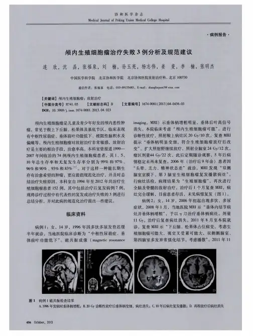
我院2011年成功开展一例生殖细胞瘤切除术,现汇报如下:一、临床资料患者男,二十岁,未婚,2011年3月3日入院。
三年前无明显诱因,出现全身乏力,食欲下降,体重共下降五公斤,无昏迷晕厥,无头痛呕吐,无明显肢体功能障碍,无肢体抽搐,无明显性格改变及多饮多尿,未予特殊诊治,半年前出现双眼视力缓慢下降伴口渴多饮,每日尿量约3000毫升,头颅CT及MRI示:鞍区占位,考虑垂体瘤可能大,来院疹治,入院后完善各项术前准备,于3月13日,在全麻下行右翼点开颅肿瘤切除术。
病理检查为鞍区生殖细胞瘤。
术后予补液抗炎止血,营养支持治疗,监测生命体征,每小时尿量、24小时出入量,3月14日,查血钾4.5mmol/L ,血钠168.1mmol/L,血氯124.10mmol/L,立即停用生理盐水,口服温开水,每2小时200毫升,3月15日查血钾4.17mmol/L,血钠143.3mmol/L,3月16日查血钾3.93mmol/L,血钠127.8mmol/L,予浓钠静滴,尿量较多,予垂体后叶素控制,诉头痛,加用甘露醇静滴,3月18日查血钾4.91mmol/L,血钠132.8mmol/L,继续予抗炎消肿,控制尿量,维持水电平衡,患者病情逐渐好转,生命体征平稳,能自行下床活动,精神食欲好,于3月28日出院,口服甲状腺素片,弥凝片剂,医师建议行放射治疗。
二、护理措施1术前护理1.1心理护理由于头痛、呕吐,视力下降等不适,病人存在有严重的焦虑情绪。
⑴要了解病人产生不良情绪的原因,主动关心安慰病人,在生活上给予无微不至的照顾,使病人感受到亲情般的温暖。
⑵与医生联系,对于病人出现的头痛、呕吐,给予相应的治疗。
1.2饮食给予流质或半流质饮食,必要时少量多餐,预防饮食过饱,导致呕吐造成误吸的发生。


颅内生殖细胞瘤如何治疗日本患者10年无病生存率超80%颅内生殖细胞肿瘤是多发于儿童的性腺外的罕见肿瘤。
与欧美国家相比,颅内胚细胞肿瘤在亚洲的发病率更高。
在日本,颅内生殖细胞瘤占所有脑肿瘤的2.3%,但是在美国是0.4%。
颅内生殖细胞瘤的高发年龄段在10-20岁之间。
男性多于女性(肿瘤多发生于男性青少年,位于鞍上生殖细胞瘤则以女性多见)。
本文将为大家介绍一下日本治疗颅内生殖细胞瘤的现状。
图源:创客贴一、颅内生殖细胞瘤的症状:肿瘤位置不同,导致患者出现的临床症状也不尽相同。
脑下垂体机能低下的情况较多,鞍上部肿瘤中60 ~ 90%的患者会出现尿崩症。
在松果体部肿瘤中,即使没有发现鞍上部的病变,也有尿崩症的发生。
鞍上部肿瘤有视觉功能下降,生长激素缺乏和性早熟的症状。
松果体部肿瘤导致中脑水道阻塞,引起水头症,出现头痛、呕吐、意识障碍等症状。
进而,压迫盖板呈现Parinaud症候群(由于会聚反射麻痹而产生的假Argyll-Robertson瞳孔或伴随对光反射减退的双侧性上方注视麻痹)。
在基底神经节肿瘤中,发生脑功能降低,由于锥体束障碍引起的偏瘫以及智力功能障碍。
颅内压亢进症状、视功能障碍为初发症状时,诊断所需的时间较短,但如果是食思不佳、精神症状、行动异常、夜尿症等非特异性症状,诊断所需的时间较长。
二、颅内生殖细胞瘤的诊断依据:临床表现、肿瘤标志物、颅脑影像学、对试验性放疗的反应性及病理活检。
诊断鉴别:鞍上区颅内生殖细胞肿瘤应除外颅咽管瘤、垂体瘤、郎罕组织细胞增多症、淋巴性漏斗神经垂体炎、下丘脑和视神经胶质瘤;底节区需与星形细胞瘤和胶质母细胞瘤鉴别;松果体区应排除松果体细胞瘤、神经胶质瘤等。
图源:创客贴三、颅内生殖细胞瘤的治疗:1) 手术:为了确定治疗方针,有必要确定组织诊断手术的目的是对肿瘤进行活检。
近年来,出现了定位活检、神经内窥镜下活检。
但是在某些困难的情况下进行开颅肿瘤活检的情况较多。
无论采用哪种方法获取肿瘤组织,肿瘤标本都可能无法在混合生殖细胞肿瘤等中反映出整个肿瘤。

【综述】⽣殖细胞瘤《StatPearls}》⽹络版2020年6⽉12⽇在线发表由美国University of Tennessee的Qandeel Sadiq; Farhan A. Khan.更新的综述《⽣殖细胞瘤(精原细胞瘤、⽣殖细胞的癌症)Cancer, Germ Cell (Seminoma, Germinoma)》。
⽣殖细胞瘤(GCT)源⾃原始⽣殖细胞(primordial germ cells),是⼀类主要发⽣于性腺( the gonads)(睾丸testicles和卵巢ovaries)的不同类型的肿瘤,也可能发⽣于前纵隔( the anterior mediastinum)、松果体(pineal gland)和⼤脑。
它们在临床上被分为两⼤类,具有重要的临床和预后意义:精原细胞瘤( Seminomas)和⾮精原细胞瘤( non-seminomas)。
精原细胞瘤通常预后良好,⽽⾮精原细胞瘤更有可能出现转移性疾病,以及由两种或两种以上不同的⽣殖细胞瘤成分组成的混合性⽣殖细胞性肿瘤(mixed germ cell tumors)。
虽然很少见,但GCT也可发⽣于肾外部位。
在没有原发性腺肿瘤的情况下,可出现长期性腺外⽣殖细胞瘤(EGGCT),占GCT的1% - 3%。
精原细胞瘤占原发性⾻外GCT的60%,主要见于⾝体中线位置,如前纵隔、中枢神经系统和腹膜后。
除⾮另有证明,腹膜后(retroperitoneum)精原细胞瘤通常是原发性性腺精原细胞瘤( primary gonadal seminoma)的转移性疾病。
在中枢神经系统中,松果体和鞍上区域的受累更为频繁。
⽣殖细胞瘤(Germinoma)是⼀种颅内对应的(counterpart )精原细胞瘤(性腺外的精原细胞瘤extragonadal seminoma),约占所有颅内⽣殖细胞性肿瘤(GCT)的三分之⼆。
⽣殖细胞瘤可以是单纯的,也可以是混合的(与其他⽣殖细胞性肿瘤[GCT]共存,分类为⾮⽣殖细胞瘤性[non-germinomatous]⽣殖细胞性肿瘤[GCT])。
European Journal of Radiology 49(2004)204–211MRI and CT findings of neurohypophyseal germinomaMitsunori Kanagaki a ,Yukio Miki a ,∗,Jun A.Takahashi b ,Yuta Shibamoto c ,Takahiro Takahashi a ,Tetsuya Ueba b ,Nobuo Hashimoto b ,Junji Konishi aaDepartment of Nuclear Medicine and Diagnostic Imaging,Graduate School of Medicine,Kyoto University,54Shogoin Kawahara-cho,Sakyo-ku,Kyoto 606-8507,JapanbDepartment of Neurosurgery,Graduate School of Medicine,Kyoto University,54Shogoin Kawahara-cho,Sakyo-ku,Kyoto 606-8507,Japanc Department of Radiology,Nagoya City University Graduate School of Medical Sciences,1Kawasumi,Mizuho-cho,Mizuho-ku,Nagoya 467-8601,JapanReceived 22January 2003;received in revised form 30May 2003;accepted 2June 2003AbstractObjective:Magnetic resonance (MR)imaging and computed tomography (CT)findings of neurohypophyseal germinoma have not previously been described in detail.The purpose of the present study was to establish the spectrum of MR imaging and CT findings in neurohypophyseal germinomas.Materials and methods:MR and CT images of 13consecutive patients (seven males,six females;mean age:15years;range:6–31years)with neurohypophyseal germinoma were retrospectively analyzed.The diagnosis had been made either histologically (n =8)or clinically according to established criteria (n =5).All patients had been examined using MR imaging and CT before treatment.Results:On MR imaging,infundibular thickening (up to 16mm)was observed in all 13cases.Hyperintensity of the posterior pituitary on T1-weighted image was absent in all 13cases (100%)and 12of the 13displayed central diabetes insipidus.Ten germinomas (77%)were isointense to cerebral cortex on T1-weighted image,but variable intensities were exhibited on T2-weighted image.MR images revealed intratumoral cysts in six cases (46%),most of which demonstrated intra-third ventricular extension.Eleven of the 13cases (85%)revealed hyperdense solid components on unenhanced CT.Calcification was absent in all cases (100%).Conclusion:Infundibular thickening,absence of the posterior pituitary high signal on T1-weighted image,lack of calcification and hyperdensity on unenhanced CT are common imaging features of neurohypophyseal germinoma.©2003Elsevier Ireland Ltd.All rights reserved.Keywords:Germ cell neoplasm;Magnetic resonance imaging;Computed tomography;Neurohypophysis1.IntroductionGerm cell tumors are relatively rare in Western countries,and constitute only 0.3–0.5%of all primary intracranial tumors [1–3].However,these tumors are far more common in Northeast Asia,accounting for ≈3.0%of all primary intracranial tumors [4].Approximately 90%of germ cell tu-mors occur in patients under 20-years-old.The pineal gland is the most common site of origin (≈50%),followed by the suprasellar region (20–30%).Other sites include the basal ganglia,thalamus,brainstem and spinal cord [1–3,5–7].Germ cell tumors can be divided into germinomatous and non-germinomatous germ cell tumors.The former type is∗Corresponding author.Tel.:+81-75-751-3790;fax:+81-75-751-4353.E-mail address:mikiy@kuhp.kyoto-u.ac.jp (Y .Miki).further classified into pure germinoma and germinoma with syncytiotrophoblastic giant cells (STGC),while the latter comprises teratoma,embryonal carcinoma,yolk sac tumor and choriocarcinoma [8].According to pathological examination of autopsy cases,germinomas of the suprasellar region (suprasellar germino-mas)involve the hypothalamo–neurohypophyseal axis (hy-pothalamus,infundibulum and posterior lobe of the pituitary gland),which is related to the development of diabetes in-sipidus [2,9].MR findings also suggest that suprasellar ger-minomas primarily arise from the posterior pituitary to the infundibulum [10,11].On the basis of these pathological and imaging findings,suprasellar germinoma is also called neurohypophyseal germinoma [10,12].Although germinomas are fatal if untreated,they dif-fer from other suprasellar neoplasms in that the tumors are highly susceptible to irradiation and chemotherapy and0720-048X/$–see front matter ©2003Elsevier Ireland Ltd.All rights reserved.doi:10.1016/S0720-048X(03)00172-4M.Kanagaki et al./European Journal of Radiology49(2004)204–211205are potentially curable.If the possibility of germinoma can be determined prior to surgical intervention,biopsy and histopathological diagnosis would allow the avoidance of dissemination or hematogenous metastasis of tumor due to aggressive surgery[13].Knowledge of the full spectrum of MR imaging and computed tomography(CT)findings of neurohypophyseal germinoma is therefore vital.To the best of our knowledge,detailed evaluation of both MR imaging and CTfindings of neurohypophyseal germi-noma has yet to be reported.The purpose of the present study was to establish the spectrum of MR imaging and CT findings of neurohypophyseal germinoma correlating with clinical information,particularly central diabetes insipidus.2.Materials and methodsThirteen consecutive patients with neurohypophyseal germinoma hospitalized in the neurosurgical department of our institution from January1987to December2002 were retrospectively enrolled in this study.Patients included seven males and six females with a mean age of15(range: 6–31years).Diagnosis of neurohypophyseal germinoma was made histologically in eight cases.In the remaining five cases,diagnosis was made on the basis of clinical fea-tures including age,serum and/or cerebrospinalfluid(CSF) tumor markers and rapid tumor response to irradiation or chemotherapy,according to established criteria[14]. Cases diagnosed histologically as non-germinomatous germ cell tumor,cases positive for␣-fetoprotein(AFP)and cases with markedly elevated serum and/or CSF concen-trations of human chorionic gonadotropin(HCG)(>2000 mIU/ml)were excluded from the study.Elevated AFP is generally restricted to yolk sac tumors and some special types of teratoma[15].Marked increases in serum or CSF HCG above2000mIU/ml are characteristic of choriocarci-noma,while moderate increases in serum or CSF HCG can be associated with germinoma containing HCG-producing STGCs(germinoma with STGC)with no definite evidence of choriocarcinoma[7].The majority of germinomas with STGC can be clinically diagnosed when serum HCG con-centration is elevated but below2000mIU/ml[16].Seven of the13cases in the present study met this criterion,display-ing moderately elevated concentrations of HCG suggestive of germinoma with STGC.All patients had been examined using CT and MR imag-ing before treatment.Axial or coronal unenhanced CT scans were obtained with slice thicknesses of5–10mm. MR imaging studies were performed using a1.5-T super-conducting magnet.Both sagittal and coronal T1-weighted images(spin-echo;repetition time/echo time/excitations: 400-630/8-35/1-2)and axial and coronal T2-weighted im-ages(spin-echo or fast spin-echo;2000-7800/80-126/1-2) were obtained.Additional MR imaging parameters included 3–5mm slice thickness,20–24cmfield of view and a 192–256×256matrix.Sagittal,in addition to axial or coro-nal contrast-enhanced T1-weighted images,were obtained in11patients(85%)after intravenous injection of either gadodiamide(Gd-DTPA-BMA)or gadopentetate dimeglu-mine(Gd-DTPA)at a dose of0.1mmol/kg bodyweight. The results of MR imaging and CT were reviewed in a non-blinded manner by three experienced neuroradiologists. Tumor location,CT density,MR signal intensity and en-hancement patterns were evaluated,as was the presence of calcification,infundibular thickening,posterior lobe hyper-intensity and cystic components.Tumor location was evaluated regarding the following four regions:(1)intrasellar;(2)infundibulum;(3)third ven-tricle;and(4)basal ganglia or lateral ventricles.Intrasel-lar involvement was determined by anterior displacement of the anterior pituitary or enlargement of the sella turcica. Infundibular involvement was defined when the maximum diameter of the infundibulum was equal to or>4mm[17]. Intra-third ventricular extension was defined as protrusion of the tumor into the third ventricle.According to medical charts,central diabetes insipidus was present in12of the13patients(92%).These12patients required desmopressin acetate(DDA VP)to control urinary volume.In the remaining case,clinical evaluation for dia-betes insipidus was not possible due to the presence of blad-der disturbance.3.ResultsMR imaging and CTfindings are summarized in Table1.3.1.MRIfindingsInfundibular thickening(up to16mm)was observed in all cases(Fig.1c,Fig.3b,Fig.4b).Six of the13cases (46%)also displayed an intrasellar component(Fig.1c). Intra-third ventricular extension was identified infive cases (38%)(Fig.2b).Three tumors(23%)had infiltrated the basal ganglia,one of which had invaded the wall of the lateral ventricle(Fig.4a).Four of the six cases displaying an in-trasellar component revealed anterior pituitaries that were compressed anterior to the tumor(Fig.1c).Three cases dis-played enlargement of the sella turcica.Hyperintensity of the posterior pituitary on T1-weighted images was absent in all cases(100%)(Fig.1a,Fig.3a). On T1-weighted images,signal intensity of the solid por-tion was slightly hyperintense to cerebral cortex in two cases (15%),isointense in ten(77%)(Fig.1a)and hypointense in one(8%).Five cases showed inhomogeneous signal intensity on the T2-weighted images.On T2-weighted im-ages,signal intensity of the dominant solid portion was hyperintense to cerebral cortex in three cases(23%),isoin-tense in eight(62%)(Fig.1b)and hypointense in two (15%).Contrast-enhanced T1-weighted imaging was performed for11patients.All tumors displayed significant contrastM.Kanagaki et al./European Journal of Radiology49(2004)204–211207Fig.1.Case8.Neurohypophyseal germinoma in an11-year-old girl.Intra-and suprasellar tumor components are homogeneous and isointense to cerebral cortex on both T1-weighted(SE400/15)(a)and T2-weighted(FSE5000/130)MR images(b).Sagittal T1-weighted MR image(SE400/15)(a)shows absence of normal signal hyperintensity in the posterior pituitary(arrow).Sagittal post-gadolinium T1-weighted MR image(SE400/15)(c)shows heterogeneously enhancing tumor involving the intrasellar region and infundibulum.Thickened infundibulum is observed(arrow).The anterior pituitary is compressed anteroinferiorly(arrowhead)[10,18,25].The tumor is less enhancing than the anterior pituitary.Unenhanced CT reveals a hyperdense tumor with no calcification(d).enhancement:six(55%)revealed homogeneous enhance-ment(Fig.3b)andfive(45%)showed heterogeneous en-hancement(Fig.1c,Fig.2b,Fig.4a).Four of thefive cases with heterogeneous enhancement demonstrated involve-ment of the third ventricularfloor or infiltration to the basal ganglia,while only one of the six cases with homogeneous enhancement displayed intra-third ventricular extension or infiltration to the basal ganglia.Intratumoral cysts were seen in six of the13cases(46%), andfive of these displayed intra-third ventricular extension (Fig.2a,Fig.4a).None of the remaining seven tumors with-out intratumoral cysts demonstrated intra-third ventricular extension.Multifocal lesions were observed in four of the13cases (31%)and pineal lesions were observed in all four cases. In addition to the pineal lesions,two cases demonstrated ventricular wall dissemination and one displayed a spinal cord lesion.3.2.CTfindingsIn11of the13cases(85%),the solid portion of the tumor was hyperdense to cerebral cortex on unenhanced CT (Fig.1d,Fig.2c).In the remaining two cases(15%),CT density could not be evaluated due to the small size of the lesion.Calcification was absent in all cases(100%).208M.Kanagaki et al./European Journal of Radiology 49(2004)204–211Fig.2.Case 12.Neurohypophyseal germinoma in a 26-year-old man.Axial T2-weighted MR image (FSE 4100/80)displays multiple intratumoral cysts (arrowheads)(a).Coronal post-gadolinium T1-weighted MR image (SE 400/14)shows tumor protruding into the third ventricle (b).Tumor is hyperdense to cerebral cortex on unenhanced CT (arrows)(c).4.DiscussionTo the best of our knowledge,few papers have evalu-ated infundibular thickening of neurohypophyseal germi-noma [10,18].All 13cases in the present study displayed infundibular thickening.Fujisawa et al.[10]reported that six of their seven cases showed infundibular thickening and Liang et al.[18]described thickening of the infundibulum as the only abnormal imaging finding for small tumors.This high frequency of infundibular thickening is not unexpected given the predominant localization of neurohypophyseal ger-minoma in the hypothalamo–neurohypophyseal axis [10,11].We consider infundibular thickening as a typical finding for neurohypophyseal germinoma.The hyperintense signal of the posterior pituitary was ab-sent in all 13cases (100%)in our series,confirming the re-sults of previous reports [10,19,20].This absence of normal hyperintensity in the posterior pituitary is closely related to the loss of hypothalamo-hypophyseal function,particularly diabetes insipidus [21–23].In the present series,12of the 13patients (92%)showed evidence of diabetes insipidus.MR signal intensity of the solid portion is non-specific on both T1-and T2-weighted images.Ten of the 13ger-minomas (77%)were isointense to cerebral cortex on T1-weighted images,but intensities on T2-weighted images were variable.Typically,germinoma is iso-or slightly hy-pointense on T1-weighted images and iso-or hyperintense on T2-weighted images [18,24–26].In our series,all 11tumors examined under gadolinium administration revealed intense enhancement.Marked con-trast enhancement is a common finding for neurohypophy-seal germinoma [18,25].Five of the 11cases displayedM.Kanagaki et al./European Journal of Radiology 49(2004)204–211209Fig.3.Case 9.Neurohypophyseal germinoma in a 15-year-old boy.Sagittal T1-weighted MR image (SE 400/20)(a)shows the absence of normal signal hyperintensity in the posterior pituitary (arrow).Sagittal post-gadolinium T1-weighted MR image (SE 400/20)(b)shows a small tumor involving the upper portion of the infundibulum (arrow).heterogeneous enhancement.A recent study reported that heterogeneous enhancement is commonly seen in relatively large neurohypophyseal germinoma [18].Large tumors tend to exhibit heterogeneous enhancement,probably due to in-homogeneous blood supply,microcyst formation or presence of necrosis [18].To the best of our knowledge,frequency of intratumoral cysts in neurohypophyseal germinoma has not been evalu-ated.Intratumoral cysts were seen in six of our 13cases (46%).Of these six cases,five displayed intra-third ventric-ular extension,while none of other seven cases without in-tratumoral cysts demonstrated either intra-thirdventricular Fig.4.Case 7.Neurohypophyseal germinoma with STGC in a 16-year-old girl.Axial post-gadolinium T1-weighted MR image (SE 630/15)reveals a tumor with multiple intratumoral cysts extending into the third ventricle,basal ganglia and lateral ventricle (a).Another axial post-gadolinium image shows the thickened infundibulum (arrow)(b).extension or infiltration to the basal ganglia.Germinomas arising from the basal ganglia or thalamus reportedly tend to contain multiple cysts of varying size [27–29].Large tumor size and presence of brain parenchymal involvement could be related to intratumoral cyst formation also in neurohy-pophyseal germinoma.Multifocal germ cell tumors usually involve the pineal and suprasellar compartments simultaneously or sequen-tially,and account for 6–13%of all intracranial germ cell tumors [1,3,30].In our series,four of the 13cases (31%)dis-played a synchronous pineal lesion.If restricted to cases of neurohypophyseal germinoma,this rate is within the range210M.Kanagaki et al./European Journal of Radiology49(2004)204–211of those reported previously[18,25,31,32].In general,syn-chronous mass lesions in these regions can be diagnosed as primary germ cell tumors.The origins of multifocal lesions as either metastatic spread from one location to another or as lesions of true multicentric origin remain controversial [30].In our series,11of the13tumors(85%)were hyperdense to cerebral cortex on unenhanced CT.In the remaining two cases,CT density could not be evaluated due to the small size of the lesion.Hyperdensity on unenhanced CT is probably attributable to their hypercellularity[24,33,34].No cases in the present study displayed calcification,again confirming the results of previous studies[24,34].Infundibular thickening and absence of normal signal hy-perintensity in the posterior pituitary on T1-weighted MR images and relative hyperdensity without calcification on unenhanced CT are characteristic but not specific for neu-rohypophyseal germinoma.Fifteen to40%of patients with Langerhans cell histiocytosis(LCH)manifest with diabetes insipidus due to histiocytic infiltration of the neurohypoph-ysis and may show pituitary stalk thickening in the absence of the posterior pituitary bright signal[35,36].Diabetes insipidus may present asfirst manifestation in patients with LCH,but the majority of these patients develop diseases outside of the hypothalamo–neurohypophyseal axis during the follow-up course[37].Lymphocytic infundibuloneuro-hypophysitis(LIN),an autoimmune-mediated inflamma-tory disorder which causes central diabetes insipidus,may also shows thickening of the pituitary stalk and absence of normal hyperintense signal of the posterior pituitary on T1-weighted images[38].LIN can be differentiated from neurohypophyseal germinoma by the following:it usu-ally occurs in adults;the natural course of this disorder is self-limited;and thickening of the pituitary stalk will disap-pear on follow-up course[23,38].It might be difficult to dif-ferentiate lymphoma,leukemia and metastasis,which may localize in the neurohypophyseal region[39],from neurohy-pophyseal germinoma on the basis of radiological imaging alone.Many other clinical features of these diseases may aid in differentiating them from neurohypophyseal germinoma; lymphomas are predominantly seen in adults and their only involvement of the neurohypophysis is extremely rare [40],metastases are also common in adults and leukemias are usually seen with bone marrow lesions.Besides these, some other granulomatous diseases,such as tuberculosis, sarcoidosis,Wegener’s granulomatosis and granulomatous hypophysitis may mimic neurohypophyseal germinomas [41–44].Central nervous system(CNS)involvement of tu-berculosis almost always occurs secondary to a non-CNS focus of infection and usually shows basal meningeal en-hancement or enhancing nodules in the brain parenchyma [45].Approximately5%of patients with sarcoidosis may present with intracranial involvement and some may have infundibulo–neurohypophyseal axis involvement[46].Most of patients with CNS sarcoidosis develop manifestations at non-CNS regions and demonstrate distinctivefindings on chest X-ray,high titer of ACE,as well as the presence of uveitis[42].5.ConclusionAlthough MRIfindings of neurohypophyseal germino-mas except those involving the pineal gland are rather non-specific,infundibular thickening and absence of normal sig-nal hyperintensity in the posterior pituitary on T1-weighted MR images and relative hyperdensity without calcification on unenhanced CT represent common imaging features for neurohypophyseal germinomas.References[1]Jennings MT,Gelman R,Hochberg F.Intracranial germ-cell tumors:natural history and pathogenesis.J Neurosurg1985;63:155–67. [2]Bjornsson J,Scheithauer BW,Okazaki H,Leech RW.Intracranialgerm cell tumors:pathobiological and immunohistochemical aspects of70cases.J Neuropathol Exp Neurol1985;44:32–46.[3]Hoffman HJ,Otsubo H,Hendrick EB,et al.Intracranial germ-celltumors in children.J Neurosurg1991;74:545–51.[4]The Committee of Brain Tumor Registry of Japan.Frequency ofPrimary Brain Tumors.Report of Brain Tumor Registry of Japan (1969–1993).Neurol Med Chir(Tokyo)2000;40:Suppl5.[5]Korogi Y,Takahashi M.Current concepts of imaging in patients withpituitary/hypothalamic dysfunction.[Review].Semin Ultrasound CT MR1995;16:270–8.[6]Matsutani M,Sano K,Takakura K,et al.Primary intracranial germcell tumors:a clinical analysis of153histologically verified cases.J Neurosurg1997;86:446–55.[7]Ho DM,Liu HC.Primary intracranial germ cell tumor.Pathologicstudy of51patients.Cancer1992;70:1577–84.[8]Brandes AA,Pasetto LM,Monfardini S.The treatment of cranialgerm cell tumours.Cancer Treat Rev2000;26:233–42.[9]Jellinger K.Primary intracranial germ cell tumours.Acta Neuropathol(Berl)1973;25:291–306.[10]Fujisawa I,Asato R,Okumura R,et al.Magnetic resonance imagingof neurohypophyseal germinomas.Cancer1991;68:1009–14. [11]Oishi M,Morii K,Okazaki H,Tamura T,Tanaka R.Neurohypophy-seal germinoma traced from its earliest stage via magnetic resonance imaging:case report.Surg Neurol2001;56:236–41.[12]Abe T,Tanioka D,Izumiyama H,Matsumoto K,Shinozuka e-fulness of gallium-67scintigraphy in neurohypophyseal germinoma.Acta Neurochir(Wien)2000;142:655–60.[13]Ono N,Kakegawa T,Zama A,et al.Suprasellar germinomas;rela-tionship between tumour size and diabetes insipidus.Acta Neurochir (Wien)1992;114:26–32.[14]Shibamoto Y,Takahashi M,Sasai K.Prognosis of intracranial ger-minoma with syncytiotrophoblastic giant cells treated by radiation therapy.Int J Radiat Oncol Biol Phys1997;37:505–10.[15]Rosenblum MK,Matsutani M,Van Meir S germ cell tumours.In:Kleihues P,Cavenee W,editors.Pathology and genetics tumours of the nervous system.Lyon:IARC Press;2000,208–14.[16]Utsuki S,Kawano N,Oka H,et al.Cerebral germinoma with syn-cytiotrophoblastic giant cells:feasibility of predicting prognosis us-ing the serum hCG level.Acta Neurochir(Wien)1999;141:975–7, discussion7–8.[17]Simmons GE,Suchnicki JE,Rak KM,Damiano TR.MR imagingof the pituitary stalk:size,shape,and enhancement pattern.Am J Roentgenol1992;159:375–7.[18]Liang L,Korogi Y,Sugahara T,et al.MRI of intracranial germ-celltumours.Neuroradiology2002;44:382–8.M.Kanagaki et al./European Journal of Radiology49(2004)204–211211[19]Mootha SL,Barkovich AJ,Grumbach MM,et al.Idiopathic hypotha-lamic diabetes insipidus,pituitary stalk thickening,and the occult in-tracranial germinoma in children and adolescents.J Clin Endocrinol Metab1997;82:1362–7.[20]Tamura T,Tanaka R,Morii K,Okazaki H,Kawasaki T.Doeshypopituitarism due to neurohypophyseal germinoma recover after chemotherapy?Endocr J1999;46:S109–11.[21]Fujisawa I,Nishimura K,Asato R,et al.Posterior lobe of thepituitary in diabetes insipidus:MRfindings.J Comput Assist Tomogr 1987;11:221–5.[22]Moses AM,Clayton B,Hochhauser e of T1-weighted MRimaging to differentiate between primary polydipsia and central di-abetes insipidus.Am J Neuroradiol1992;13:1273–7.[23]Maghnie M,Cosi G,Genovese E,et al.Central diabetes insipidusin children and young adults.New Engl J Med2000;343:998–1007.[24]Fujimaki T,Matsutani M,Funada N,et al.CT and MRI features ofintracranial germ cell tumors.J Neurooncol1994;19:217–26. [25]Sumida M,Uozumi T,Kiya K,et al.MRI of intracranial germ celltumours.Neuroradiology1995;37:32–7.[26]Saeki N,Nagano O,Sakaida T,et al.Recurrent neurohypophy-seal germinoma causing invasion localized to temporal bone marrow-unreported neuroimaging studies compared to autopsyfind-ings.Acta Neurochir(Wien)2001;143:407–11.[27]Higano S,Takahashi S,Ishii K,et al.Germinoma originating in thebasal ganglia and thalamus:MR and CT evaluation.Am J Neuroradiol 1994;15:1435–41.[28]Moon WK,Chang KH,Kim IO,et al.Germinomas of the basalganglia and thalamus:MRfindings and a comparison between MR and CT.Am J Roentgenol1994;162:1413–7.[29]Kim DI,Yoon PH,Ryu YH,Jeon P,Hwang GJ.MRI of germino-mas arising from the basal ganglia and thalamus.Neuroradiology 1998;40:507–11.[30]Sugiyama K,Uozumi T,Kiya K,et al.Intracranial germ-cell tumorwith synchronous lesions in the pineal and suprasellar regions:report of six cases and review of the literature.Surg Neurol1992;38:114–20.[31]Saeki N,Murai H,Kubota M,Fujimoto N,Yamaura A.Long-termKarnofsky performance status and neurological outcome in patients with neurohypophyseal germinomas.Br J Neurosurg2001;15:402–8.[32]Saeki N,Tamaki K,Murai H,et al.Long-term outcome of endocrinefunction in patients with neurohypophyseal germinomas.Endocr J 2000;47:83–9.[33]Ganti SR,Hilal SK,Stein BM,et al.CT of pineal region tumors.Am J Roentgenol1986;146:451–8.[34]Chang T,Teng MM,Guo WY,Sheng WC.CT of pineal tumorsand intracranial germ-cell tumors.Am J Roentgenol1989;153:1269–74.[35]Rosenzweig KE,Arceci RJ,Tarbell NJ.Diabetes insipidus secondaryto Langerhans’cell histiocytosis:is radiation therapy indicated?Med Pediatr Oncol1997;29:36–40.[36]Maghnie M,Arico M,Villa A,et al.MR of the hypothalamic–pituitary axis in Langerhans cell histiocytosis.Am J Neuroradiol 1992;13:1365–71.[37]Kaltsas GA,Powles TB,Evanson J,et al.Hypothalamo-pituitaryabnormalities in adult patients with Langerhans cell histiocytosis: clinical,endocrinological,and radiological features and response to treatment.J Clin Endocrinol Metab2000;85:1370–6.[38]Imura H,Nakao K,Shimatsu A,et al.Lymphocytic infundibuloneu-rohypophysitis as a cause of central diabetes insipidus.New Engl J Med1993;329:683–9.[39]Kimmel DW,O’Neill BP.Systemic cancer presenting as diabetesinsipidus.Clinical and radiographic features of11patients with a review of metastatic-induced diabetes insipidus.Cancer1983;52: 2355–8.[40]Kaufmann TJ,Lopes MB,Laws Jr ER,Lipper MH.Primary sellarlymphoma:radiologic and pathologicfindings in two patients.Am J Neuroradiol2002;23:364–7.[41]Altunbasak S,Baytok V,Alhan E,Yuksel B,Aksaray N.Suprasellartuberculoma causing endocrinologic disorders and imitating cranio-pharyngioma.Pediatr Neurosurg1995;23:328–31.[42]Dumas JL,Valeyre D,Chapelon-Abric C,et al.Central nervoussystem sarcoidosis:follow-up at MR imaging during steroid therapy.Radiology2000;214:411–20.[43]Czarnecki EJ,Spickler EM.MR demonstration of Wegener granu-lomatosis of the infundibulum,a cause of diabetes insipidus.Am J Neuroradiol1995;16:968–70.[44]Endo T,Kumabe T,Ikeda H,Shirane R,Yoshimoto T.Neurohy-pophyseal germinoma histologically misidentified as granulomatous hypophysitis.Acta Neurochir(Wien)2002;144:1233–7.[45]Jinkins JR,Gupta R,Chang KH,Rodriguez-Carbajal J.MR imag-ing of central nervous system tuberculosis.Radiol Clin North Am 1995;33:771–86.[46]Stern BJ,Krumholz A,Johns C,Scott P,Nissim J.Sarcoidosis andits neurological manifestations.Arch Neurol1985;42:909–17.。
最新:中国肿瘤整合诊治指南—中枢神经系统生殖细胞肿瘤要点(全文)前言中枢神经系统生殖细胞肿瘤(CNS GCTs)是儿童及青少年中枢神经系统常见的恶性肿瘤,占儿童原发性神经系统肿瘤的8.1%(中国)~15.3%(日本),好发于3~15岁,常发生于松果体区、鞍上区或丘脑基底节区、少数可发生在三脑室、脑干、胼胝体等中线部位。
生殖细胞肿瘤(GCTs)包括生殖细胞瘤(germinoma)和非生殖细胞瘤性生殖细胞肿瘤(NGGCT)两大类。
NG‐GCT包括胚胎癌、卵黄囊瘤、绒毛膜细胞癌、畸胎瘤(成熟型和未成熟型)和畸胎瘤伴恶性转化和混合型生殖细胞肿瘤。
其中由两种或两种以上不同生殖细胞肿瘤成分构成的肿瘤称为混合性生殖细胞肿瘤。
在生殖细胞肿瘤中,除成熟型畸胎瘤属良性外,其余均为恶性肿瘤。
颅内生殖细胞肿瘤中以生殖细胞瘤最多见,占半数以上。
目前国际上治疗CNS GCTs均采用放疗、化疗和手术等整合治疗手段。
第一章概述第一节发病率第二节病理1 颅内GCTs的WHO 2021分类表2 生殖细胞瘤2.1 大体所见生殖细胞瘤约占iGCTs的2/3,色灰红,大多呈浸润性生长,与周围脑组织边界中枢神经系统生殖细胞肿瘤第一章概述不清,质软而脆,结节状,肿瘤组织易于脱落,也有肿瘤呈胶冻状,瘤内可出血、坏死和囊性变。
2.2 镜下观察2.3 免疫组化胎盘碱性磷酸酶(PLAP)在大多数生殖细胞瘤的细胞膜和细胞浆中存在(70%~100%)。
半数生殖细胞瘤对人绒毛促性腺激素(HCG)表达阳性。
OCT4可在生殖细胞瘤细胞核中表达阳性。
3 畸胎瘤与未成熟畸胎瘤畸胎瘤由2种或3种胚层分化而成,这些组织虽同时存在,但排列无序,外观上不像正常可辨的组织器官。
3.1 大体所见3.2 镜下观察3.3 免疫组化畸胎瘤结构复杂,免疫组化也呈多样性。
4 卵黄囊瘤4.1 大体所见4.2 镜下观察4.3 免疫组化部分卵黄囊瘤对PLAP呈阳性表达,多数内胚窦瘤对AFP,Keratin呈阳性表达。
生殖细胞瘤确诊标准
生殖细胞瘤的确诊标准主要包括以下几个方面:
1. 临床症状和体征:患者可能出现与生殖细胞瘤相关的症状,如腹部肿块、头痛、视力障碍、内分泌异常等。
2. 影像学检查:如CT、MRI 等影像学检查可以发现肿瘤的存在、大小、位置以及与周围组织的关系。
3. 病理检查:对肿瘤组织进行病理活检是确诊生殖细胞瘤的重要依据。
通过手术或穿刺等方式获取肿瘤组织,进行病理学检查,以确定肿瘤的类型和性质。
4. 生化检查:某些生殖细胞瘤可能会导致特定的生化指标异常,如血清人绒毛膜促性腺激素(β-HCG)、甲胎蛋白(AFP)等的升高。
5. 排除其他疾病:排除其他可能引起类似症状的疾病,以确保诊断的准确性。
6. 医生综合判断:医生会综合考虑患者的临床表现、影像学检查结果、病理检查结果以及其他相关因素,进行综合分析和判断,最终确诊生殖细胞瘤。
需要注意的是,确诊生殖细胞瘤可能需要结合多种检查方法和医生的经验判断。
此外,对于一些疑难病例,可能需要进行专家会诊或进一步的检查。
如果怀疑有生殖细胞瘤,应及时就医并遵循医生的建议进行相关检查和治疗。
Advances in Clinical Medicine 临床医学进展, 2020, 10(3), 270-278Published Online March 2020 in Hans. /journal/acmhttps:///10.12677/acm.2020.103043Study Progress of Intracranial Germ CellTumorsXiaohong Gao1, Yaming Wang2, Mangmang Bai1*1Yanan University Affiliated Hospital Neurosurgery, Yan’an Shaanxi2Xuanwu Hospital Capital Medical University Neurosurgery, BeijingReceived: Feb. 27th, 2020; accepted: Mar. 13th, 2020; published: Mar. 20th, 2020AbstractIntracranial germ cell tumors (IGCTs) are uncommon tumours occurring in children and young adults with a tendency to appear in pineal, suprasellar and basal ganglia region. They are usually segregated into germinomas and nongerminomatous tumours (NGGCTs). The main clinical ma-nifestations of the tumors depend on their size and location. The diagnosis has been divided into clinical diagnosis and surgical biopsy pathology diagnosis. The tumor therapy regimen is mostly chemoradiotherapy and surgery. New tumor markers microRNA (MiRNA) may be potentially val-uable biomarkers in the diagnosis and evaluation of treatment of intracranial germ cell tumors in the future. Histopathology and molecular analyses are attempting to further specify the different IGCTs subtypes and are helping to direct the development of distinct therapeutic targets to im-prove treatment and prognosis. Here is to make a review of the current status and management in intracranial germ cell tumorsKeywordsIntracranial Germ Cell Tumors, Diagnosis, Therapy, MiRNA颅内生殖细胞肿瘤研究进展高晓红1,王亚明2,白茫茫1*1延安大学附属医院神经外科,陕西延安2首都医科大学宣武医院神经外科,北京收稿日期:2020年2月27日;录用日期:2020年3月13日;发布日期:2020年3月20日*通讯作者。
颅内生殖细胞瘤的临床与影像探讨【摘要】目的:探讨颅内生殖细胞瘤的特征和临床及其影像学的特征。
方法:结合38例颅内生殖细胞瘤患者的临床治疗情况进行影像学分析。
结论:儿童、青少年易发颅内生殖细胞瘤,一般发病的区域多数时候在松果体区、鞍区或基底节区。
患者临床常常表现为颅内压升高、尿崩症、视功能障碍或偏瘫等等。
在治疗上影像学可有效帮助颅内生殖细胞瘤的治疗。
【关键词】颅内生殖细胞瘤;磁共振影像1临床资料1.1一般资料本文的38例案例男性33人,女性5人,在年龄分布上,位于7~32岁,平均年龄14岁,其中未成年人(小于18岁)的有32例,占总案例的84%。
病程15日~30个月,平均病程在11个月。
有11位病人病变区位于鞍区,占总案例29%;13位患者病变区位于松果体区,占总案例34%;14位患者病变区位于基底节区,占总案例37%;1.2临床表现在临床表现上:①11位病变区位于鞍区的患者中,其中11人患有尿崩症,有7人视力减退,有4人复视,还有3人性早熟和1人患有癫痫症状。
②13位病变区位于松果体区的患者的临床表现为,8人有头痛和呕吐症状以及视乳头水肿,7位患者表现佩里诺综合症,有1例视力减退,癫痫1例和1人耳鸣。
③14位病变区位于基底节区患者的临床表现为:14人均有偏瘫,耳鸣1例。
1.3实验室及辅助检查在本文的案例中,所有患者的生化检查表现正常,其中5名患者进行了人绒毛膜促性腺激素和甲胎蛋白检查,检查结果正常。
所有患者都进行了脑部ct检查,检查结果表明11例病变位于鞍区的患者表现为圆形、椭圆形混合,在影像的边缘很清晰,对患者进行ct增强扫描后,发现病灶呈现均匀一致,范围比一般的扫描时增大;其中3例肿瘤体积较大累及第三脑室前部,出现中、重度脑积水,且1例伴瘤内钙化。
病变位于松果体区的13例也表现为圆形、椭圆形病灶,其中表现为等密度病灶伴斑点状钙化改变5例;表现为高密度边缘清晰无钙化病灶8例,13例ct增强扫描均显示病灶呈不均匀强化。