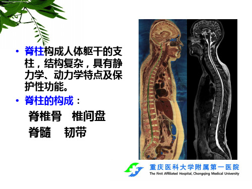
最新脊柱外伤影像学表现
- 格式:ppt
- 大小:2.82 MB
- 文档页数:7

脊柱创伤患者MRI及CT表现特点【摘要】目的研究脊柱创伤患者磁共振成像(MRI)及计算机断层扫描(CT)表现特点。
方法回顾性分析2018年6月到2021年6月在本院诊治的62例脊柱创伤患者的临床资料,全部患者都进行MRI及CT检查,比较MRI及CT脊柱创伤检出情况,并总结MRI及CT表现特点。
结果MRI脊椎骨折、韧带损伤检出率明显高于CT,碎骨片检出状况明显低于CT,差异有统计学意义(P<0.05);MRI共检出脊椎骨折62例,均伴有后缘碎骨片移向椎管,椎体中均出现T2WI高信号、T1WI低信号或等信号,其中,椎管容积改变有28例,韧带损伤有44例,神经根损伤有12例,椎旁软组织损伤有46例;CT共检出脊椎骨折56例,均伴有椎体楔状压缩变化或椎体变形,有3例没有显著骨折征象、3例表现为颈椎滑脱,其中,椎管容积改变有28例,韧带损伤有7例,神经根损伤有10例。
结论MRI在韧带损伤与脊椎骨折检查方面优势显著,CT在脊柱创伤患者碎骨片移位状况的检查方面优势显著,临床实践中可按照患者的实际状况使用MRI及CT 这两种检查方式。
【关键词】脊柱创伤;磁共振成像;计算机断层扫描;表现特点脊柱创伤属于骨科常见疾病,会造成残废,甚至威胁生命[1]。
临床一般通过计算机断层扫描(ComputedTomography,CT)、X线、磁共振成像(MagneticReso-nanceImaging,MRI)、颈静脉加压试验等方式检查此病,其中比较常见的有MRI及CT检查[2]。
MRI能准确鉴别脊髓损伤情况,可将早期脊髓损伤的出血、水肿等状况及诸多病理变化显示出来[3]。
CT在移位骨折块侵犯椎管程度判定方面优势显著。
基于此,本研究回顾性分析在本院诊治的62例脊柱创伤患者的临床资料,研究脊柱创伤患者MRI及CT表现特点。
现报道如下。
1资料与方法1.1一般资料:回顾性分析2018年6月到2021年6月在杭州市临平区第一人民医院诊治的62例脊柱创伤患者的临床资料。

110例脊柱外伤的CT和MRI影像诊断对比分析目的探讨脊柱外伤的CT和MRI影像的表现以及特点,并研究两者在脊柱外伤诊断中的价值与临床意义。
方法收集脊柱外伤患者的CT、MRI资料110例,并对其进行分析。
对比分析两者对损伤脊柱的附件骨折、椎体骨折,碎骨片位移、脊髓损伤、脊柱曲度改变、椎管容积改变、韧带损伤、软组织损伤、神经根损伤、椎间盘损伤的诊断效果。
结果(1)CT对发现单纯无变形椎体裂纹骨折与特殊部位的骨折效果明显,对显示椎体、椎弓骨折线和骨折片的移位优于MRI;(2)MRI则对显示脊髓损伤(P<0.05)、韧带损伤(P<0.01)、神经根损伤(P<0.05)、软组织损伤(P<0.05)比CT更敏感、更清晰,与CT比较显示损伤范围更大、更明确。
结论CT、MRI对显示脊柱外伤都有各自的优势和劣势,但MRI对脊柱外伤空间定位比较准确,并可检查出外伤导致的脊髓病变和损伤,因此,联合两种方法能够有效的避免假阴性和假阳性的出现。
标签:脊柱外伤;CT;MRI;脊髓损伤Comparative study on the diagnose of CT and MRI in 110 cases with spinal trauma HUANG Jian-feng, DANG Ye-tian, NING Jin-long, HUANG De-an, GUO Wei. Hezhou Chinese Medicine Hospital, Hezhou 542800, China【Abstract】Objective To explore the diagnostic value of two routine checking methods, CT and MRI, on Spinal Trauma.Methods To observe and analysis 110 caseswith Spinal Trauma s CT and MRI data accepted and cured. Furthermore, thefollowing items would be compared and analyzed such as accessories fracturerate, centrum fracture, the displacement of the broken bone, spinal cord damage, the change of spinal column curvature, the change of spinal canal volume, ligament injury, soft tissue damage, spinal nerve root damage and intervertebral discs injury.Results (1)CT Imaging was more effective in the discovering the pure no-deformation vertebral crack fracture and special parts fracture than MRI. The centrum, vertebral arch fracture line and fracture bone displacement in CT Imaging was superior to that of MRI imaging. (2)The MRI imaging was more sensitive and clearer than CT Imaging in showing spinal cord damage(P<0.05), ligation damage(P<0.01), nerve root damage(P<0.05), soft tissue damage(P<0.05) than CTimaging. Meanwhile, compared with CT imaging, the display range was wide and explicit.Conclusion There are both advantages and disadvantages in the display of spinal trauma in CT and MRI imaging. MRI imaging is good at orientation of spinal trauma, which is the best method for checking the spinal disease. The combination of two methods can make up for the deficiencies.【Key words】Spinal trauma; CT;MRI;Spinal cord damage脊柱外伤是外科损伤中的常见疾病,常见的病因有高处坠落、车祸、暴力冲击脊柱、以及弯腰弓背时受到较强的挤压力等[1],轻者会出现椎骨裂纹骨折、软组织损伤、韧带损伤,重者会出现碎骨片移位、椎管容积和脊柱曲度的改变以及神经根损伤等[2]。