syntax评分
- 格式:doc
- 大小:512.00 KB
- 文档页数:7

08年长城会上听专家讲了Syntax score评分方法,这评分方法比较复杂,主要是依据冠脉造影的结果来判断。
在准备阅读之前,先要熟悉冠脉解剖的节段分布情况:(上图为左优势型冠脉解剖分布,下图为右优势型解剖分布)Figure 1. Definition of the coronary tree segments(冠脉束血管段的识别)1. RCA proximal: From the ostium to one half the distance to the acute margin of the heart.2. RCA mid: From the end of first segment to acute margin of heart.3. RCA distal: From the acute margin of the heart to the origin of the posterior descending artery.4. Posterior descending artery: Running in the posterior interventricular groove.16. Posterolateral branch from RCA: Posterolateral branch originating from the distal coronary artery distal to the crux.16a. Posterolateral branch from RCA: First posterolateral branch from segment 16.16b. Posterolateral branch from RCA: Second posterolateral branch from segment 16.16c. Posterolateral branch from RCA: Third posterolateral branch from segment 16.5. Left main: From the ostium of the LCA through bifurcation into left anterior descending and left circumflex branches.6. LAD proximal: Proximal to and including first major septal branch.7. LAD mid: LAD immediately distal to origin of first septal branch and extending to the point where LAD forms an angle (RAO view). If thisangle is not identifiable this segment ends at one half the distance from the first septal to the apex of the heart.8. LAD apical: Terminal portion of LAD, beginning at the end of previous segment and extending to or beyond the apex.9. First diagonal: The first diagonal originating from segment 6 or 7.9a. First diagonal a: Additional first diagonal originating from segment 6 or 7, before segment 8.10. Second diagonal: Originating from segment 8 or the transition between segment 7 and 8. 10a. Second diagonal a: Additional second diagonal originating from segment 8.11. Proximal circumflex artery: Main stem of circumflex from its origin of left main and including origin of first obtuse marginal branch.12. Intermediate/anterolateral artery: Branch from trifurcating left main other than proximal LAD or LCX. It belongs to the circumflex territory.12a. Obtuse marginal a: First side branch of circumflex running in general to the area of obtuse margin of the heart.12b. Obtuse marginal b: Second additional branch of circumflex running in the same direction as 12.13. Distal circumflex artery: The stem of the circumflex distal to the origin of the most distal obtuse marginal branch, and running along the posteriorleft atrioventricular groove. Caliber may be small or artery absent.14. Left posterolateral: Running to the posterolateral surface of the left ventricle. May be absent or a division of obtuse marginal branch.14a. Left posterolateral a: Distal from 14 and running in the same direction.14b. Left posterolateral b: Distal from 14 and 14 a and running in the same direction.15. Posterior descending: Most distal part of dominant left circumflex when present. It givesorigin to septal branches. When this arteryis present, segment 4 is usually absent。


Gensini评分和SYNTAX评分--冠状动脉病变积分冠脉病变的形态和狭窄的程度决定了治疗方案的选择,目前以Gensini评分和SYNTAX评分系统较为常用,两者侧重点不同,各有优缺点。
Gensini评分反映的是斑块的负荷情况,但没有涉及分叉、钙化和扭曲病变特征;SYNTAX评分反映的是斑块的类型及PCI的复杂程度,不仅能描述冠脉病变解剖结构,还可以在临床医生为高危患者制定最佳治疗方案时提供指导。
对于病变既适于PCI又适于CABG、且预期外科手术病死率低的患者,可用SYNTAX评分帮助制定治疗决策。
Gensini评分根据造影结果,对每支冠脉血管病变狭窄程度进行定量评定,狭窄程度以最严重处为标准,狭窄直径<25%计1分,25%≤直径<50%计2分,50%≤直径<75%计4分,75%≤直径<90%计8分,90%≤直径<99%计16分,≥99%计32分。
根据不同冠脉分支将以上得分乘以相应系数:左主干病变,得分x5;左前降支近段x2.5,中段得分x1.5,远段得分x1;第一对角支x1,第二对角支x0.5;左回旋支近段×2.5,远段和后降支均x1,后侧支×0.5;右冠近、中、远段和后降支均x1。
各病变支得分总和即为患者的冠状动脉病变狭窄程度总积分。
SYNTAX评分SYNTAX评分采用冠状动脉树16分段法,综合考虑冠状动脉的优势分型、病变部位、狭窄程度以及病变特征,对直径≥1.5mm的血管进行评分。
该评分系统共包括12个问题,内容包括优势分型、病变数、累及节段和病变特征(完全闭塞、三分叉、分叉、主动脉冠状动脉开口病变、严重扭曲、病变长度>20mm、严重钙化、血栓、弥漫/小血管病变),对每一病变进行评分后的总分值即为 SYNTAX积分。
冠状动脉树16分段法冠状动脉树16分段法各节段权重病变特征及加分SYNTAX评分系统中除根据优势分型、病变数和累及节段进行评分外,同时需考虑是否存在完全闭塞、三分叉、分叉、主动脉-开口病变、严重扭曲、病变长度>20mm、严重钙化、血栓、弥漫/小血管病变,如有则需根据病变特征进行相应的加分。

冠状动脉病变SYNTAX 评分体系(附图)一、冠状动脉树注:1. 右冠状动脉近段11. 回旋支近段2. 右冠状动脉中段12. 中间支3. 右冠状动脉远段12a. 第一钝缘支4. 右冠-后降支12b. 第二钝缘支16. 右冠-后侧支13. 回旋支远段16a. 右冠-后侧支第一分支14. 左后侧支16b. 右冠-后侧支第二分支14a. 左后侧支a16c. 右冠-后侧支第三分支14b. 左后侧支b5. 左主干15. 回旋支-后降支6. 前降支近段7. 前降支中段8. 前降支心尖段9. 第一对角支9a. 第一对角支a10. 第二对角支10a. 第二对角支a二、各节段的权重因数冠脉节段右优势型冠脉左优势型冠脉1. 右冠状动脉近段 1 02. 右冠状动脉中段 1 03. 右冠状动脉远段 1 04. 右冠-后降支 1 /16. 右冠-后侧支0.5 /16a. 右冠-后侧支第一分支0.5 /16b. 右冠-后侧支第二分支0.5 /16c. 右冠-后侧支第三分支0.5 /5. 左主干 5 66. 前降支近段 3.5 3.57. 前降支中段 2.5 2.58. 前降支心尖段 1 19. 第一对角支 1 19a. 第一对角支a 1 110. 第二对角支0.5 0.510a. 第二对角支a 0.5 0.511. 回旋支近段 1.5 2.512. 中间支 1 112a. 第一钝缘支 1 112b. 第二钝缘支 1 113. 回旋支远段0.5 1.514. 左后侧支0.5 114a. 左后侧支a 0.5 114b. 左后侧支b 0.5 115. 回旋支-后降支/ 1三、病变不良特征评分血管狭窄-完全闭塞×5-50-99%狭窄×2完全闭塞-大于3个月或闭塞时间不祥+1-钝型残端+1-桥侧枝+1-闭塞后的第一可见节段+1/每一不可见节段-边支-边支小于1.5mm +1三叉病变-1个病变节段+3-2个病变节段+4-3个病变节段+5-4个病变节段+6分叉病变-A、B、C型病变+1-E、D、F、G型病变+2-角度小于70°+1开口病变+1严重扭曲+2长度大于20mm +1严重钙化+2血栓+1弥漫病变/小血管病变+1/每一节段四、SYNTAX评分系统SYNTAX积分通过计算机程序计算得出。
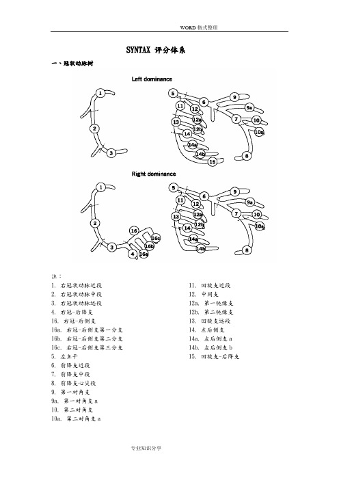
SYNTAX 评分体系一、冠状动脉树注:1. 右冠状动脉近段 11. 回旋支近段2. 右冠状动脉中段 12. 中间支3. 右冠状动脉远段 12a. 第一钝缘支4. 右冠-后降支 12b. 第二钝缘支16. 右冠-后侧支 13. 回旋支远段16a. 右冠-后侧支第一分支 14. 左后侧支16b. 右冠-后侧支第二分支 14a. 左后侧支a 16c. 右冠-后侧支第三分支 14b. 左后侧支b5. 左主干 15. 回旋支-后降支6. 前降支近段7. 前降支中段8. 前降支心尖段9. 第一对角支9a. 第一对角支a10. 第二对角支10a. 第二对角支a二、各节段的权重因数冠脉节段右优势型冠脉左优势型冠脉1. 右冠状动脉近段 1 02. 右冠状动脉中段 1 03. 右冠状动脉远段 1 04. 右冠-后降支 1 /16. 右冠-后侧支 0.5 /16a. 右冠-后侧支第一分支 0.5 /16b. 右冠-后侧支第二分支 0.5 /16c. 右冠-后侧支第三分支 0.5 /5. 左主干 5 66. 前降支近段 3.5 3.57. 前降支中段 2.5 2.58. 前降支心尖段 1 19. 第一对角支 1 19a. 第一对角支a 1 110. 第二对角支 0.5 0.510a. 第二对角支a 0.5 0.511. 回旋支近段 1.5 2.512. 中间支 1 112a. 第一钝缘支 1 112b. 第二钝缘支 1 113. 回旋支远段 0.5 1.514. 左后侧支 0.5 114a. 左后侧支a 0.5 114b. 左后侧支b 0.5 115. 回旋支-后降支 / 1三、病变不良特征评分-完全闭塞×5-50-99%狭窄×2完全闭塞-大于3个月或闭塞时间不祥 +1-钝型残端 +1-桥侧枝 +1-闭塞后的第一可见节段 +1/每一不可见节段-边支 -边支小于1.5mm +1三叉病变-1个病变节段 +3-2个病变节段 +4-3个病变节段 +5-4个病变节段 +6分叉病变-A、B、C型病变 +1-E、D、F、G型病变 +2-角度小于70° +1开口病变 +1严重扭曲 +2长度大于20mm +1严重钙化 +2血栓 +1弥漫病变/小血管病变 +1/每一节段四、SYNTAX评分系统SYNTAX积分通过计算机程序计算得出。

s y n t a x评分系统方法及意义(总7页)-CAL-FENGHAI.-(YICAI)-Company One1-CAL-本页仅作为文档封面,使用请直接删除08年长城会上听专家讲了Syntax score评分方法,这评分方法比较复杂,主要是依据冠脉造影的结果来判断。
在准备阅读之前,先要熟悉冠脉解剖的节段分布情况:(上图为左优势型冠脉解剖分布,下图为右优势型解剖分布)Figure 1. Definition of the coronary tree segments(冠脉束血管段的识别)1. RCA proximal: From the ostium to one half the distance to the acute margin of the heart.2. RCA mid: From the end of first segment to acute margin of heart.3. RCA distal: From the acute margin of the heart to the origin of the posterior descending artery.4. Posterior descending artery: Running in the posterior interventricular groove.16. Posterolateral branch from RCA: Posterolateral branch originating from the distal coronary artery distal to the crux.16a. Posterolateral branch from RCA: First posterolateral branch from segment 16. 16b. Posterolateral branch from RCA: Second posterolateral branch from segment 16. 16c. Posterolateral branch from RCA: Third posterolateral branch from segment 16.5. Left main: From the ostium of the LCA through bifurcation into left anterior descending and left circumflex branches.6. LAD proximal: Proximal to and including first major septal branch.7. LAD mid: LAD immediately distal to origin of first septal branch and extending to the point where LAD forms an angle (RAO view). If thisangle is not identifiable this segment ends at one half the distance from the first septal to the apex of the heart.8. LAD apical: Terminal portion of LAD, beginning at the end of previous segment and extending to or beyond the apex.9. First diagonal: The first diagonal originating from segment 6 or 7.9a. First diagonal a: Additional first diagonal originating from segment 6 or 7, before segment 8.10. Second diagonal: Originating from segment 8 or the transition between segment 7 and 8.10a. Second diagonal a: Additional second diagonal originating from segment 8.11. Proximal circumflex artery: Main stem of circumflex from its origin of left main and including origin of first obtuse marginal branch.12. Intermediate/anterolateral artery: Branch from trifurcating left main other than proximal LAD or LCX. It belongs to the circumflex territory.12a. Obtuse marginal a: First side branch of circumflex running in general to the area of obtuse margin of the heart.12b. Obtuse marginal b: Second additional branch of circumflex running in the same direction as 12.13. Distal circumflex artery: The stem of the circumflex distal to the origin of the most distal obtuse marginal branch, and running along the posteriorleft atrioventricular groove. Caliber may be small or artery14. Left posterolateral: Running to the posterolateral surface of the left ventricle. May be absent or a division of obtuse marginal branch.14a. Left posterolateral a: Distal from 14 and running in the same direction.14b. Left posterolateral b: Distal from 14 and 14 a and running in the same direction.15. Posterior descending: Most distal part of dominant left circumflex when present. It gives origin to septal branches. When this arteryis present, segment 4 isusually absent。
冠状动脉病变SYNTAX评分体系(附图)、冠状动脉树注:1. 右冠状动脉近段2. 右冠状动脉中段3. 右冠状动脉远段4. 右冠-后降支16.右冠-后侧支16a.右冠-后侧支第一分支16b.右冠-后侧支第二分支16c.右冠-后侧支第三分支5. 左主干6. 前降支近段7. 前降支中段8. 前降支心尖段9. 第一对角支9a.第一对角支a10. 第二对角支10a.第二对角支aLeft dominanceRight dominance11. 回旋支近段12. 中间支12a.第一钝缘支12b.第二钝缘支13. 回旋支远段14. 左后侧支14a.左后侧支a14b.左后侧支b15. 回旋支-后降支、各节段的权重因数冠脉节段右优势型冠脉左优势型冠脉1.右冠状动脉近段102.右冠状动脉中段103.右冠状动脉远段104.右冠-后降支1/16.右冠-后侧支0.5/16a.右冠-后侧支第一分支0.5/16b.右冠-后侧支第二分支0.5/16c.右冠-后侧支第三分支0.5/5.左主干566.前降支近段 3.5 3.57.前降支中段 2.5 2.58.前降支心尖段119.第一对角支119a.第一对角支a1110.第二对角支0.50.510a.第二对角支a0.50.511.回旋支近段 1.5 2.512.中间支1112a.第一钝缘支1112b.第二钝缘支1113.回旋支远段0.5 1.514.左后侧支0.5114a.左后侧支a0.5114b.左后侧支b0.5115.回旋支-后降支/ 1、病变不良特征评分血管狭窄-完全闭塞X 5-50-99% 狭窄X 2完全闭塞-大于3个月或闭塞时间不祥+ 1-钝型残端+ 1-桥侧枝+ 1-闭塞后的第一可见节段+ 1/每一不可见节段-边支-边支小于1.5mm+1三叉病变-1个病变节段+3-2个病变节段+4-3个病变节段+5-4个病变节段+6分叉病变-A、B、C型病变+1-E、D、F、G型病变+2-角度小于70°+1开口病变+ 1严重扭曲+2长度大于20mm+1严重钙化+2血栓+ 1弥漫病变/小血管病变+1/每•节段四、SYNTAX评分系统SYNTAX积分通过计算机程序计算得出。
《冠脉SYNTAX评分及其衍生评分与肾功能相关性研究》篇一一、引言随着社会发展和人口老龄化进程,心血管疾病的发病率逐渐升高,冠状动脉疾病成为影响人们健康的重要问题。
冠脉SYNTAX评分作为一种评估冠状动脉病变复杂程度的工具,在临床实践中得到了广泛应用。
同时,随着医学研究的深入,一系列衍生评分也应运而生。
本文旨在探讨冠脉SYNTAX评分及其衍生评分与肾功能的相关性,以期为临床治疗和预后评估提供更多依据。
二、研究背景冠脉SYNTAX评分是一种基于冠状动脉病变的解剖学评分系统,主要用于评估病变的复杂程度和手术风险。
该评分系统的应用,有助于医生制定更合理的治疗方案和评估患者预后。
然而,SYNTAX评分与其他临床指标如肾功能的关系尚未完全明确。
因此,本研究将关注冠脉SYNTAX评分及其衍生评分与肾功能的相关性。
三、研究方法本研究采用回顾性分析方法,收集了某大型医院心血管内科的冠状动脉疾病患者资料。
所有患者均接受了冠脉SYNTAX评分及肾功能相关指标的检测。
根据患者的SYNTAX评分和其他临床指标,我们将患者分为不同组别,并分析各组间肾功能指标的差异。
同时,我们还对SYNTAX评分的衍生评分与肾功能的关系进行了探讨。
四、研究结果1. 冠脉SYNTAX评分与肾功能相关性分析本研究发现,随着冠脉SYNTAX评分的增加,患者肾功能指标(如肾小球滤过率、血尿素氮等)呈现出逐渐恶化的趋势。
在多因素回归分析中,冠脉SYNTAX评分被证实为肾功能恶化的独立危险因素。
2. 衍生评分与肾功能相关性分析除了SYNTAX评分外,我们还探讨了其他衍生评分与肾功能的关系。
结果显示,某些衍生评分与肾功能恶化也存在一定相关性,但具体关系需结合其他临床指标综合分析。
五、讨论本研究表明,冠脉SYNTAX评分与肾功能之间存在显著相关性。
随着冠脉病变的复杂程度增加,患者肾功能可能逐渐恶化。
这可能与冠状动脉病变导致的血液循环障碍、肾脏缺血等因素有关。
SYNTAX 评分体系一、冠状动脉树注:1. 右冠状动脉近段 11. 回旋支近段2. 右冠状动脉中段 12. 中间支3. 右冠状动脉远段 12a. 第一钝缘支4. 右冠-后降支 12b. 第二钝缘支16. 右冠-后侧支 13. 回旋支远段16a. 右冠-后侧支第一分支 14. 左后侧支16b. 右冠-后侧支第二分支 14a. 左后侧支a 16c. 右冠-后侧支第三分支 14b. 左后侧支b5. 左主干 15. 回旋支-后降支6. 前降支近段7. 前降支中段8. 前降支心尖段9. 第一对角支9a. 第一对角支a10. 第二对角支10a. 第二对角支a二、各节段的权重因数冠脉节段右优势型冠脉左优势型冠脉1. 右冠状动脉近段 1 02. 右冠状动脉中段 1 03. 右冠状动脉远段 1 04. 右冠-后降支 1 /16. 右冠-后侧支 0.5 /16a. 右冠-后侧支第一分支 0.5 /16b. 右冠-后侧支第二分支 0.5 /16c. 右冠-后侧支第三分支 0.5 /5. 左主干 5 66. 前降支近段 3.5 3.57. 前降支中段 2.5 2.58. 前降支心尖段 1 19. 第一对角支 1 19a. 第一对角支a 1 110. 第二对角支 0.5 0.510a. 第二对角支a 0.5 0.511. 回旋支近段 1.5 2.512. 中间支 1 112a. 第一钝缘支 1 112b. 第二钝缘支 1 113. 回旋支远段 0.5 1.514. 左后侧支 0.5 114a. 左后侧支a 0.5 114b. 左后侧支b 0.5 115. 回旋支-后降支 / 1三、病变不良特征评分-完全闭塞×5-50-99%狭窄×2完全闭塞-大于3个月或闭塞时间不祥 +1-钝型残端 +1-桥侧枝 +1-闭塞后的第一可见节段 +1/每一不可见节段-边支 -边支小于1.5mm +1三叉病变-1个病变节段 +3-2个病变节段 +4-3个病变节段 +5-4个病变节段 +6分叉病变-A、B、C型病变 +1-E、D、F、G型病变 +2-角度小于70° +1开口病变 +1严重扭曲 +2长度大于20mm +1严重钙化 +2血栓 +1弥漫病变/小血管病变 +1/每一节段四、SYNTAX评分系统SYNTAX积分通过计算机程序计算得出。
08年长城会上听专家讲了Syntax score评分方法,这评分方法比较复杂,主要是依据冠脉造影的结果来判断。
在准备阅读之前,先要熟悉冠脉解剖的节段分布情况:(上图为左优势型冠脉解剖分布,下图为右优势型解剖分布)Figure 1. Definition of the coronary tree segments(冠脉束血管段的识别)1. RCA proximal: From the ostium to one half the distance to the acute margin of the heart.2. RCA mid: From the end of first segment to acute margin of heart.3. RCA distal: From the acute margin of the heart to the origin of the posterior descending artery.4. Posterior descending artery: Running in the posterior interventricular groove.16. Posterolateral branch from RCA: Posterolateral branch originating from the distal coronary artery distal to the crux.16a. Posterolateral branch from RCA: First posterolateral branch from segment 16.16b. Posterolateral branch from RCA: Second posterolateral branch from segment 16.16c. Posterolateral branch from RCA: Third posterolateral branch from segment 16.5. Left main: From the ostium of the LCA through bifurcation into left anterior descending and left circumflex branches.6. LAD proximal: Proximal to and including first major septal branch.7. LAD mid: LAD immediately distal to origin of first septal branch and extending to the point where LAD forms an angle (RAO view). If thisangle is not identifiable this segment ends at one half the distance from the first septal to the apex of the heart.8. LAD apical: Terminal portion of LAD, beginning at the end of previous segment and extending to or beyond the apex.9. First diagonal: The first diagonal originating from segment 6 or 7.9a. First diagonal a: Additional first diagonal originating from segment 6 or 7, before segment 8.10. Second diagonal: Originating from segment 8 or the transition between segment 7 and 8. 10a. Second diagonal a: Additional second diagonal originating from segment 8.11. Proximal circumflex artery: Main stem of circumflex from its origin of left main and including origin of first obtuse marginal branch.12. Intermediate/anterolateral artery: Branch from trifurcating left main other than proximal LAD or LCX. It belongs to the circumflex territory.12a. Obtuse marginal a: First side branch of circumflex running in general to the area of obtuse margin of the heart.12b. Obtuse marginal b: Second additional branch of circumflex running in the same direction as 12.13. Distal circumflex artery: The stem of the circumflex distal to the origin of the most distal obtuse marginal branch, and running along the posteriorleft atrioventricular groove. Caliber may be small or artery absent.14. Left posterolateral: Running to the posterolateral surface of the left ventricle. May be absent or a division of obtuse marginal branch.14a. Left posterolateral a: Distal from 14 and running in the same direction.14b. Left posterolateral b: Distal from 14 and 14 a and running in the same direction.15. Posterior descending: Most distal part of dominant left circumflex when present. It gives origin to septal branches. When this arteryis present, segment 4 is usually absent。
SYNTAX 评分方法是基于下面的分级系统发展出来的:1.根据ARTS研究修改的关于冠脉树血管段的AHA分级;2.Leaman评分;3. ACC/AHA 病变分级系统;4.完全闭塞分型系统;5.Duke 和ICPS分支病变分型系统;6.专家的意见。
现在介绍这个评分系统的具体内容,并结合病例来说明,注意一定要结合上述图上的血管段分级:Syntax score临床意义:1)SYNTAX评分值较低的组:0 到22分:研究数据显示通过PCI和CABG实现再血管化有相近的预后。
12个月CABG组累积MACCE 率为14.4%(n=247),TAXUS支架组为13.5%(n=299; P=0.71).SYNTAX评分值<22分的患者的治疗策略可以基于患者的个体病情特点、患者自己的意愿以及医生的判断,此时PCI和CABG均可。
2)SYNTAX评分值在23至32之间:12个月累积MACCE率在CABG组为11.7%(n=300),TAXUS组为16.6%(n=310; P=0.10)。
从中可见这些不良事件在PCI组轻微增加,但不显著。
这表明PCI在SYNTAX评分中等的患者仍然是可以选择的。
最终选择何种治疗策略取决于患者的特点和伴随疾病情况。
3)SYNTAX评分值高(≥33):直接导致12个月时PCI组明显增高的MACCE率。
CABG组为10.7%(n=316),TAXUS组为23.3%(n=290; P.001)这些患者解剖上十分复杂,从MACCE率表明PCI对于这群患者并不是个好选择,他们应该作为外科的候选者,此时应选择CABG。
SYNTAX实验的结果表明55%的患者仍然最好应选择CABG,然而,对于余下的患者采用PCI也是非常好的。
具体举例:见病变1:1)为左主干病变,对应血管段为5,对应权重为5分,由于为严重狭窄病变,应乘于2才是最后得分:5*2=10分;2)这个左主干病变为分叉前的病变,判断为type A分叉病变,得1分;3)存在严重的钙化,2分此病变共:10+1+2=13分。
病变2:1)为前降支近段重度狭窄,对应血管段为6,对应权重为3.5分,由于为严重狭窄病变,应乘于2才是最后得分:3.5*2=7分;2)这个左前降支病变为分叉前的病变,判断为type A分叉病变,得1分;3)成角<70度,1分;4)存在严重的钙化,2分此病变共:7+1+1+2=11分。
病变3:1)为回旋支病变,对应血管段为11,对应权重为1.5分,由于为闭塞病变,应乘于5才是最后得分:1.5*5=7.5分;2)闭塞时间不明,1分;3)闭塞残断钝,1分;4)边支血管累及,1分;5)闭塞以远段侧枝显影后可见血管段13(回旋支远段)显影,1分;6)存在严重的钙化,2分;7)闭塞长度大于20mm,1分;此病变共:7.5+1+1+1+1+2+1=14.5分。
病变4:1)为右冠近段病变,对应血管段为1,对应权重为1分,由于为闭塞病变,应乘于5才是最后得分:1*5=5分;2)闭塞时间不明,1分;3)闭塞残断钝,1分;4)边支血管累及,1分;5)闭塞以远段侧枝显影后可见血管段4(后降支)显影,3分;6)扭曲病变,2分;7)闭塞长度大于20mm,1分;8)存在严重的钙化,2分;此病变共:5+1+1+1+3+2+2+1=16分。
一共:13+11+14.5+16=54.5分。
大于33分,适合CABG,如果行PCI的话,那么可能增加不良事件发生。
有个网页,在这里面它有个计算器。