2013-cancer cell-Chromatin-Bound IκBα RegulatesPolycomb Target Genes in Differentiation and Cancer
- 格式:pdf
- 大小:6.05 MB
- 文档页数:16

Cell Cycle 1657Cell Cycle 12:11, 1657–1658; June 1, 2013; © 2013 Landes BioscienceEditoriaLs: CELL CyCLE FEaturEs EditoriaLs: CELL CyCLE FEaturEs PIWI proteins and their associated smallRNAs (PIWI-interacting RNAs, piR-NAs) are essential for fertility in mam-mals.1,2 piRNAs (24–35 nt) are longerthan miRNAs (21–23 nt) and have2'-O -methyl-modified 3' termini.3-6Like miRNAs, piRNAs bind membersof the Argonaute family of proteins, butpiRNAs are unique in that they guidePIWI proteins, a specialized subfamilyof Argonaute proteins that are expressedmainly in germ cells.1 The sequences ofpiRNAs are more diverse than any otherknown class of cellular RNAs. For exam-ple, our 8.8 million piRNA reads in adeep sequencing library from adult mousetestis comprised 2.7 million differentpiRNAs; > 90% of piRNA species weresequenced just once.piRNAs map to large blocks ofgenomic sequence called clusters.4,5 Thearchitecture of piRNA clusters suggeststhat mature piRNAs derive from precur-sor transcripts via multiple RNA process-ing steps. While > 80% of piRNA readsin flies map to transposons, ~93% ofadult mouse piRNA reads map to a singlesite in the genome. That so many mousepiRNAs map uniquely to the genomefacilitates the study of their precursortranscripts. Using total RNA sequencing,we detected long RNAs (> 100 nt) frompiRNA clusters. Chromatin immunopre-cipitation of RNA polymerase II and IIIsuggests that these transcripts are tran-scribed by RNA polymerase II. Consistentwith mouse piRNA precursors being RNApolymerase II transcripts, the long RNAsbear a standard cap structure (detected bycap analysis of gene expression; CAGE), and their 3' ends possess a poly(A) tail Defining piRNA primary transcriptsXin Zhiguo Li, Christian K. roy, Melissa J. Moore and Phillip d. Zamore*Howard Hughes Medical institute; rNa therapeutics institute and department of Biochemistry and Molecular Pharmacology; university of Massachusetts Medical school; Worcester, Ma usa*Correspondenceto:PlillipD.Zamore;Email:***************************Submitted: 04/22/13; Accepted: 04/23/13/10.4161/cc.24989Comment on: Li XZ, et al. Mol Cell 2013; 50:67-81; PMID:23523368; /10.1016/j.molcel.2013.02.016(detected by polyadenylation site sequenc-ing; PAS-seq).7Historically, piRNA clusters have been defined computationally using mature piRNA sequences. Thus, clus-ters are not transcriptional units. When we began our studies, it was unknown whether piRNAs originated from sin-gle continuous transcripts or multiple short transcripts, as they do in nema-todes. The location of piRNA precursor transcription start sites (TSSs) and pro-moters or whether they are spliced was also unknown. We discovered the first transcription factor regulating piRNA biogenesis by analyzing a subset of 15 clusters that appeared to be transcribed bidirectionally. In these clusters, a short piRNA region lacking piRNA separates piRNAs mapping to the genomic minus strand from those mapping to the plus strand. These putative bidirectional pro-moter regions contain binding motifs for the MYB family of transcription fac-tors (E = 8.3 × 10⁻¹²). We used the sub-set of clusters for motif finding, because the computationally defined bound-ary of most clusters is more than 3 kbp away from experimentally determined TSSs. Accurately annotating the TSSs of piRNA cluster transcripts allowed us to discover that MYB motifs are signifi-cantly enriched (E = 9.1 × 10⁻²⁸) in 100 piRNA promoters, including both bidi-rectional and unidirectional transcribed clusters.7 This allowed us to define the transcription units of piRNA-producing loci and replace the previous computa-tionally defined cluster annotations.We identified primary piRNA tran-scripts by a combination of de novo and reference-based transcript assembly using paired-end total RNA sequencing. We further corrected the ends of these tran-scripts using CAGE and PAS. Our data allowed us to define 467 piRNA precursor transcripts derived from 214 piRNA-pro-ducing loci.7 The 214 loci constitute just 0.33% of the mouse genome yet account for 95% of piRNAs in the adult mouse testis.The 214 loci can be divided into two different types of piRNA-producing genes. Genic piRNA loci are known pro-tein-coding genes that also produce piR-NAs and are mostly expressed during early spermatogenesis before the pachytene stage of meiosis. Historically, piRNAs mapping to these genes have been referred to as pre-pachytene piRNAs.8 Intergenic piRNA loci lie far in the genome from other annotated genes. Most piRNAs from these loci are detected after 12.5 days postpartum and have been called pachy-tene piRNA clusters.The transcription factor A-MYB binds at the promoters of the pachytene piRNA-producing loci, and the loss of A-Myb depleted both piRNA precursor tran-scripts as well as mature piRNAs across the length of the loci.7 Thus, our data represent the first formal demonstration that long RNA polymerase II transcripts are the precursors of mature piRNAs in mammals. That is, piRNAs are derived from long, continuous RNAs that are subsequently fragmented and processed into piRNAs (Fig. 1). The set of piRNA loci and well-annotated piRNA precursor transcripts defined by our work provide an invaluable resource for further study of piRNA biogenesis and function.1. Deng W, et al. Dev Cell 2002; 2:819-30;PMID:12062093; /10.1016/S1534-5807(02)00165-X2. K uramochi-Miyagawa S, et al. Development 2004;131:839-49; PMID:14736746; http://dx.doi.org/10.1242/dev.009733. Aravin A, et al. Nature 2006; 442:203-7;PMID:167517774. Lau NC, et al. Science 2006; 313:363-7;PMID:16778019; /10.1126/sci-ence.11301645. Girard A, et al. Nature 2006; 442:199-202;PMID:167517766. Grivna ST, et al. Genes Dev 2006; 20:1709-14;PMID:16766680; /10.1101/gad.14344067. Li XZ, et al. Mol Cell 2013; 50:67-81; PMID:23523368;/10.1016/j.molcel.2013.02.0168. Aravin AA, et al. Science 2007; 316:744-7;PMID:17446352; /10.1126/sci-ence.1142612Figure 1. a model for pirNa biogenesis. Primary pirNa transcripts are transcribed by rNa poly-merase ii and contain 5' caps, exons and introns and poly(a) tails. the transcription of pachytenepirNa genes is controlled by a-MyB; transcription factor(s) (tF) controlling pre-pachytene pirNagenes remain to be discovered. Current models of pirNa biogenesis propose that PLd6 deter-mines the 5' end of pirNa intermediates with lengths > 30 nt. these intermediates are proposedto then be loaded into PiWi proteins. after PiWi binding, a nuclease is thought to trim the 3' endof the pirNa to the length characteristic of the particular bound PiWi protein. Finally, further trim-ming is prevented by addition of a 2'-O-methyl group to the 3' end of the mature pirNa by the-adenosylmethionine-dependent methyltransferase Hen1.1658 Cell Cycle Volume 12 issue 11Below is given annual work summary, do not need friends can download after editor deleted Welcome to visit againXXXX annual work summaryDear every leader, colleagues:Look back end of XXXX, XXXX years of work, have the joy of success in your work, have a collaboration with colleagues, working hard, also have disappointed when encountered difficulties and setbacks. Imperceptible in tense and orderly to be over a year, a year, under the loving care and guidance of the leadership of the company, under the support and help of colleagues, through their own efforts, various aspects have made certain progress, better to complete the job. For better work, sum up experience and lessons, will now work a brief summary.To continuously strengthen learning, improve their comprehensive quality. With good comprehensive quality is the precondition of completes the labor of duty and conditions. A year always put learning in the important position, trying to improve their comprehensive quality. Continuous learning professional skills, learn from surrounding colleagues with rich work experience, equip themselves with knowledge, the expanded aspect of knowledge, efforts to improve their comprehensive quality.The second Do best, strictly perform their responsibilities. Set up the company, to maximize the customer to the satisfaction of the company's products, do a good job in technical services and product promotion to the company. And collected on the properties of the products of the company, in order to make improvement in time, make the products better meet the using demand of the scene.Three to learn to be good at communication, coordinating assistance. On‐site technical service personnel should not only have strong professional technology, should also have good communication ability, a lot of a product due to improper operation to appear problem, but often not customers reflect the quality of no, so this time we need to find out the crux, and customer communication, standardized operation, to avoid customer's mistrust of the products and even the damage of the company's image. Some experiences in the past work, mentality is very important in the work, work to have passion, keep the smile of sunshine, can close the distance between people, easy to communicate with the customer. Do better in the daily work to communicate with customers and achieve customer satisfaction, excellent technical service every time, on behalf of the customer on our products much a understanding and trust.Fourth, we need to continue to learn professional knowledge, do practical grasp skilled operation. Over the past year, through continuous learning and fumble, studied the gas generation, collection and methods, gradually familiar with and master the company introduced the working principle, operation method of gas machine. With the help of the department leaders and colleagues, familiar with and master the launch of the division principle, debugging method of the control system, and to wuhan Chen Guchong garbage power plant of gas machine control system transformation, learn to debug, accumulated some experience. All in all, over the past year, did some work, have also made some achievements, but the results can only represent the past, there are some problems to work, can't meet the higher requirements. In the future work, I must develop the oneself advantage, lack of correct, foster strengths and circumvent weaknesses, for greater achievements. Looking forward to XXXX years of work, I'll be more efforts, constant progress in their jobs, make greater achievements. Every year I have progress, the growth of believe will get greater returns, I will my biggest contribution to the development of the company, believe inyourself do better next year!I wish you all work study progress in the year to come.。
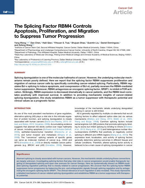
Cancer CellArticleThe Splicing Factor RBM4ControlsApoptosis,Proliferation,and Migrationto Suppress Tumor ProgressionYang Wang,1,2,*Dan Chen,3Haili Qian,4Yihsuan S.Tsai,2Shujuan Shao,5Quentin Liu,1Daniel Dominguez,2and Zefeng Wang2,*1Institute of Cancer Stem Cell,Second Affiliated Hospital,Cancer Center,Dalian Medical University,Dalian116044,China2Department of Pharmacology and Lineberger Comprehensive Cancer Center,University of North Carolina,Chapel Hill,NC27599,USA3Department of Pathology,First Affiliated Hospital.Dalian Medical University,Dalian116001,China4State Key Laboratory of Molecular Oncology,Peking Union Medical College and Chinese Academy of Medical Sciences,Beijing100021, China5Key Laboratory of Proteomics of Liaoning Province,Dalian Medical University,Dalian116044,China*Correspondence:yangwang@(Y.W.),zefeng@(Z.W.)/10.1016/r.2014.07.010SUMMARYSplicing dysregulation is one of the molecular hallmarks of cancer.However,the underlying molecular mech-anisms remain poorly defined.Here we report that the splicing factor RBM4suppresses proliferation and migration of various cancer cells by specifically controlling cancer-related splicing.Particularly,RBM4reg-ulates Bcl-x splicing to induce apoptosis,and coexpression of Bcl-xL partially reverses the RBM4-mediated tumor suppression.Moreover,RBM4antagonizes an oncogenic splicing factor,SRSF1,to inhibit mTOR acti-vation.Strikingly,RBM4expression is decreased dramatically in cancer patients,and the RBM4level corre-lates positively with improved survival.In addition to providing mechanistic insights of cancer-related splicing dysregulation,this study establishes RBM4as a tumor suppressor with therapeutic potential and clinical values as a prognostic factor.INTRODUCTIONAs one of the most prevalent mechanisms of gene regulation, alternative splicing(AS)plays a vital role in the intricate regula-tion of protein function,and splicing dysregulation is closely associated with human cancers(David and Manley,2010;Ol-tean and Bates,2013;Venables,2006).Accumulating evidence suggests that aberrant AS elicits control over major hallmarks of cancer,including apoptosis(Schwerk and Schulze-Osthoff, 2005),epithelial-mesenchymal transition(Warzecha et al., 2010),and tumor invasion and metastasis(Ghigna et al., 2008).The‘‘cancerous’’splicing variants of specific genes can serve as molecular markers of cancer(e.g.,CD44and WT1)(Venables et al.,2008)or directly mediate cancer patho-genesis(e.g.BRCA1and p53)(Venables,2006).However,knowledge of the mechanistic details underlying deregulated splicing in cancer is still limited.AS is generally regulated by multiple cis-elements that recruit splicing factors to affect adjacent splice sites(ss)via various mechanisms(Matera and Wang,2014;Matlin et al.,2005; Wang and Burge,2008).Common splicing factors include serine/arginine-rich(SR)proteins that promote splicing by bind-ing to exons but inhibit splicing by binding to introns(Erkelenz et al.,2013;Wang et al.,2013)and heterogeneous nuclear ribo-nucleoproteins(hnRNPs)that positively or negatively control splicing in different pre-mRNA regions(Wang et al.,2012).The expression level,localization,and activity of splicing factors generally determine splicing outcomes in different tissues and cellular conditions.Therefore,altered splicing factor activity is believed to be a main cause of splicing dysregulation incancer374Cancer Cell26,374–389,September8,2014ª2014Elsevier Inc.(Bechara et al.,2013;Shkreta et al.,2013).For example,SRSF1 is a proto-oncogene that controls splicing of several cancer-related genes,including those in the mammalian target of rapa-mycin(mTOR)pathway(Blaustein et al.,2005;Karni et al.,2007). Because splicing dysregulation is one of the molecular hallmarks of cancer(Oltean and Bates,2013),specifically targeting splicing factors opens potential new avenues for cancer therapy(Dehm, 2013).We have previously identified RNA-binding motif4(RBM4)as a binding factor for a group of intronic splicing regulatory ele-ments that control the AS of human genes(Wang et al.,2012). Initially identified by sharing the nuclear import pathway with SR proteins,RBM4shuttles between the cytoplasm and nucleus but is mostly found in nuclear speckles(Lai et al.,2003),where most splicing events occur.RBM4has been shown consistently to control the AS of Tau and a-tropomyosin(Kar et al.,2006;Lin and Tarn,2005).In addition,RBM4has been found to affect translation(Lin and Tarn,2009;Uniacke et al.,2012).Multiple physiological functions have been reported for RBM4,including mediating differentiation of muscle and pancreas cells(Lin et al., 2007;Lin et al.,2013).However,the involvement of RBM4in tumorigenesis has not been reported.Here we systematically analyzed RBM4-mediated changes of the transcriptome and as-sessed the role of RBM4in cancer progression.RESULTSRBM4Is a Sequence-Specific Splicing Inhibitor that Regulates Various AS EventsPreviously we identified several groups of intronic splicing regu-latory elements and their cognate splicing factors(Wang et al., 2012,2013).We demonstrated that,of those factors,RBM4spe-cifically binds to the GTAACG motif to inhibit splicing from in-trons(Wang et al.,2012).In addition,another RBM4binding motif(CGG repeats)was also identified with crosslinking immu-noprecipitation sequencing(Uniacke et al.,2012).Because AS is usually regulated in a context-dependent manner,we sought to examine how RBM4controls splicing when bound to distinct RNA motifs in different pre-mRNA contexts.We generated four splicing reporters with candidate RBM4-binding motifs(GTAACG or CGGCGG)inserted in different re-gions to examine whether RBM4can specifically alter their splicing(Figure1).First,we found that RBM4specifically in-hibited the inclusion of a cassette exon containing its cognate binding sites,whereas the control reporter was not affected(Fig-ure1A).Furthermore,RBM4specifically suppressed the inclu-sion of a reporter exon with a downstream RBM4binding site (Figure1B).These results suggest that RBM4functions as a gen-eral splicing inhibitor to specifically suppress splicing from both exonic and intronic contexts.Such activities are in contrast to DAZAP1,a splicing factor that recognizes the same GTAACG site but functions as a splicing activator(Choudhury et al., 2014).Interestingly,DAZAP1does not affect splicing of exons containing a nearby CGGCGG site(Figures S1A and S1B avail-able online),suggesting a partial overlap of binding specificity and an incomplete functional competition between RBM4and DAZAP1.Using splicing reporters containing RBM4-binding motifs be-tween alternative50ss or30ss,we found that RBM4reduced the use of the downstream50ss(Figure1C)or upstream30ss (Figure1D).The inhibition of distal alternative ss is again sequence-specific because RBM4showed no effect on the con-trol reporters(Figures1C and1D).Consistently,knockdown of RBM4with small hairpin RNA had opposite effects by increasing exon inclusion of the same splicing reporters that contain RBM4-binding sites in various locations(Figures S1C–S1F).In addition, similar results were obtained in a different cell type(e.g.HeLa cells),indicating that the splicing regulation activity of RBM4is not limited to a specific cell line(Figures S1G–S1J).Together, these data demonstrate that RBM4is a general splicing inhibitor that controls different types of AS when specifically binding to pre-mRNA.Like many canonical splicing factors,RBM4has a modular domain configuration.The N terminus contains two RNA recog-nition motifs(RRMs)and a CCHC-type zincfinger that can specifically bind RNAs,whereas the C terminus contains a low-complexity region(i.e.Ala-rich stretches)that can interact with other proteins(Lin and Tarn,2009)(Figure1E).To examine whether RBM4has a modular role in splicing regulation,we fused the full-length N-or C-terminal fragments of RBM4to another RNA binding domain,Pumilio/FBF(PUF)(Wang et al., 2009).We coexpressed the fusion proteins with splicing re-porters containing cognate PUF targets inside an alternative exon(Figure1F)or at a downstream intron(Figure1G)and measured how splicing is affected.As expected,tethering the full-length RBM4to a target site inside an alternative exon sup-pressed exon inclusion.Surprisingly,tethering either the N-or C-terminal domain of RBM4partially inhibited exon inclusion (Figure1F),suggesting that the RNA binding fragment and the low-complexity domain both serve as functional modules. Such an effect is sequence-specific because these fusion pro-teins had no effect on control reporters with a noncognate target. Consistently,the full-length RBM4inhibited exon inclusion when tethered downstream of a cassette exon(Figure1G).Interest-ingly,the N-terminal fragment partially inhibited splicing from an intron,whereas the C terminus showed a slight splicing-inhib-itory activity(Figure1G).Together,the N-terminal RNA-binding fragment and the C-terminal low-complexity domain of RBM4 function cooperatively to control different types of AS events in a sequence-specific manner.Global Regulation of the Transcriptome by RBM4in Cancer-Related GenesTo gain further insights into RBM4-regulated AS events and, thereby,its physiological functions,we conducted high-throughput sequencing of mRNA(mRNA-seq)with H157cells expressing RBM4.With$80million100-nt paired-end reads, we identified473RBM4-regulated AS events with an obvious change of percent-spliced-in(PSI)values(PSI R0.15).Figure2A shows the read tracks of two examples.We found that various types of AS can be regulated by RBM4,including skipped exon(SE),alternative50ss exon(A5E),alternative30ss exon (A3E),retained intron(RI),mutually exclusive exons(MXE),and tandem UTR(TUTR)(Figure2B;Table S1).Subsequent analysis indicated that most of the AS events were negatively regulated by RBM4(decreased PSI value by RBM4expression)(Figure2C), consistent with ourfinding that RBM4suppressed splicing when binding directly to its pre-mRNA targets(Figures1A–1D).Cancer CellRBM4Inhibits Tumorigenesis via Splicing ControlCancer Cell26,374–389,September8,2014ª2014Elsevier Inc.375(legend on next page)Cancer CellRBM4Inhibits Tumorigenesis via Splicing Control376Cancer Cell 26,374–389,September 8,2014ª2014Elsevier Inc.We further analyzed RNA motifs in RBM4-regualted pre-mRNAs by extracting the sequences near the RBM4-regulated SEs or between alternative 50ss of A5E.The relative abundance of RBM4binding motifs (GTAACG and CGGCGG)in these re-gions was compared with control exons unaffected by RBM4(Fairbrother et al.,2002).We found that RBM4-binding motifs are enriched near the SEs or A5Es negatively regulated by RBM4(Figure 2D),consistent with the model that RBM4directly recognizes these pre-mRNAs to control splicing.The AS events apparently promoted by RBM4are likely due to indirect effects because these exons lack known RBM4binding motifs (Figure 2D).When analyzing cellular functions of RBM4-regulated AS events using gene ontology,we found that RBM4affects genes in the RNA processing pathway,including translation control,RNA processing,and the mRNA metabolic process (Figure 2E).Such functional enrichment is not surprising because RBM4is an RNA binding factor known to regulate splicing and translation.Intriguingly,RBM4targets are also enriched with cancer-related functions such as regulation of the NF-k B cascade and cell cy-cle.In addition,several RBM4-regulated AS events were found to regulate the apoptotic pathway.Although this enrichment of apoptosis is slightly below our significance cutoff,the changes of PSI value are fairly large and,therefore,may have significant functional consequences.Many of the RBM4-regulated splicing targets were functionally connected into well linked interaction networks,as judged by the Search Tool for the Retrieval of Inter-acting Genes/Proteins (STRING)(Figure 2F).As expected,two large subgroups of RBM4targets contain genes involved in translation control and RNA processing.Surprisingly,the other subgroup includes many genes involved in cell migration and adhesion (Figure 2F).Taken together,these results suggest that the biological processes affected by RBM4are related to apoptosis,proliferation,migration,and tumorigenesis.We subsequently validated mRNA-seq results by measuring the splicing change of ten newly identified targets that were selected arbitrarily to include genes with a cancer-related func-tion.We confirmed that RBM4either positively or negatively con-trols all endogenous AS events tested (Figure 2G)and that the relative changes of PSIs obtained from RT-PCR are highly corre-lated to those observed by mRNA-seq (Figure S2A;R 2=0.6).These events were also validated in another cell line (HeLa)(Fig-ure S2B),suggesting that RBM4can regulate AS with consistent activity across different cell types.In addition,we found that knockdown of RBM4caused opposite changes of splicing inendogenous RBM4targets,further confirming the reliability of our analyses (Figures S2C and S2D).We also analyzed how RBM4affects global gene expression.We identified 185genes with significant expression change (>2-fold with adjusted p <0.05)(Table S2).These genes are associ-ated significantly with cancer-related functions,as judged by gene ontology (including DNA replication,chemotaxis,cell pro-liferation,response to wounding,cell cycle,and cell migration;Figure 2H),again suggesting that RBM4is involved in cancer cell proliferation and migration.Many RBM4-regulated genes were also connected functionally into a densely linked network that contains genes involved in regulating cell proliferation,wound healing,cell cycle,and DNA damage (Figure 2I).The selected RBM4targets were further validated with real-time RT-PCRs (Figure 2J).Taken together,these data imply that RBM4may be a key regulator of cell proliferation and migration,therefore controlling cancer progression.RBM4Inhibits Cancer Cell Proliferation and Migration To examine this possible role of RBM4in cancer progression,we stably expressed RBM4in a panel of human cancer cells,including H157(lung cancer),MDA-MB-231(breast cancer),SKOV3(ovarian cancer),Panc-1(pancreatic cancer),HepG2(liver cancer),and PC-3(prostate cancer)(Figure S3A).Strikingly,in all cancer cells tested,RBM4inhibited anchorage-dependent or anchorage-independent growth,as judged by colony forma-tion or soft agar assay (Figure 3A).In addition,RBM4inhibited migration of these cells in a wound healing assay (Figure 3B).Together,the inhibition of cancer cell proliferation and migration by RBM4suggests that it may function as a tumor suppressor.We further analyzed how RBM4affects cancer progression using non-small cell lung carcinoma (NSCLC)cells,which repre-sent one of the most prevalent human cancers.The RBM4levels were decreased markedly in a panel of NSCLC cells compared with normal bronchial cells (Figure 3C).Consistently,when re-expressed in a NSCLC cell line,H157,RBM4significantly in-hibited cell growth (Figure 3D;p =0.02by t test).Similar growth inhibition by RBM4was observed in 293T cells (Figures S3B and S3C).Interestingly,although both the N-and C termini of RBM4partially regulate splicing,lung cancer cells expressing either domain (amino acids (aa)1–177or aa 178–364of RBM4)dis-played normal growth rates (Figure 3E),suggesting that both do-mains are required to suppress tumorigenesis.To further assess whether RBM4affects cancer growth in vivo,we determined whether RBM4re-expression can suppressFigure 1.Splicing Regulation by RBM4(A)The RBM4binding sites and a control (GAATTG)were inserted into splicing reporter pGZ3and cotransfected with the RBM4expression vector or an empty vector (mock)into 293T cells.Splicing changes were examined by electrophoresis of RT-PCR products.(B)The same set of sequences as analyzed in (A)was inserted downstream of a cassette exon in the pZW2C reporter to measure splicing changes as in (A).(C and D)The same set of RBM4-binding sequences as analyzed in (A)was inserted into the splicing reporters between two tandem sites with competing 50(C)or 30ss (D),and splicing changes were measured as in (A).(E)Schematics of RBM4domains.The R1R2Z fragment contains two RRM domains and a zinc finger domain.The polyalanine fragment contains a polyalanine stretch.(F and G)Different RBM4fragments were fused to a PUF domain,PUF(3-2),that specifically binds to its target RNA.The fusion proteins were cotransfected with a splicing reporter containing a PUF binding site or a control (Ctl)site in a cassette exon (F)or at a downstream intron (G),and splicing changes were measured as in (A).The arrowhead indicates a nonspecific product (F).In panels measuring changes in splicing,expression of exogenous protein was confirmed by western blot analyses.Tubulin served as a protein loading control.Three independent experiments were conducted,with the mean ±SD of PSIs plotted below the representative gels.*p <0.05as calculated by Student’s t test.See also Figure S1.Cancer CellRBM4Inhibits Tumorigenesis via Splicing ControlCancer Cell 26,374–389,September 8,2014ª2014Elsevier Inc.377(legend on next page)Cancer CellRBM4Inhibits Tumorigenesis via Splicing Control378Cancer Cell 26,374–389,September 8,2014ª2014Elsevier Inc.tumor growth in a xenograft mouse model.We generated H157-luc-RBM4cells and control cells with lentiviral vectors and injected them subcutaneously into the flanks of nude mice (left flank,RBM4;right flank,control).The growth of tumors was measured every 3days for 5weeks,and xenograft tumors were removed for final analysis.Consistent with the in vitro re-sults,cells expressing RBM4developed smaller tumors compared with control cells (Figures 3F and 3G).In addition,the xenograft tumors with RBM4re-expression grew much slower than controls (Figure 3H),suggesting that RBM4substan-tially inhibits cancer progression in vivo.Together,these findings indicate that RBM4is a potent tumor suppressor that inhibits lung cancer progression both in cultured cells and in a tumor xenograft model.RBM4Induces Cancer Cell Apoptosis via Regulating AS of Bcl-xTo determine the mechanisms of how RBM4affects cancer pro-gression,we focused on an RBM4target gene,Bcl-x,an apoptosis regulator that produces two splicing isoforms with opposite functions.By alternative use of 50ss,Bcl-x is spliced as an antiapoptotic isoform (Bcl-xL)or a proapoptotic isoform (Bcl-xS)(Adams and Cory,2007).RBM4expression appeared to shift Bcl-xL into Bcl-xS (Figure 2G).Such a shift requires an entire RBM4because neither the N terminus nor the C terminus can affect Bcl-x splicing by itself (Figure S4A).We identified a po-tential RBM4binding site (CGGCGG)between the two alterna-tive 50ss (Figure 4A),implying that RBM4may control splicing through binding directly to Bcl-x pre-mRNA.Consistently,with an RNA immunoprecipitation assay,we found that RBM4indeed binds directly to the endogenous Bcl-x pre-mRNA but not the control pre-mRNA of another alternatively spliced apoptotic gene (Mcl1)(Figure 4B).Using a splicing reporter containing Bcl-x pre-mRNA,we found that RBM4binding is indeed depen-dent on the CGGCGG site because mutation of this site abol-ished RNA-protein interaction (Figure 4C).Replacing the mutated sequence with the other RBM4-binding site (GTAACG)restored the interaction,confirming that RBM4directly recog-nizes the exon extension region of Bcl-x.In addition to H157cells,an inducible expression of RBM4also shifted splicing of Bcl-x in 293cells (Figure 4D).This shift caused a rapid and robust decrease of Bcl-xL protein,as judgedby western blot analysis (Figure S4B).To determine whether the binding by RBM4is responsible for the observed splicing shift,we cotransfected RBM4with a series of Bcl-x reporters contain-ing various mutations near the alternative 50ss (Figure 4A).We found that RBM4shifted the splicing of the wild-type reporter by reducing Bcl-xL and that such a regulation was not affected by three exonic mutations (mutations 1–3)(Figure 4E).However,the mutation of the RBM4binding site (mut 4)completely abol-ished the splicing regulation through RBM4,indicating that the RBM4binding motif (CGGCGG)is indeed responsible for the Bcl-x splicing switch.Importantly,replacing CGGCGG with another RBM4binding site (mut 5)restored the regulation by RBM4(Figure 4E),suggesting that binding of RBM4to Bcl-x pre-mRNA is sufficient to shift splicing.The two splicing isoforms of Bcl-x have opposite functions in controlling apoptosis (Adams and Cory,2007).Bcl-xL is the pre-dominant isoform in cancer,and RNAi of Bcl-xL has been shown to induce apoptosis in several cancer cell lines (Mercatante et al.,2001;Zhu et al.,2005).We found that expression of RBM4in H157cells substantially reduced the level of Bcl-xL protein,re-sulting in the cleavage of caspase 3and poly-ADP-ribose poly-merase (PARP),two molecular markers of apoptosis (Figure 4F).Consistently,RBM4dramatically increased spontaneous apoptosis,as judged by flow cytometry (Figure 4G;Figure S4C).These results support the model that sequence-specific binding of RBM4to Bcl-x pre-mRNA shifts its splicing from antiapoptotic Bcl-xL to proapoptotic Bcl-xS,thereby promoting cancer cell death.RBM4Suppresses Tumor Progression in Part through Bcl-xBecause RBM4may inhibit cancer proliferation through modu-lating Bcl-x splicing,we next examined whether coexpression of Bcl-xL,but not other similar apoptotic regulators,can overturn the tumor suppressor activity of RBM4.We stably transfected the parental H157line containing re-expressed RBM4with Bcl-xL or another apoptotic inhibitor,Mcl-1(Figure 5A),generating a cell line with a partially restored Bcl-xL/Bcl-xS ratio and reduced PARP cleavage (Figure 5B).We found that cells expressing RBM4/Bcl-xL grew much faster than those ex-pressing RBM4alone,although the growth rate was not fully restored compared with the control (Figure 5C).However,cellsFigure 2.Global Splicing and Transcriptional Regulation by RBM4(A)Examples of alternative exons affected by RBM4.Genes were chosen to represent both an increase and a decrease of PSI,and the numbers of exon junction reads are indicated.(B)Quantification of the different AS events affected by RBM4.(C)The relative fraction of each AS event affected positively or negatively by RBM4.(D)Relative enrichment of the indicated RNA motifs bound by RBM4.Enrichment scores were computed by comparing RBM4-regulated SEs or A5Es with control AS events unaffected by RBM4.AS events with increased or decreased PSI values upon RBM4expression were analyzed separately.(E)Gene ontology of RBM4-regulated AS targets.Fisher exact p values were plotted for each enriched functional category.(F)Functional association network of RBM4-regulated AS targets.The genes in (E)were analyzed using the STRING database,and subgroups are marked according to their functions.(G)Validation of different types of RBM4-regulated AS events by semiquantitative RT-PCR using H157cells transfected with RBM4or control vectors.The mean ±SD of PSIs from three experiments were plotted (p values were calculated by paired Student’s t test).(H)Gene ontology analyses of RBM4-regulated gene expression events.Fisher exact p values were plotted for each category.(I)The functional association networks of RBM4-regulated genes were analyzed using the STRING database,with subgroups marked by their functions.(J)Validation of gene expression changes by real-time RT-PCR.The mean ±SD of relative fold changes from triplicate experiments were plotted,with p values calculated by paired Student’s t test.See also Figure S2and Tables S1and S2.Cancer CellRBM4Inhibits Tumorigenesis via Splicing ControlCancer Cell 26,374–389,September 8,2014ª2014Elsevier Inc.379(legend on next page)Cancer CellRBM4Inhibits Tumorigenesis via Splicing Control380Cancer Cell 26,374–389,September 8,2014ª2014Elsevier Inc.expressing RBM4/Mcl-1showed a similar growth rate compared with cells expressing RBM4alone (Figure 5C),indicating that such phenotypical rescue is specific for Bcl-xL.In addition,can-cer cells expressing RBM4/Bcl-xL migrated significantly faster than cells expressing RBM4alone or RBM4/Mcl-1(Figure 5D),again suggesting that restoring the Bcl-xL level partially reversed the RBM4phenotype.Consistently,the xenograft tumors gener-ated from RBM4/Bcl-xL cells were significantly larger than those from RBM4/vector cells,indicating that reducing the Bcl-xL level is partially responsible for RBM4-mediated tumor suppression in vivo (Figure 5E).This phenotypic rescue is robust and statisti-cally significant,although it could not fully restore tumor progres-sion,probably because of the partial reversal of the Bcl-xL/Bcl-xS ratio (Figure 5B).We further applied a specific Bcl-xL inhibitor (WEHI-539)in cells expressing RBM4and examined its effect on cell growth.Consistent with a previous report (Lessene et al.,2013),WEHI-539did not significantly affect the viability of control cells.However,WEHI-539treatment inhibited the proliferation of RBM4-expressing cancer cells compared with untreated cells (Figures 5F and 5G).Such an apparent synergistic effect may reflect two mechanisms that are not mutually exclusive:(1)Through splicing regulation,RBM4reduces the level of Bcl-xL to the extent where the WEHI-539can have a detectable effect;(2)RBM4inhibits cell proliferation through other mecha-nisms in addition to reducing antiapoptotic Bcl-xL,whereas WEHI-539specifically inhibits Bcl-xL.By targeting parallel pro-survival pathways,the combination of RBM4and WEHI-539syn-ergistically suppressed cancer cell proliferation.Consistently,we found an increased expression of Bcl-xL in lung cancers,breast cancers,and pancreatic cancers,which is correlated inversely to the RBM4level (Figure 5H;Figures S5A and S5B).This finding further supports the hypothesis that RBM4inhibits tumor progression (at least partially)via controlling Bcl-x splicing.RBM4Antagonizes Oncogenic SRSF1to Inhibit mTOR ActivationAlthough our data clearly demonstrate that RBM4suppresses cancer progression by modulating Bcl-x splicing,this may not be the only mechanism because coexpression of Bcl-xL partially reversed the phenotype of RBM4.To eliminate the apoptosis ef-fect,we treated cells with a pan-caspase inhibitor,carboben-zoxy-valyl-alanyl-aspartyl (Z-VAD).We found that,even when the apoptosis was inhibited strongly (Figure 6A),proliferation and migration of cancer cells were still suppressed significantly by RBM4(Figure 6B).This observation suggests that RBM4might also inhibit cancer progression through other mechanisms besides regulating apoptosis.It has been reported previously that the general splicing factor SRSF1functions as a proto-oncogene to transform rodent fibro-blasts (Karni et al.,2007).We found that RBM4interacted with SRSF1in a coimmunoprecipitation assay (Figure S6A).Remark-ably,RBM4can reduce the protein level of SRSF1in a dose-dependent manner (Figure 6C).Such inhibition is specific to SRSF1because two other splicing factors,DAZAP1and hnRNPA1,were not affected (Figure 6C).Similar results were also obtained in a cell line with inducible expression of RBM4(Figure S6B).Since SRSF1is a well characterized oncogenic factor to promote tumorigenesis through multiple pathways (An-czuko´w et al.,2012;Karni et al.,2007),our observation suggests that RBM4may also inhibit cancer progression by antagonizing SRSF1.SRSF1is known to control multiple AS events that promotetumorigenesis (Anczuko´w et al.,2012;Karni et al.,2007).For example,BIN1is a tumor suppressor that binds to MYC (Saka-muro et al.,1996),and SRSF1promotes inclusion of BIN1exon 12a to generate a BIN1+12isoform that lacks tumor suppressor activity (Karni et al.,2007).SRSF1also inhibits the exclusion of exon 11in RON,generating RON D 11,which promotes cellmigration and invasion (Anczuko´w et al.,2012).We examined whether RBM4could affect the splicing of cancer-related SRSF1targets using cells stably expressing SRSF1,RBM4,or SRSF1/RBM4.As expected,RBM4regulated splicing of both BIN1and RON in an opposite fashion as SRSF1,shifting their splicing toward antioncogenic isoforms (Figure 6D;Figure S6C).SRSF1has also been reported to activate the mTOR pathway by increasing phosphorylation of S6K1and 4E-BP1as well as by promoting oncogenic S6K1splicing isoform 2(Karni et al.,2007;Karni et al.,2008).Coexpression of RBM4with SRSF1substan-tially inhibited SRSF1-induced mTOR activation,as judged by the dramatic reduction in the phosphorylation of S6K1and 4E-BP1(Figure 6E).However,phosphorylation of two upstreamFigure 3.RBM4Inhibits Cancer Progression(A)RBM4effects on the proliferation of various cancer cells,including H157,MDA-MB-231,SKOV3,Panc-1,HepG2,and PC-3cells.The cells were stably transfected with RBM4or a vector control and analyzed by colony formation (top panels)or soft agar (bottom panels)assays.All experiments were performed in triplicate,with mean ±SD of relative colony numbers plotted (p values were calculated by Student’s t test).Images of the whole plate are shown in the top panels.Scale bars,100m m.(B)Different cancer cell lines expressing RBM4or a vector control were analyzed by wound healing assay.Percent of wound closure was measured in triplicate experiments,with mean ±SD plotted (p values were calculated by Student’s t test).Scale bar,200m m.(C)Levels of RBM4in the indicated NSCLC cell lines and normal bronchial cells were measured by western blot analysis.(D)H157cells stably expressing RBM4or a vector control were grown for 9days,with cell numbers counted every 2days.The changes of cell numbers were compared to day 0.The mean ±SD from three experiments was plotted.(E)H157cells expressing full-length (FL)RBM4or the N-terminal (N-term)or C-terminal (C-term)fragments of RBM4were analyzed by colony formation assay.Representative pictures of the whole plates from triplicate experiments are shown.The mean ±SD of relative colony numbers were plotted,with p values calculated by Student’s t test.(F)H157-luc-RBM4and control cells were injected subcutaneously into the left and right flanks of seven nude mice.The growth of xenograft tumors was monitored by bioluminescence imaging on days 3and 35,and pictures of two representative mice are shown.(G)Pictures of the tumors removed after 35days.(H)The average sizes of xenograft tumors measured every 3days (n =7,error bars indicate SD,p <0.05by Student’s t test).See also Figure S3.Cancer CellRBM4Inhibits Tumorigenesis via Splicing ControlCancer Cell 26,374–389,September 8,2014ª2014Elsevier Inc.381。

Chapter 6 Circulating Methylated DNA as Biomarkers for Cancer DetectionHongchuan Jin, Yanning Ma, Qi Shen andXian WangAdditional information is available at the end of the chapter/10.5772/514191. IntroductionIn addition to genetic alterations including deletion or point mutations, epigenetic changes such as DNA methylation play an important role in silencing tumor suppressor genes dur‐ing cancer development. By adding a methyl group from S-adenosyl-L-methionine to the cy‐tosine pyrimidine or adenine purine ring, DNA methylation is important to maintain genome structure and regulate gene expression. In mammalian adult tissues, DNA methyla‐tion occurs in CpG dinucleotides that often cluster in the genome as CpG islands in the 5’regulatory regions of the genes. Through recruiting transcriptional co-repressors including methyl-CpG-binding domain proteins (MBDs) and chromatin remodeling proteins like his‐tone deacetylases (HDACs) or impeding the binding of transcriptional activators, DNA methylation could suppress the transcription of many tumor suppressor genes critical to cancer initiation and progression [1-3].More and more results confirmed that cancer is a multi-stage process fuelled by many epige‐netic changes in addition to genetic changes in DNA sequence [4]. Chemical molecules like Trichostatin A (TSA) and 5-aza-2'-deoxycytidine (5-Aza-CdR) targeting epigenetic regula‐tors such as histone modifications and DNMTs (DNA methyltransferases) have been found to inhibit tumor growth both in vitro and in vivo. By reversing the epigenetic silencing of important tumor suppressor genes, an increasing number of epigenetic drugs such as 5-Aza-CdR, 5-Aza-CR and Vorinostat (SAHA) are currently investigated in the clinical trials for cancer treatment as a single drug or in combination with other epigenetic drugs or other ap‐proaches such as chemotherapy and showed very promising activities by offering signifi‐cant clinical benefits to cancer patients [5-13].© 2013 Jin et al.; licensee InTech. This is an open access article distributed under the terms of the CreativeCommons Attribution License (/licenses/by/3.0), which permits unrestricted use,distribution, and reproduction in any medium, provided the original work is properly cited.As one of the major epigenetic changes to inactivate tumor suppressor genes critical to hu‐man cancer development, DNA methylation was recognized as the biomarker for cancer de‐tection or outcome prediction in addition to the identification of novel tumor suppressor genes. DNA mutations will occur randomly in any nucleotides of one particular gene and the comprehensive determination of DNA mutations is thus very difficult and time-consum‐ing. In contrast, aberrant DNA hypermethylation usually takes place in defined CpG Islands within the regulatory region of the genes and it is much more convenient to detect DNA methylation in a quantitatively manner. In addition, DNA methylation can be amplified and is thus easily detectable using PCR-based approaches even when the DNA concentration af‐ter sample extraction is relatively low. Due to such advantages over DNA mutation- or pro‐tein-based biomarkers, DNA methylation-based biomarkers have been intensively investigated in the recent years. A large body of research reports has proved the value of DNA methylations in the prognosis prediction and detection of various cancers. DNAs used for such methylation analyses are usually extracted from tumor tissues harvested after sur‐gical operation or biopsy, thus limiting its wide application as the biomarkers for the early detection or screening of human cancers. Recently, it has been reported that there are certain amount of circulating DNAs in the peripheral blood of cancer patients, providing an ideal source to identify novel biomarkers for non-invasive detection of cancers. Both genetic and epigenetic changes found in the genomic DNAs extracted from primary tumor cells could be detected in the circulating DNAs, indicating that the detection of methylated DNAs in the circulation represents a new direction to develop novel biomarkers for cancer detection or screening in a non-invasive manner.2. Cell free DNA in the circulationAccording to the origin of circulating tumor-related DNA, it could be grouped into circulat‐ing cell free DNA or DNA from cells in the blood such as circulating tumor cells (CTC) in cancer patients (Figure 1).In 1869, the Australian physician Thomas Ashworth observed CTCs in the blood of a cancer patient. Therefore, it was postulated that CTCs were responsible for the tumor metastases in distal sites and should have important prognostic and therapeutic implications [14-16].However, the number of CTCs is very small compared with blood cells. Usually around 1-10CTCs together with several million blood cells could be found in 1 ml of whole blood, mak‐ing the specific and sensitive detection of CTCs very difficult [17-18]. Until recently, technol‐ogies with the requisite sensitivity and reproducibility for CTC detection have been developed to precisely analyze its biological and clinical relevance. The US Food and Drug Administration (FDA) approved the test for determining CTC levels in patients with meta‐static breast cancer in 2004. Currently, it has been expanded to other cancer types such as advanced colorectal cancer and prostate cancer. Although CTCs-counting based test have proven its value in predicting prognosis and monitoring therapeutic effects, the number of CTCs per ml of blood limited its sensitivity greatly [19]. With the development of high-sen‐sitive PCR-based methods, the detection of gene mutations or epigenetic changes such asMethylation - From DNA, RNA and Histones to Diseases and Treatment138DNA methylation within small amount of CTCs could be the next generation of CTC-based test for cancer detection. However, the cost of such tests will be greatly exacerbated, thuslimiting its wide application in the clinic [20-22].Figure 1. Circulating tumor cells and cell free DNA. Circulating Tumor cells (CTC) escape from primary sites and spread into the vessel to form metastases in the distal organs with. Cell free DNAs (cf-DNAs) are released into the circulation from dead cancer cells or proliferating tumor cells. RBC: red blood cell; WBC: white blood cell.Although its origin and biological relevance remains unknown, circulating cell free DNA (cf-DNA) is supposed to be valuable source to identify cancer markers with ideal sensitivity and specificity for non-invasive detection of cancer [23-24]. Early in 1948, two French scientists Mandel and Metais firstly reported the presence of cf-DNAs in human plasma [25]. Such an important discovery has been unnoticed for a long time until cell-free circulating nucleic acid was found to promote the spread and metastasis of crown gall tumor in plants [26]. Subse‐quently, increased level of cf-DNAs was found in patients with various diseases such as lupus erythematosus and rheumatoid arthritis cancer [27-28]. In 1977, Leon et al. reported that higher level of circulating DNA in the plasma of cancer patients when compared to healthy con‐trols. Moreover, greater amounts of cf-DNA were found in the peripheral blood of cancer patients with tumor metastases and cf-DNA levels decreased dramatically after radiothera‐py while persistently high or increasing DNA concentrations were associated with a lack of response to treatment [29], clearly revealing the potential value of cf-DNA as biomarker for cancer detection. Following studies confirmed that cf-DNAs in the plasma contains genetic and epigenetic changes specific to DNAs within the tumor cells from primary tissues, indicat‐ing that tumor specific cf-DNAs are originated from tumor cells rather than lymphocytes reacting towards the disease [30-31]. For example, K-Ras mutation was found in cf-DNA from 17 out of 21 patients with pancreatic adenocarcinoma and mutations were similar in corre‐sponding plasma and tissues samples. Importantly, such DNA alterations were found inCirculating Methylated DNA as Biomarkers for Cancer Detection/10.5772/51419139patients with pancreatitis who were diagnosed as pancreatic cancer 5-14 months later, indi‐cating that release of tumor-specific DNA into the circulation is an early event in cancer development and cf-DNA could be used as the biomarkers for early cancer detection [32].Treatment resulted in disappearance of K-Ras mutations in plasma DNA in six of nine pa‐tients. Three patients with a persistently positive K-Ras gene mutation in plasma samples from patients before and after treatment showed early recurrence or progression and pancreatic carcinoma patients with the mutant-type K-ras gene in plasma DNA exhibited a shorter survival time than patients with the wild-type gene, indicating the cf-DNA could be of value in monitoring disease progression or evaluating treatment response [31, 33].Through quantitatively analyzing plasma DNAs from patients with organ transplantation,Lo et al found that the majority of plasma DNAs was released from the hematopoietic sys‐tem. However, donor DNA could be detected in the plasma of recipients suffering from the graft rejection because of the large amount of cell death which promotes the release of donor DNAs into the peripheral blood of the recipients [34]. Therefore, it was postulated that cell-free tumor related DNA could originate from the apoptotic tumor cells since high-rate of apoptosis indeed occurs in primary and metastatic tumor tissues. However, cf-DNA quanti‐ties are significantly reduced in cancer patients after radiotherapy when a great number of tumor cells were believed to undergo apoptotic cell death and cf-DNAs in supernatants of cultured cancer cells increases with cell proliferation rather than apoptosis or necrosis, indi‐cating that proliferating tumor cells could actively release cf-DNA into the tumor microen‐vironment and circulation.In contrast to labile RNAs that were included into the actively secreted exosomes, the nature of cf-DNAs remains to be clarified. As negatively charged molecules, cf-DNA was bound by plasma proteins to escape from endonuclease-mediated degradation. Unfortunately, plasma proteins bound to cf-DNAs was not well characterized yet. Meanwhile, secreted exosomes could remodel microenviroments and promote tumor metastasis since RNAs within exo‐somes especially microRNA with high stability may influence gene expression in neighbor cells. The biological relevance of cf-DNAs remains unknown. DNA was believed to be more structural rather than functional. However, it was supposed that cf-DNA could play a role as vaccine in tumor microenvironment.3. Methods for the detection of methylated DNAIt is unclear so far whether serum or plasma is better for cf-DNA extraction. Although the DNA amount is significantly higher in the serum, the majority of the increase was due to the release of nuclear acids from destroyed blood cells during blood clotting [35]. In addition,the time gap between blooding drawing and DNA extraction as well as the methodologies used for DNA isolation contribute greatly to the amount of cf-DNA harvested. On an aver‐age, around 30 ng cf-DNA could be extracted from one ml of blood sample [36]. Therefore,in order to determine the quantity of potential cf-DNA-based biomarkers precisely and pro‐mote its wide application for cancer detection, it is very important to unify the source asMethylation - From DNA, RNA and Histones to Diseases and Treatment140well as the methodologies for cf-DNA extraction and use various internal controls to adjustpossible inter-laboratory variations.Figure 2. Schematic introductions of various methods for methylation analyses. MSP, BGS and COBRA are based on bisulfite-mediated conversion of unmethylated cytosines into uracils. CpG methylation could block DNA digestion by some restriction enzymes, making it possible to determine methylation status independent of bisulfite treatment by analyzing digestion products. Alternatively, DNA fragments containing methylated CpG sites could be enriched by an‐ti-methylcytosine antibody or methylation binding proteins. Advances in next generation genome sequencing tech‐nology led to the development of noel techniques such as SMRT which can specially analyze 5-methylcytosines with genome wide coverage.In general, the detection of DNA methylation could be bisulfite-dependent or -independent (Figure 2).The chemical reaction of sodium bisulfite with DNA could convert unmethylated cytosine of CpG into uracil or UpG but leave methylated cytosine of CpG unchanged. The following analyses such as methylation-and unmethylation specific polymerase chain reaction (M- and U-SP), bisulfite genome sequencing (BGS) or combined bisulfite restriction analysis (CO‐BRA) could determine the conversion of CpG sites of interest, thus reflecting their methyla‐tion status as methylated or unmethylated [37]. With varied resolution levels, different bisulfite-dependent DNA methylation analysis methods detect the conversion after bisulfite treatment of genomic DNA, which could have certain artificial effects such as incomplete conversion of unmethylated CpG into UpG, leading to high rate of false negative conclusion of DNA methylation status.Recently, some new modifications of cytosine in CpG dinucleotides have been discovered such as 5-hydoxymethylcytosine which was called the sixth base since 5-methylcytosine was named as the fifth base [38]. Generated from the oxidation of 5-methylcytosine by the Tet family of enzymes, 5-hydoxymethylcytosine was first found in bacteriophages and recentlyCirculating Methylated DNA as Biomarkers for Cancer Detection/10.5772/51419141shown to be abundant in human and mouse brains as well as in embryonic stem cells [39-40]. Although the exact relevance of 5-hydoxymethylcytosine in the genome is still not fully clarified, it has been found to regulate gene expression or promote DNA demethyla‐tion. The in vitro synthesized artificial oligonucleotides containing 5-hydoxymethylcyto‐sines can be converted into unmodified cytosines when introduced into mammalian cells,indicating that 5-hydoxymethylcytosine might be one of intermediate products during ac‐tive DNA demethylation [41]. Therefore, the increase of 5-hydoxymethylcytosine might re‐flect the demethylation of CpG dinucleotides. Unfortunately, 5-hydoxymethylcytosines,similar to 5-methylcytosines, appear to be resistant to bisulfite-mediated conversion and PCR could amplify DNA fragments containing 5-hydoxymethylcytosines or 5-methylcyto‐sines with similar efficiency [42-43]. Therefore, bisulfite-dependent methylation analyses could produce false positive results by counting 5-hydoxymethylcytosines into 5-methylcy‐tosines. In addition to 5-hydroxymethylcytosines, some forms of DNA modifications such as the seventh base, 5-formylcytosine and the eighth base, 5-carboxylcytosine, have been found in mammalian cells recently [44-47]. As the products of 5-hydoxymethylcytosine oxidation through TET hydroxylases, both 5-formylcytosine and 5-carboxylcytosine will be read as the uracil after bisulfite conversion, thus making it impossible for bisulfite-dependent analyses to distinguish unmodified cytosines from 5-formylcytosines and 5-carboxylcytosines.Bisulfite independent analyses such as MedIP (methylated DNA immunoprecipitation)could more or less detect DNA methylation specifically. In bisulfite independent analyses, 5-methylcytosines are differentiated from unmethylated cytosine by either enzyme digestion or affinity enrichment. DNA methylation analysis using restriction enzyme digestion is based on the property of some methylation-sensitive and -resistant restriction enzymes such as HpaII and MspI that target CCGG for digestion. HpaII fails to cut it once the second cyto‐sine was methylated while MspI-mediated digestion is not affected by DNA methylation,thus making it possible to determine the methylation status of CpG in the context of CCGG tetranucleotides by analyzing the products of DNAs digested by HpaII and MspI respective‐ly. As a primary method to analyze DNA methylation, it can only determine the methyla‐tion of CpG in the context of CCGG tetranucleotides and will overlook the majority of CpG dinucleotides in the genome.The development of monoclonal antibody specific to 5-methylcytosines revolutionized the analyses of DNA methylation [48-49]. Immunoprecipitated DNA by this antibody could be subject to DNA microarray or even deep sequencing to reveal novel sequences or sites con‐taining 5-methylcytosines [50]. This antibody specifically recognizes 5-methylcytosines but not 5-hydoxymethylcytosines. However, 5-methylcytosines could present not only in CpG dinucleotides but also in CHH or CHG trinucleotides, especially in plants, human embryon‐ic stem cells and probably cancer cells as well. CHH methylation indicates a 5-methylcyto‐sine followed by two nucleotides that may not be guanine and CHG methylation refers to a 5-methylcytosine preceding an adenine, thymine or cytosine base followed by guanine. Such non-CpG DNA methylations were enriched at transposons and repetitive regions, although the exact biological relevance remains unknown. However, antibody against 5-methylcyto‐Methylation - From DNA, RNA and Histones to Diseases and Treatment142sine may precipitate methylated CHH and CHG trinucleotide containing DNA fragments in addition to DNA sequences with methylated CpG sites.DNA methylation functions as the signal for DNA-interacting proteins to maintain genome structure or regulate gene expression. The proteins such as MBD1 (methyl-CpG binding do‐main protein 1), MeCP2 (methyl CpG binding protein 2) and MBD4 (methyl-CpG binding domain protein 4) bind methylated CpG specifically to regulate gene expression [51-52].Therefore, methyl-CpG binding domain could specifically enrich differentially methylated regions (DMRs) of physiological relevance [53]. Similar to MeDIP, MBD capture specifically enrich methylated CpG sites rather than hydroxymethlated CpG sites. The detailed analysis to compare MeDIP and MBD capture revealed that both enrichment techniques are sensitive enough to identify DMRs in human cancer cells. However, MeDIP enriched more methylat‐ed regions with low CpG densities while MBD capture favors regions of high CpG densities and identifies the greater proportion of CpG islands [49].Recently, the advance of next generation sequencing led to the development of several novel techniques, making it possible to quantitatively analyze DNA methylation at single nucleo‐tide resolution with genome wide coverage. Both the single molecule real time sequencing technology (SMRT) and the single-molecule nanopore DNA sequencing platform could dis‐criminate 5-methylcytosines from other DNA bases including 5-hydroxymethylcytosines even methyladenine independent of bisulfite conversion [54-55]. With many advantages such as less bias during template preparation, lower cost and better accuracy, such new techniques could offer more methods to detect DNA methylation with high specificity and sensitivity in addition to more potential DNA methylation based biomarkers for cancer de‐tection and screening.4. Potential DNA methylation biomarkers for cancer detectionIt has been questioned whether the methylated DNA in the circulation is sensitive to detect cancers early enough for curative resection. However, the development of sensitive detection methods confirmed the potential value of DNA methylation in cancer detection (Table 1).Most of DNA methylation biomarkers are well-known tumor suppressor genes silenced in primary tumor tissues. However, the biomarks do not have to be functional relevant. For ex‐ample, currently well-used biomarkers such as AFP (Alpha-Fetal Protein), PSA (Prostate-specific antigen) and CEA (Carcinoembryonic antigen) are not tumor suppressor genes with important biological functions. Profiling of methylated DNA in the circulation instead of primary tumor tissues with MeDIP or MBD capture or other methylation specific analyses methods would identify more potential biomarks rather than functional important tumor suppressor genes.Circulating Methylated DNA as Biomarkers for Cancer Detection/10.5772/51419143Cancer Markers Sensitivity Specificity Methods Ref.Bladder cancer CDKN2A (ARF) CDKN2A(INK4A)CDKN2A (INK4A)13/27 (48%)2/27 (7%)19/86 (22%)N/AN/A31/31 (100%)MSPMSPMSP[58][59]Breast cancer CDKN2A (INK4A)CDKN2A (INK4A)5/35 (14%)6/43 (14%)N/AN/AMS-AP-PCRMS-AP-PCR[56][57]Colorectal cancerMLH1CDKN2A (INK4A) CDKN2A(INK4A) CDKN2A (INK4A)ALX4CDH4NGFRRUNX3SEPT9TMEFF23/18 (17%)14/52 (27%)13/94 (11%)21/58 (36%)25/30 (83%)32/46 (70%)68/133 (51%)11/17 (65%)92/133 (69%)87/133 (65%)N/A44/44 (100%)N/AN/A36/52 (70%)17/17 (100%)150/179 (84%)10/10 (100%)154/179 (86%)123/179 (69%)MSPMSPMSPMSPMSPMSPMSPMSPMSPMSP[60][61][62][63][64][65][66][67][66]Esophageal cancer APCAPCCDKN2A (INK4A)13/52 (25%)2/32 (6%)7/38 (18%)54/54 (100%)54/54 (100%)N/AMSPMSPMSP[68][69]Gastric cancer CDH1CDKN2A (INK4A)CDKN2B (INK4B)DAPK1GSTP1Panel of five 31/54 (57%)28/54 (52%)30/54 (56%)26/54 (48%)18/54 (15%)45/54 (83%)30/30 (100%)30/30 (100%)30/30 (100%)30/30 (100%)30/30 (100%)30/30 (100%)MSPMSPMSPMSPMSPMSP[70]Head and neck cancer CDKN2A (INK4A)DAPK1MGMTPanel of threeDAPK18/95 (8%)3/95 (3%)14/95 (15%)21/95 (22%)N/AN/AN/AN/AN/AN/AMSPMSPMSPMSPMSP[71][72]Liver cancer CDKN2A (INK4A) CDKN2B(INK4B)13/22 (45%)4/25 (16%)48/48 (100%)35/35 (100%)MSPMSP[73][74]Lung cancer CDKN2A (INK4A)DAPK1GSTP1MGMTPanel of fourCDKN2A (INK4A)APC 3/22 (14%)4/22 (18%)1/22 (5%)4/22 (18%)11/22 (50%)N/A42/89 (47%)N/AN/AN/AN/AN/AN/A50/50 (100%)MSPMSPMSPMSPMSPMSPMSP[75][76][77]Methylation - From DNA, RNA and Histones to Diseases and Treatment 144Cancer Markers Sensitivity Specificity Methods Ref.CDKN2A (INK4A)CDKN2A (INK4A)77/105 (73%)12/35 (34%)N/A15/15 (100%)MSP MSP [78][79]Prostate cancer GSTP1GSTP123/33 (70%)25/69 (36%)22/22 (100%)31/31 (100%)MSP MSP[80][81]Table 1. Methylated DNA biomarkers in the literature.Most of the methods used for methylation biomarkers analyses are still bisulfite dependent.Few reports used MS-AP-PCR (methylation-sensitive arbitrarily primed PCR) which takes the advantage of methylation sensitive restriction endonucleases to distinguish methylated CpG from unmethylated form, although the sensitivity seems to be lower than MSP [56-57].Interestingly, combination of more than one methylated DNA as a methylation panel could great increase the sensitivity for cancer detection without significant reduction of specificity.Unfortunately, most of studies were performed in a retrospective manner. More prospective studies with large sample sizes will be warranted to compare different approaches especial‐ly bisulfite-independent methods in addition to confirm the value of DNA methylation for cancer detection.5. Conclusion and PerspectivesWith the development of the next generation genome sequencing as well as single molecular PCR, it became possible to analyze trace amount of DNAs including circulating cell-free DNA. Circulating tumor cells have been proven its value in prognosis predication even ear‐ly detection of various cancers. The analyses of methylated DNAs in the circulating will be the next promising epigenetic biomarkers for cancer detection. As one of the intermediate products of DNA demethylation, 5-hydroxymethlcytosines are resistant to bisulfite conver‐sion. Therefore, it should be carefully to interpret the data of methylation analyses based on bisulfite treatment due to potentially high rate of false positive results. Although some me‐thylated DNAs were found to valuable as a single biomarker for cancer detection, more po‐tential DNA methylations will be found after the wide application of SMRT and other sequencing platforms with high speed, depth and accuracy. DNA methylation signatures in‐cluding a panel of methylated DNAs will show the potential in the early diagnosis or screening and prognosis or therapy response prediction of many cancers. In addition, such DNA methylation biomarkers could be more sensitive and specific for cancer detection when combined with well-used biochemical biomarkers. However, unified methods with gold standards will be warranted to promote the development and clinical application of DNA methylation biomarkers.Circulating Methylated DNA as Biomarkers for Cancer Detection/10.5772/51419145AcknowledgementsThis work was supported by the National Natural Science Foundation of China (81071963;81071652), Program for Innovative Research Team in Science and technology of Zhejiang Province (2010R50046) and Program for Qianjiang Scholarship in Zhejiang Province (2011R10061; 2011R10073).Author detailsHongchuan Jin, Yanning Ma, Qi Shen and Xian Wang **Address all correspondence to: wangx118@Department of Medical Oncology, Laboratory of Cancer Epigenetics, Biomedical Research Center, Sir Runrun Shaw Hospital, Zhejiang University, ChinaReferences[1]Jones, P. A., & Baylin, S. B. (2007). The epigenomics of cancer. Cell , 128, 683-692.[2]Jones, P. A., & Baylin, S. B. (2002). The fundamental role of epigenetic events in can‐cer. Nat Rev Genet , 3, 415-428.[3]Baylin, S. B., Esteller, M., Rountree, M. R., Bachman, K. E., Schuebel, K., & Herman, J.G. (2001). Aberrant patterns of DNA methylation, chromatin formation and gene ex‐pression in cancer. Hum Mol Genet , 10, 687-692.[4]Baylin, S. B., & Herman, J. G. (2000). DNA hypermethylation in tumorigenesis: epige‐netics joins genetics. Trends Genet , 16, 168-174.[5]Oki, Y., & Issa, J. P. (2006). Review: recent clinical trials in epigenetic therapy. Rev Re‐cent Clin Trials , 1, 169-182.[6]Kelly, T. K., De Carvalho, D. D., & Jones, P. A. (2010). Epigenetic modifications astherapeutic targets. Nat Biotechnol , 28, 1069-1078.[7]Ramalingam, S. S., Maitland, M. L., Frankel, P., Argiris, A. E., Koczywas, M., Gitlitz,B., Thomas, S., Espinoza-Delgado, I., Vokes, E. E, Gandara, D. R., & Belani,C. P.(2010). Carboplatin and Paclitaxel in combination with either vorinostat or placebo for first-line therapy of advanced non-small-cell lung cancer. J Clin Oncol , 28, 56-62.[8]Braiteh, F., Soriano, A. O., Garcia-Manero, G., Hong,D., Johnson, MM, Silva Lde, P.,Yang, H., Alexander, S., Wolff, J., & Kurzrock, R. (2008). Phase I study of epigenetic modulation with 5-azacytidine and valproic acid in patients with advanced cancers.Clin Cancer Res , 14, 6296-6301.Methylation - From DNA, RNA and Histones to Diseases and Treatment146/10.5772/51419 [9]Font, P. (2011). Azacitidine for the treatment of patients with acute myeloid leukemiawith 20%-30% blasts and multilineage dysplasia. Adv Ther, 3(28), 1-9.[10]Fu, S., Hu, W., Iyer, R., Kavanagh, J. J., Coleman, R. L., Levenback, C. F., Sood, A. K.,Wolf, J. K., Gershenson, D. M., Markman, M., Hennessy, B. T., Kurzrock, R., & Bast, R. C., Jr. (2011). Phase 1b-2a study to reverse platinum resistance through use of a hypomethylating agent, azacitidine, in patients with platinum-resistant or platinum-refractory epithelial ovarian cancer. Cancer, 117, 1661-1669.[11]Silverman, L. R., Fenaux, P., Mufti, G. J., Santini, V., Hellstrom-Lindberg, E., Gatter‐mann, N., Sanz, G., List, A. F., Gore, S. D., & Seymour, J. F. (2011). Continued azaciti‐dine therapy beyond time of first response improves quality of response in patients with higher-risk myelodysplastic syndromes. Cancer.[12]Sonpavde, G., Aparicio, A. M., Zhan, F., North, B., Delaune, R., Garbo, L. E., Rousey,S. R., Weinstein, R. E., Xiao, L., Boehm, K. A., Asmar, L., Fleming, M. T., Galsky, M.D., Berry, W. R., & Von Hoff, D. D. (2011). Azacitidine favorably modulates PSA ki‐netics correlating with plasma DNA LINE-1 hypomethylation in men with chemo‐naive castration-resistant prostate cancer. Urol Oncol, 29, 682-689.[13]Keating, G. M. (2012). Azacitidine: a review of its use in the management of myelo‐dysplastic syndromes/acute myeloid leukaemia. Drugs, 72, 1111-1136.[14]Alix-Panabieres, C., Schwarzenbach, H., & Pantel, K. (2012). Circulating tumor cellsand circulating tumor DNA. Annu Rev Med, 63, 199-215.[15]Zhe, X., Cher, M. L., & Bonfil, R. D. (2011). Circulating tumor cells: finding the needlein the haystack. Am J Cancer Res, 1, 740-751.[16]Fidler, I. J. (2003). The pathogenesis of cancer metastasis: the ‘seed and soil’ hypothe‐sis revisited. Nat Rev Cancer, 3, 453-458.[17]Ghossein, RA, Bhattacharya, S, & Rosai, J. (1999). Molecular detection of micrometa‐stases and circulating tumor cells in solid tumors. Clin Cancer Res, 5, 1950-1960. [18]Pelkey, TJ, Frierson, H. F., Jr, & Bruns, D. E. (1996). Molecular and immunological de‐tection of circulating tumor cells and micrometastases from solid tumors. Clin Chem, 42, 1369-1381.[19]Mocellin, S., Keilholz, U., Rossi, C. R., & Nitti, D. (2006). Circulating tumor cells: the‘leukemic phase’ of solid cancers. Trends Mol Med, 12, 130-139.[20]Chimonidou, M., Strati, A., Tzitzira, A., Sotiropoulou, G., Malamos, N., Georgoulias,V., & Lianidou, E. S. (2011). DNA methylation of tumor suppressor and metastasis suppressor genes in circulating tumor cells. Clin Chem, 57, 1169-1177.[21]Garcia-Olmo, D. C., Gutierrez-Gonzalez, L., Ruiz-Piqueras, R., Picazo, M. G., & Gar‐cia-Olmo, D. (2005). Detection of circulating tumor cells and of tumor DNA in plas‐ma during tumor progression in rats. Cancer Lett, 217, 115-123.。

NF-κB信号通路促进乳腺癌细胞增殖和转移机制的研究进展黄环静【期刊名称】《天津医科大学学报》【年(卷),期】2016(022)003【总页数】3页(P270-272)【关键词】乳腺癌;NF-κB;增殖;凋亡;转移【作者】黄环静【作者单位】天津医科大学肿瘤医院肿瘤研究所,国家肿瘤临床医学研究中心,天津市“肿瘤防治”重点实验室,乳腺癌防治教育部重点实验室,天津300060【正文语种】中文【中图分类】R730.2黄环静综述,冯玉梅审校(天津医科大学肿瘤医院肿瘤研究所,国家肿瘤临床医学研究中心,天津市“肿瘤防治”重点实验室,乳腺癌防治教育部重点实验室,天津300060)乳腺癌是在全球女性中发病率及致死率最高的恶性肿瘤之一,并且近几年的发病率呈上升趋势。
研究证实炎症促进乳腺癌的发生,炎症因子IL-1β、IL-8和TNF-α是乳腺癌发生和发展的重要因素[1]。
NF-κB信号通路通常能够被这些炎症因子诱导激活,并且参与炎症反应,促进肿瘤细胞的增殖、转移,与多种肿瘤的发生发展相关。
大量研究证实激活的NF-κB信号通路,可以通过调节增殖、转移、生存相关的一些基因导致肿瘤的发生或者促进肿瘤的恶化[2-3]。
同时NF-κB上游信号分子、调节蛋白对其进行一系列调控,影响下游靶基因的表达。
NF-κB信号通路存在于多种类型肿瘤细胞中,NF-κB家族成员能两两结合成同源性或异源性二聚体,最为常见的是p50/p65异源二聚体,能迅速被多种刺激激活[4]。
这些蛋白中只有RelA、RelB、c-Rel含有转录激活区,行使转录因子的作用[5-6]。
当细胞受到刺激时,激活的IKKβ使NF-κB抑制剂IκB-α的Ser32和Ser36磷酸化,从而使IκB-α降解,释放p50/p65二聚体,二聚体入核以后结合到靶基因的DNA启动子区,实现对靶基因的调控。
这些靶基因包括与增殖相关基因:细胞周期蛋白D1(CCND1)、c-MYC;与血管生成相关基因:血管内皮生长因子(VEGF)、IL-6等;细胞生存相关:X连锁凋亡抑制蛋白(XIAP)、BCL-xL、c-IAP2;与侵袭转移相关基因:基质金属蛋白酶(MMP9)、Snail、E-钙黏着蛋白(E-Cad)[7-9]。

Xiaomei Tong and Xin YeJunhui Li, Min Deng, Qian Wei, Ting Liu,Regulates Its Function in Cell Cycle (Cdk2)Cyclin E/Cyclin-dependent Kinase 2 Phosphorylation of MCM3 Protein byCell Biology:doi: 10.1074/jbc.M111.226464 originally published online September 30, 20112011, 286:39776-39785.J. Biol. Chem.10.1074/jbc.M111.226464Access the most updated version of this article at doi:.JBC Affinity Sites Find articles, minireviews, Reflections and Classics on similar topics on theAlerts:When a correction for this article is posted •When this article is cited • to choose from all of JBC's e-mail alertsClick here Supplemental material:/content/suppl/2011/09/30/M111.226464.DC1.html/content/286/46/39776.full.html#ref-list-1This article cites 38 references, 24 of which can be accessed free at by guest on December 13, 2013/Downloaded fromPhosphorylation of MCM3Protein by Cyclin E/Cyclin-depen-dent Kinase 2(Cdk2)Regulates Its Function in Cell Cycle *□SReceived for publication,February 4,2011,and in revised form,September 22,2011Published,JBC Papers in Press,September 30,2011,DOI 10.1074/jbc.M111.226464Junhui Li ‡§,Min Deng ‡§,Qian Wei ‡§,Ting Liu ‡§,Xiaomei Tong ‡,and Xin Ye ‡1From the ‡Center for Molecular Immunology,Chinese Academy of Sciences Key Laboratory of Pathogenic Microbiology and Immunology,Institute of Microbiology,Chinese Academy of Sciences,Beijing 100101,China and the §Graduate University of Chinese Academy of Sciences,Beijing 100101,ChinaMCM2–7proteins form a stable heterohexamer with DNA helicase activity functioning in the DNA replication of eukary-otic cells.The MCM2–7complex is loaded onto chromatin in a cell cycle-dependent manner.The phosphorylation of MCM2–7proteins contributes to the formation of the MCM2–7complex.However,the regulation of specific MCM phosphorylation still needs to be elucidated.In this study,we demonstrate that MCM3is a substrate of cyclin E/Cdk2and can be phosphoryl-ated by cyclin E/Cdk2at Thr-722.We find that the MCM3T722A mutant binds chromatin much less efficiently when compared with wild type MCM3,suggesting that this phospho-rylation site is involved in MCM3loading onto chromatin.Interestingly,overexpression of MCM3,but not MCM3T722A mutant,inhibits the S phase entry,whereas it does not affect the exit from mitosis.Knockdown of MCM3does not affect S phase entry and progression,indicating that a small fraction of MCM3is sufficient for normal S phase completion.These results sug-gest that excess accumulation of MCM3protein onto chromatin may inhibit DNA replication.Other studies indicate that excess of MCM3up-regulates the phosphorylation of CHK1Ser-345and CDK2Thr-14.These data reveal that the phosphorylation of MCM3contributes to its function in controlling the S phase checkpoint of cell cycle in addition to the regulation of forma-tion of the MCM2–7complex.During each cell cycle,vast amounts of genetic information must be faithfully duplicated once and only once before cell division.This remarkable accomplishment is ensured by theDNA replication licensing system.Two fundamental stages central to this system are the assembly of pre-replicative com-plexes (pre-RCs)2in late M and G 1phases and their subsequent activation at the G 1/S boundary (1,2).A core component of pre-RC is the putative DNA helicase,known as the MCM2–7complex,which loads onto chromatin in an origin recognition complex-,CDC6-,and CDT1-dependent manner (3).The recruitment of the MCM2–7complex to an origin signals the completion of pre-RC assembly and renders the competence of the origin to initiate DNA synthesis.Conversion of the MCM2–7complex into an active helicase is triggered by a con-certed activity of S phase kinase cyclin E/Cdk2and CDC7/DBF4(4),which ultimately results in recruitment of CDC45and initiation of DNA replication.Thus,tight regulation of DNA replication can be divided into two discrete steps,which are closely correlated with oscillations in Cdk activity.That is,the pre-RC complex can only assemble during late M and G 1phases when Cdk activity is low (5–7).Then,increased Cdk activity at the G 1/S boundary triggers the activation of MCM2–7complex helicase activity.In addition to triggering initiation,high levels of Cdk persist throughout S and G 2phases until the cyclin-dependent kinases are inactivated at the end of M phase,ensuring that origins initiate only once per cell cycle.However,the mechanism by which Cdks prevent re-replication and identity of their inhibitory targets are poorly understood.In mammals,some studies have focused on origin recognition complex and CDC6as targets of negative regula-tion by Cdks,possibly by means of proteolytic destruction or nuclear export (8–10).However,these results are not mutually consistent,and there is significant controversy regarding pre-cise mechanisms regulating the licensing of replication origins (11,12).In recent years,there are experimental evidence emerged to suggest that MCM2–7is not only involved in DNA unwinding but also subject to direct regulation in DNA synthe-sis and transcription (13–18).Multiple MCM2–7phosphoryl-ation sites were found to be scattered in each of the protein*This work was supported by National Natural Science Foundation of ChinaGrant 30871238,The Ministry of Science and Technology of China Grants 2009ZX10004-101,2008ZX10002-009,2011CB504705,2012CB519003,and Chinese Academy of Sciences Innovation Projects KSCX2-EW-J-6.□SThe on-line version of this article (available at )contains supplemental Table S1.1A principal investigator of the Innovative Research Group of the National Natural Science Foundation of China (81021003).To whom correspond-ence should be addressed:Center for Molecular Immunology,Chinese Academy of Science Key Laboratory of Pathogenic Microbiology and Immunology,Institute of Microbiology,Chinese Academy of Sciences,1Beichen West Rd.,Chaoyang District,Beijing 100101,China.Tel.:86-10-64807508;Fax:86-10-64807513;E-mail:yex@.2The abbreviations used are:pre-RC,pre-replication complex;aa,amino acids;MCM,minichromosome maintenance;Cdk2,cyclin-dependent kinase 2;ATM,ataxia telangiectasia-mutated;ATR,ATM and Rad3related.THE JOURNAL OF BIOLOGICAL CHEMISTRY VOL.286,NO.46,pp.39776–39785,November 18,2011©2011by The American Society for Biochemistry and Molecular Biology,Inc.Printed in the U.S.A.39776JOURNAL OF BIOLOGICALCHEMISTRYVOLUME 286•NUMBER 46•NOVEMBER 18,2011by guest on December 13, 2013/Downloaded fromsubunits,which provides a point of potential regulation by kinase signaling pathways.Phosphorylation of Xenopus MCM4 by CDC2inhibits the activity of MCM2–7complex and pre-vents illegitimate DNA replication between late S phase and mitosis(14).Hisao et al.(19)found that phosphorylation of MCM4by CDC7kinase facilitates its interaction with CDC45 on the chromatin to initiate DNA replication.Stillman and co-workers(15)demonstrated that the CDC7kinase can promote S phase by alleviating an inhibitory activity in MCM4.Cortez et al.(17)reported that MCM2and MCM3are substrates for ATM and ATR checkpoint kinases,respectively,but the biolog-ical consequence remains to be assessed.In a work by Lin et al., MCM3was found to be phosphorylated at Ser-112by Cdk1, and this modification regulates the assembly and activity of the MCM2–7complex(20).Previously,we identified MCM3as one of the Cdk2-associ-ated proteins by tandem affinity purification(21).In this report, we confirm that MCM3is the substrate of cyclin E/Cdk2,which phosphorylates MCM3at the site of Thr-722and regulates its loading onto chromatin.Moreover,overexpression of wild type MCM3but not MCM3T722A mutant inhibits the S phase entry.Finally,we demonstrate that excess of MCM3loading onto DNA will activate the phosphorylation of Chk1and cause the up-regulation of inhibitory phosphorylation of Cdk2. MATERIALS AND METHODSPlasmids and Antibodies—MCM3cDNA was amplified by RT-PCR from human embryonic kidney cDNA library and cloned into pENTR-6(modified form of pENTR-4,Invitrogen) and then into pDEST-FLAG by Gateway Recombination Clon-ing technology(Invitrogen).The truncated MCM3mutants (MCM31–194,194–490,and490–808)were cloned into pCMV FLAG.The mammalian expression vectors for cyclin E and Cdk2were used as described previously(21).The cDNAs of cyclin D and Cdk4were cloned into pCMV Myc.Full-length MCM3and Cdk2were cloned into pET41b and pET28c(Nova-gen),respectively.The polyclonal antibodies against human MCM3and MCM5were generated by immunizing the rabbits with GST-MCM3(490–807aa)or GST-MCM5(644–734aa).FLAG antibody(M2)is purchased from Sigma.Myc(9E10),His(H-3), MCM7(141–2),CDC45(H-300),proliferating cell nuclear antigen(PC-10),lamin B(M-20),Cdk2(D-12),CDC6(180.2), and-actin(1–19)antibodies are purchased from Santa Cruz Biotechnology.Phospho-CHK1(Ser-345)antibody(2341S), and phospho-threonine-proline antibody(9391)is purchased from Cell Signaling.Cdk2(phospho-Thr-14)antibody (EP2234Y)is purchased from Abcam.Mass Spectrometry—pDEST FLAG-MCM3and pCMV Myc-cyclin E/Cdk2were transfected into293T cells.The cell lysates were harvested and immunoprecipitated with anti-FLAG anti-body.The enriched FLAG-MCM3was eluted and digested with trypsin and then subjected to LC-MS/MS analysis to identify the phosphopeptides using a Thermo Fisher LTQ linear ion trap mass spectrometer.All MS/MS spectra were processed using the SEQUEST-based Bioworks3.3.1(Thermo Fisher, Inc).Co-immunoprecipitation and GST Pulldown—FLAG-tagged MCM3or its mutants with Myc-tagged cyclin E and Cdk2or Myc-tagged cyclin D and Cdk4were co-expressed in293T cells by transient transfection.The cell lysates were harvested and immunoprecipitated with FLAG or Myc antibodies and sub-jected to immunoblotting with indicated antibodies.For the GST pulldown assay,0.5g of GST-MCM3or GST as a nega-tive control were incubated with the cell lysates from293T cells overexpressing Myc-tagged cyclin E and Cdk2,or with His-Cdk2purified from BL21.The glutathione beads were then added and incubated for1h.The bound proteins were eluted with sample loading buffer and analyzed by immunoblotting with anti-Myc or Cdk2antibodies.Cell Fractionation—Cells harvested from10-cm dishes were washed with PBS and extracted with CSK buffer(10m M PIPES, 100m M NaCl,300m M sucrose,1m M MgCl2,1m M ATP,0.5% Triton X-100,protease inhibitor tablet(Roche Diagnostics)for 20min on ice and then subjected to centrifugation at1000ϫg for5min.The supernatant was collected as the CSK soluble fraction.The pellet was washed once with CSK buffer and dis-solved in SDS loading buffer as the CSK insoluble fraction. Cell Culture and Synchronization—HEK293T cells were cul-tured in DMEM containing10%fetal calf serum.T-REx TM-HeLa(Invitrogen)cell lines were maintained in DMEM con-taining10%fetal calf serum plus5g/ml of blasticidin.Thecells were synchronized at G1/S phase by double thymidine treatment as described in a previous report(21).To synchro-nize the cells to M phase,the cells were treated with thymidine for16h and released for6h and then treated with nocodazole (100ng/ml)for6h.The mitotic cells were collected by shaking off.In Vitro Kinase Assay—GST fused MCM3,MCM3S112A, MCM3T464A,MCM3S611A,MCM3T722A truncated forms of MCM3,cyclin E,and Cdk2proteins were all expressed in the BL21strain of Escherichia coli and then purified by standard procedures(21).1g of GST-MCM3proteins with1g of GST-cyclin E and Cdk2were incubated in kinase buffer(50m MTris,pH7.5,10m M MgCl2,0.02%BSA,0.04m M ATP)in the presence of0.5Ci of[␥-32P]ATP for30min at30°C.Samples were resolved by10%SDS-PAGE and autoradiographed to x-ray film.RNAi Treatment—The knockdown of MCM3was achieved by transfection of HeLa cells with two rounds of 100n M siRNA.Human MCM3siRNA target sequence is GCATTGTCACTAAATGTTCTCTAGT.Control sequence is GCAGTCACTCAATGTTCTATTTAGT.Flow Cytometry—For DNA content analysis,cells were fixed in ice-cold70%ethanol,washed with PBS-1%BSA,and then incubated with PBS-1%BSA containing20g/ml pro-pidium iodide and100g/ml RNase A.The percentage of cells in each phase of the cell cycle was estimated with Mod-Fit.All samples were analyzed on a FACSCalibur cytometer (BD Biosicences).Generation of Tet-On Stable Cell Lines—FLAG-tagged MCM3,MCM3T722A were cloned into the NotI-XhoI sites of pcDNA TM/TO(Invitrogen).The plasmids and empty vector were transfected into T-REx TM-HeLa cells(Invitrogen), respectively.48h after transfection,the cells were selectedFunction of MCM3in Cell CycleNOVEMBER18,2011•VOLUME286•NUMBER46JOURNAL OF BIOLOGICAL CHEMISTRY39777by guest on December 13, 2013/Downloaded fromwith 5g/ml of blasticidin and 250g/ml of zeocin for 3weeks.The individual clones were picked,and MCM3expression was analyzed by immunoblotting after tetracy-cline treatment.RESULTSMCM3Interacts with Cyclin E/Cdk2—We previously have identified a number of novel Cdk2-associated proteins by tan-dem affinity purification (21).One of these is the MCM3pro-tein,a subunit of the MCM2–7complex known as replicative DNA helicase in eukaryotes.To confirm whether MCM3is a bona fide Cdk2-interacting partner,we further analyzed the association between MCM3and Cdk2.FLAG-tagged MCM3and Myc-tagged cyclin E/Cdk2constructs were co-trans-fected into 293T cells.The cell lysates were subjected to immunoprecipitate with FLAG antibody and immuno-blotted with Myc antibody.As shown in Fig.1A ,Myc-tagged cyclin E/Cdk2were co-immunoprecipitated with FLAG-MCM3.GST pulldown assay with GST-MCM3also showed MCM3could interact with cyclin E and Cdk2from cell lysates (Fig.1B ).To verify the direct interaction between MCM3and Cdk2,GST-MCM3and His-Cdk2were used for the GST pulldown assay.The data showed that MCM3inter-acts with Cdk2in vitro (Fig.1C ).There are eight R X L motifs in MCM3,which are potentially important for its interaction with cyclin E/Cdk2.To map whichregion of MCM3is critical for its binding with cyclin E/Cdk2,we generated a set of truncated MCM3mutants for co-immu-noprecipitation assay.The truncated FLAG-tagged MCM3and Myc-tagged cyclin E/Cdk2were co-expressed in 293T cells.The cell lysates were immunoprecipitated with FLAG antibody and immunoblotted with Myc antibody.As shown in Fig.1D ,MCM3(1–194aa)with four of R X L motifs and MCM3(1–490aa)can interact with cyclin E/Cdk2,whereas MCM3(194–490aa)and MCM3(490–808aa)failed to bind with cyclin E/Cdk2.These data demonstrate that the N-terminal region of MCM3is essential for its interaction with cyclin E/Cdk2.MCM3Is Phosphorylated by Cyclin E/Cdk2—To examine whether MCM3can be phosphorylated by cyclin E/Cdk2,GST-MCM3and MCM3mutants were prepared for in vitro kinase assay.As shown in Fig.2A ,MCM3is phosphorylated by cyclin E/Cdk2and the mutation at all four potential phosphorylation sites of MCM3reduced its phosphorylation.These four sites at MCM3do not strictly match the consensus sequence (S/T)P X (K/R)but fit the relaxed consensus sequence (S/T)P (Ser-112,Thr-464,Ser-611,Thr-722).To identify the phosphorylation site of MCM3by cyclin E/Cdk2,FLAG-MCM3and Myc-cyclin E/Cdk2were co-expressed in 293T cells.Affinity-purified MCM3was digested with trypsin and subjected to mass spectrometry analysis.The data showed that Ser-711and Thr-722at MCM3were phosphorylated (supplemental Table S1).By comparingtheFIGURE 1.MCM3interacts with cyclin E/Cdk2.A ,co-immunoprecipitation assay.293T cells were transfected Myc-tagged cyclin E and Cdk2or Myc-tagged cyclin D and Cdk4with pcDNA3.1or FLAG-tagged MCM3.Cell lysates were harvested for immunoprecipitation with FLAG antibody followed by immunoblot-ting with Myc antibody.B ,293T cells were transfected with Myc-tagged cyclin E and Myc-tagged Cdk2plasmids.The cell lysates were harvested and subject to GST pulldown with GST or GST-MCM3fusion proteins and then immunoblotted (IB )with Myc antibody.C ,GST protein or GST-MCM3immobilized on gluta-thione beads was incubated with purified His-Cdk2.The associated protein was eluted with SDS loading buffer and immunoblotted with His antibody.D ,293T cells were co-transfected with Myc-tagged cyclin E and Myc-tagged Cdk2with pcDNA3.1or FLAG-tagged MCM3(1–193aa),MCM3(194–490aa),MCM3(1–490aa),MCM3(490–807aa),and full-length MCM3.Cell lysates were subjected to immunoprecipitation with FLAG antibody followed by immunoblotting with Myc antibody.Arrows indicate the bands of FLAG-MCM3and its truncated forms.Function of MCM3in Cell Cycle39778JOURNAL OF BIOLOGICALCHEMISTRYVOLUME 286•NUMBER 46•NOVEMBER 18,2011by guest on December 13, 2013/Downloaded frompeptide sequence,including Ser-711and Thr-722among dif-ferent species,we found that Thr-722but not Ser-711is con-served from chicken to human (Fig.2B ).To further confirm that MCM3is phosphorylated at Thr-722in vivo ,HeLa cells were transfected with FLAG-MCM3and FLAG-MCM3T722A mutant.Cell lysates were immunoprecipitated with FLAG anti-body and immunoblotted with phospho-threonine proline antibody.As shown in Fig.2C ,the phosphorylation of MCM3T722A was reduced significantly compared with that of wild type MCM3,indicating that Thr-722of MCM3is phosphoryl-ated in vivo .To further confirm phosphorylation site of MCM3by cyclin E/Cdk2,truncated GST-MCM3(566–656,677–747)and their mutants were prepared for in vitro kinase assay.As shown in Fig.2D ,wild type MCM3(677–747)but not MCM3(566–656)can be phosphorylated by cyclin E/Cdk2,whereas the phospho-rylation of MCM3(677–747)T722A mutant was abolished entirely.These data indicated that cyclin E/Cdk2phosphory-lates the carboxyl-terminal region of MCM3exclusively at Thr-722.Phosphorylation of MCM3Affects Its Loading onto Chromatin —The MCM complex is loaded onto chromatin during late M and G 1phase before DNA replication initiation.6-dimethylaminopurine,an inhibitor of serine/threonine kinases,can block the loading of Xenopus MCM3to chroma-tin,which implies that phosphorylation of MCM3is neces-sary for its association with chromatin.We then asked whether phosphorylation of MCM3affects its loading onto chromatin.Wild type and phosphorylation site-mutated MCM3were transiently introduced into 293T cells,respec-tively.The amount of MCM3in total cell lysates and CSK-insoluble fraction was analyzed by immunoblotting.As shown in Fig.3A ,the mutations at all phosphorylation sites in MCM3cause the reduction of chromatin loading.The quantification data (Fig.3B )indicated that the MCM3T722A mutant almost entirely lost its ability to bind tochro-FIGURE 2.MCM3is phosphorylated by cyclin E/Cdk2at Thr-722.A ,in vitro kinase assay.1g of GST,GST-MCM3,GST-MCM3S112A,GST-MCM3T464A,GST-MCM3S611A,and GST-MCM3T722A were incubated with GST-cyclin E and GST-Cdk2in the presence of [␥-32P]ATP.Phosphorylation of MCM3was assessed by SDS-PAGE followed by autoradiography of the gel.B ,comparison of amino acid sequences of human and other species on MCM3peptide around Thr-722.The number at the end of each sequence represented the position of threonine in boldface .C ,MCM3is phosphorylated at Thr-722in vivo .Tet-on inducible cell lines (vector,MCM3,or MCM3T722A)were treated with tetracycline for 24h.Cell lysates were immunoprecipitated with FLAG antibody,followed by immunoblotting (IB )with phospho-threonine-proline antibody.D ,GST-MCM3(419–529aa),GST-MCM3(566–656aa),GST-MCM3(677–747aa),and their corresponding phosphorylation site mutations (either Ser or Thr to Ala substitutions)were prepared and subject to in vitro kinase assay as described in B .The arrows indicated the band of phosphorylated MCM3,and the asterisk pointed out the phosphorylated cyclin E (left panel ).The purified GST-MCM3in Coo-massie-stained gel is indicated by arrows as well (right panel ).Function of MCM3in Cell CycleNOVEMBER 18,2011•VOLUME 286•NUMBER46JOURNAL OF BIOLOGICAL CHEMISTRY39779by guest on December 13, 2013/Downloaded frommatin.These results suggested that the phosphorylation of MCM3regulates its chromatin loading.Overexpression of MCM3Blocks S Phase Entry —To unravel the biological role of MCM3,the inducible cell lines were gen-erated by transfecting FLAG-MCM3and FLAG-MCM3T722A into T-REx TM -HeLa cells.The data from immunoblot-ting with FLAG antibody showed that both MCM3and MCM3T722A were expressed upon tetracycline treatment,whereas there was no expression without tetracycline (Fig.4A ).The pat-tern of MCM3and MCM3T722A associated with chromatin in HeLa cells was similar as that in 293T cells (Fig.4B ).Immuno-blotting with anti-MCM3antibody,which recognizes both endogenous and FLAG-MCM3indicated that there is a 4–5-fold increase of induced MCM3at 16h after the addition of tetracycline (data not shown).First,we examined the cell cycle profile by flow cytometry in asynchronous MCM3-induced pared with controland MCM3T722A-overexpressed cells,MCM3-overexpressed cells had higher G 1phase and lower S phase (Fig.4C ).To ana-lyze whether excess MCM3affects cell cycle progression,cells were synchronized at the G 1/S boundary by double thymidine treatment and released for 3and 6h,respectively.Then,they were collected and analyzed by flow cytometry (Fig.4C ).As shown in Fig.4E ,all three cell lines were synchronized at the G 1/S boundary.At 3h after release,Ͼ60%of control and MCM3T722A overexpressed cells entered S phase,whereas only 45%of MCM3-overexpressed cells entered S phase.At 6h after release,there was still Ͼ27%of MCM3-overexpressed cells in G 1phase,whereas the majority of control and MCM3T722A-overexpressed cells entered G 2/M phase.These results suggested that overexpression of wild type MCM3but not the MCM3T722A mutant inhibits S phase entry or S phase pro-gression.To analyze whether overexpression of wild type MCM3affected M to G 1phase progression,cells weresynchro-FIGURE 3.Phosphorylation of MCM3affects its loading onto chromatin.A ,293T cells were transfected with indicated plasmids.Total cell lysates and chromatin-bound fraction were prepared and subjected to immunoblot with indicated antibodies.-Actin and lamin B were used as the loading controls.B ,quantitcation of chromatin-bound MCM3and its mutants.The wide-type MCM3was arbitrarily set to 100.Data are represented as mean ϮS.D.(n ϭ3).Function of MCM3in Cell Cycle39780JOURNAL OF BIOLOGICALCHEMISTRYVOLUME 286•NUMBER 46•NOVEMBER 18,2011by guest on December 13, 2013/Downloaded fromnized at M phase by thymidine treatment followed by nocoda-zole treatment and then released for3h(Fig.5A).Flow cytom-etry analysis revealed that there is no difference between control,MCM3,and MCM3T722A-overexpressed cells(Fig. 5B),which indicates that overexpression of wild type MCM3does not block M to G1phase progression.Then,we took theRNA interference approach to analyze whether knockdown ofMCM3will affect the S phase entry(Fig.6A).The immunoblot-ting data showed the MCM3in RNAi-treated cells was reducedtoϳ20%compared with control cells(Fig.6B).The flow cytom-etry data indicated that knockdown of MCM3did not block Sphase entry(Fig.6C).Similar results were obtained incellsFIGURE4.Overexpression of MCM3blocks S phase entry.A,Tet-on inducible HeLa cells(MCM3and MCM3T722A)were treated with or without tetracycline for24h.Cell extracts were prepared and subjected to immunoblot with anti-FLAG antibody.B,Tet-on inducible HeLa cells for control,FLAG-MCM3,and FLAG-MCM3T722A were treated with tetracycline.The chromatin-bound fraction and total cell lysates were prepared for immunoblotting with indicated antibodies.C,Tet-on inducible HeLa cells for control,FLAG-MCM3,and FLAG-MCM3T722A were treated with tetracycline and subject to flow cytometry analysis.D,schematic view of experimental design.Tet-on inducible cells were treated with2m M thymidine for16h and released in fresh medium for8h and then treated again with2m M thymidine and tetracycline for16h.Cells then were released for3and6h and subjected to flow cytometry analysis.E,cell cycle profiles were analyzed by flow cytometry analysis.Function of MCM3in Cell CycleNOVEMBER18,2011•VOLUME286•NUMBER46JOURNAL OF BIOLOGICAL CHEMISTRY39781by guest on December 13, 2013/Downloaded fromtreated with two additional SiRNAs (data not shown).Taken together,these results implied that certain amount of MCM3is sufficient for S phase entry,whereas excess of MCM3may exert a negative effect in S phase progression in cell cycle.Excess of MCM3Up-regulates Phosphorylation of CHK1Ser-345and Inhibitory Phosphorylation of Cdk2—It has been reported that ATM phosphorylates MCM3and regulate its chromatin loading (23).Because the main differences in MCM3and MCM3T722A mutant are their ability to associate with chromatin and influence on S phase entry,we asked whether excess of MCM3could trigger the ATM/ATR checkpoint path-way.First,we examined the CHK1phosphorylation at Ser-345,which is a marker of the activated ATM/ATR checkpoint path-way.As shown in Fig.7A ,the phosphorylation of CHK1Ser-345was increased in MCM3-overexpressed cells compared with that in control and MCM3T722A-overexpressed cells.It is known that CHK1activation will cause the ubiquitin-mediated degradation of CDC25A,so we analyzed protein level of CDC25A in control,MCM3,or MCM3T722A-overexpreesed cells.There is no significant difference observed among these cells (data not shown).It is known that activation of CHK1causes the inhibitory phosphorylation of Cdk and block the cell cycle progression (24).Therefore,we examined the phosphorylation status of Cdk2Thr-14as cells were synchro-nized at G 1/S phase by double thymidine block.The result showed that the phosphorylation of Cdk2Thr-14was much higher in MCM3-overexpressed cells than that in control and MCM3T722A cells (Fig.7B ).Taken together,our dataindi-FIGURE 5.Overexpression of MCM3does not affect M phase exit.A ,schematic view of experimental design.Tet-on inducible cells (control,MCM3,and MCM3T722A)were arrested at G 1/S phase by thymidine treatment for 16h and then released for 6h followed by treatment with nocodazole for 6h.Cells were released for 3h and subjected to flow cytometry analysis.B ,cell cycle profiles at 0and 3h after release from nocodazole treatment were analyzed by flow cytometry.Function of MCM3in Cell Cycle39782JOURNAL OF BIOLOGICALCHEMISTRYVOLUME 286•NUMBER 46•NOVEMBER 18,2011by guest on December 13, 2013/Downloaded fromcated that excess accumulation of MCM3in chromatin will cause the activation of CHK1and inhibition of Cdk2and even-tually block S phase entry.DISCUSSIONUsing tandem affinity purification and in vitro kinase assay,we identified MCM3,a subunit of the putative replicative DNA helicase,is a substrate of cyclin E/Cdk2.It is phosphorylated by cyclin E/Cdk2at Thr-722.Our data indicated that phosphoryl-ation of MCM3at Thr-722promotes loading of MCM3onto chromatin.MCM complex at replication origins is activated by the concerted actions of cyclin-dependent kinases and the CDC7protein kinase (25),which leads to initiation of DNA synthesis.These data allow us to propose a positive role of Cdk2-dependent MCM3phosphorylation in origin firing and movement of the replication forks.To reveal the biological con-sequence of Thr-722phosphorylation of MCM3,we generated Tet-on inducible HeLa cell lines expressing MCM3and MCM3T722A.Surprisingly,our data show that overexpressionofFIGURE 7.Overexpression of MCM3up-regulates the phosphorylation of Chk1Ser-345(A )and Cdk2Thr-14(B ).Tet-on inducible cells (control,MCM3,and MCM3T722A)were treated with tetracycline and synchronized at the G 1/S phase.Total cell lysates were harvested and subjected to immuno-blot with the indicatedantibodies.FIGURE 6.Knockdown of MCM3does not affect S phase entry.A ,schematic view of experimental design.HeLa cells were treated with thymidine and transfected with two rounds of siRNA against MCM3at the time indicated.B ,HeLa cell were treated as described in A .Cells were harvested at 0h (before final release into S phase)after two rounds of siRNA 1transfection,and cell lysates were subjected to immunoblot with MCM3antibody.-Actin was used as a loading control.C ,S phase dynamics after MCM3depletion.HeLa cells released at 0,3,and 6h after double thymine treatment and MCM3depletion were harvested and subjected to flow cytometry analysis.Function of MCM3in Cell CycleNOVEMBER 18,2011•VOLUME 286•NUMBER46JOURNAL OF BIOLOGICAL CHEMISTRY39783by guest on December 13, 2013/Downloaded from。

-632-Joumai l Clinicai arn Expedmenal Menmiae Vol.29,No.6Maa2701并诱导肿瘤血管新生,以确保肿瘤组织适应缺氧环境[5]。
血管新生是乳腺癌迁移、扩散的重要病理基础,但乳腺癌血管新生涉及机制复杂[7]。
本研究的优势在于探讨了乳腺癌血管新生的又一机制,发现HPSE、HP-la表达上调是乳腺癌组织MVD计数增高的重要原因,临床或可通过靶向干预该两个指标来抑制乳腺癌血管新生,进而治疗本病。
本研究虽然发现该两个指标存在正向相关性,但其相互影响的机制尚无结论,这正是本研究的不足之处,也将是笔者下一步的研究方向。
9结论综上所述,乳腺癌组织HPSE、HIP-la表达上调,与患者临床病理及MVD计数有一定关系,且两者间表达呈正相关。
参考文献[1]刘宇琼,李娜,黄会粉,等.HP-2a对乳腺癌干细胞生物学特性的影响[]中华医学杂志,2218,93(4):269-273.[4]Liang GH,Lin ZH,Tan LY,et al.HILI a-associateh cinDENND4CPromotes ProliTration of Breast Cancer Cells in Hypoxic Euvironmext [].Anticaucos Roz:2217,57(3):4357-4345.[]Lin MM,Liang YR,Zhu ZZ,et al.Discoven of Novel Apl Car/oxam-ido Derivatives as Hypoxia-Inducible Factor la Signaling Inhibitors with Potent Activities of Anticaucos Metastasis[J].J Meh Chem,296, 92(22):9299-9514.[4]Badurajeoa CP,Modao CD,Ranvappa S,et al.Ikextificat/p of NovelClass of T/azolc-Thiadiazoloz as Potent Inhibitors of Human Heparv-naso and Oeis Anticaucos Activity[J].BMC Cancer;2917,17( 1):435.[5]Spyron A,Kundu S,Haseeh L,et al.IndTition of Heparanaso in Peh/atric Brain Tumor Cells Attexuatos their ProliTration,Invasive Capaci-/,and In VTe Tumor Growth[J].Mol Cancer Thor,2217,6(3):1775-1712.[]彭大颖,姜丽红,吴迪.乙酰肝素酶mRNA及CD34在乳腺癌组织中的表达与病理特征及MVD的关系研究[]•癌症进展,2217,6(11):1325-1327.[7]Devi U,Singh M,Roy S,et al.PHD-4activation:a novel stratep tocontrol HP-la and miTchcxUPai stress to modulate mammap gland patUoyPysiolop in ER+suUtypo[J].Naunyy SchmieXeXenz AnhPhaniacoi,2219,394(6):1039-1256.[3]Stone JK,Kim JH,VuUadid L,et al.Hypoxia inkucoz cancer cell-speci/c chromatin interactions and increases MALAT1expression in b/ah cancer cells[J].J Biol Chem,226,294(29):11215-11220.[]潘永杰,庞文会,孙彦,等.乙酰肝素酶和D0-42在儿童甲状腺癌中的表达及临床意义[]•山东大学耳鼻喉眼学报,2217,51(9):29-34.[12]Zhang Y,Zhang GL,Suu X,et al.GuUexyilid II Inhibits Breast TumorGrowth and Metastasis Associateh with Decmaseh Heparanaso Expression and PhosphopOt/y of ERK and AKT Pathways[J].Molecules: 2217,20(5):737.[1]Morot b M,Bridges E,Val l i A,et al.Hypoxia-inUuceX soitch6-SNAT4/SLC33A9mvuOt/n gexeratoz exkocriuo resistance6-breast caucer[J].Pre Nat Acad SO USA,226,16(25):6054-12091.[10]刘艳秋,解长银,王志伟,等.糖尿病合并冠心病患者血清乙酰肝素酶水平变化及意义[]重庆医学,2217,49(5):379-372 [3]Wei RR,Suu DN,Yang H,et al.CTC clusters inUuceX bp hepapnaseexUaucc breast cancer metastasis[J].Acta Pha/nacol Sin,2918,39(3):1549-1557.[10]Lang T,Ran W,Dong XY,et al.Tumor Cells-Selective Bionic Nanodevice Exploiting Heparanaso Combats Me/static Breast Cancer[J].Aka Funct Matos,2218,23(17):627239.[15]邵为,林涛,仲海燕,等.缺氧微环境对乳腺癌骨转移机制的研究进展[]•医学研究杂志,226,43(5):19-12,54.[6]Ll HM,Miao J,Zhu ML,et al.Bishonodiol A inhibits breast cancercell invasion and migration bp suppnssTa hypoxia TUucTie factor-la [J].J Bioexerg Biomembs,226,51(5):439-K.[7]边青召,李镇伽,崔海滨.高压氧降低缺氧诱导因子-la的表达抑制乳腺癌MCF-7细胞转移的研究[].中华航海医学与高气压医学杂志,2218,25(4):225-032.[3]潘国友,董康迪,谢德天,等.miR-357-3p对胃癌细胞SGC7791中HPSE表达的调控及其对细胞增殖的影响[]•中国现代普通外科进展,226,20(4):259-291,295.[6]Yang H,Geng YH,Wang P,et al.Extracellular ATP promotoz breastcancer invasion and epithelial-mesenchymal Pansition via hypoxia-in-kucibio factor4a signaling[J].Cancer SO,226,16(3):2059-2077.[22]黄玉钿,郑曦,吴钦穗,等.缺氧诱导因子-la与非特殊型浸润性乳腺癌分子分型及血管生成的关系分析[]•中国免疫学杂志, 226,35(11):1551-1337.(收稿日期:2229-11-23)DOI:10.3969/j.issu.1671-4999.0091.02.029文章编号:1071-4999(2991)02-0232-09C-myc、Bet-2基因在弥漫性大B细胞淋巴瘤中的表达及临床意义吴亮刘香杨辉[南方医科大学顺德医院(佛山市顺德区第一人民医院)血液内科广东佛山525300]【摘要】目的探讨B细胞淋巴瘤-2基因(Bel-2)、原癌基因C-myo在弥漫性大B细胞淋巴瘤(DLBCL)中的表达及其临床意义。
Gene regulatory networkFrom Wikipedia, the free encyclopedia(Redirected from Genetic regulatory network) A gene regulatory network or genetic regulatory network (GRN) is a collection of DNA segments in a cell which interact with each other (indirectly through their RNA and protein expression products) and with other substances in the cell, thereby governing the rates at which genes in the network are transcribed into mRNA. In general, each mRNA molecule goes on to make a specific protein (or set of proteins). In some cases this protein will be structural, and will accumulate at the cell-wall or within the cell to give it particular structural properties. In other cases the protein will be an enzyme; a micromachine that catalyses a certain reaction, such as the breakdown of a food source or toxin. Some proteins though serve only to activate other genes, and these are the transcription factors that are the main players in regulatory networks or cascades. By binding to the promoter region at the start of other genes they turn them on, initiating the production of another protein, and so on. Some transcription factors are inhibitory. In single-celled organisms regulatory networks respond to the external environment, optimising the cell at a given time for survival in this environment. Thus a yeast cell, finding itself in a sugar solution, will turn on genes to make enzymes that process the sugar to alcohol.[1] This process, which we associate with wine-making, is how the yeast cell makes its living, gaining energy to multiply, which under normal circumstances would enhance its survival prospects.Structure of a Gene Regulatory Network.Control process of a Gene Regulatory Network.In multicellular animals the same principle has been put in the service of gene cascades that control body-shape. [2] Each time a cell divides, two cells result which, although they contain the same genome in full, can differ in which genes are turned on and making proteins. Sometimes a 'self-sustaining feedback loop' ensures that a cell maintains its identity and passes it on. Less understood is the mechanism of epigenetics by which chromatin modification may provide cellular memory by blocking or allowing transcription. A major feature of multicellular animals is the use of morphogen gradients, which in effect provide a positioning system that tells a cell where in the body it is, and hence what sort of cell to become. A gene that is turned on in one cell may make a product that leaves the cell and diffuses through adjacent cells, entering them and turning on genes only when it is present above a certain threshold level. These cells are thus induced into a new fate, and may even generate other morphogens that signal back to the original cell. Over longer distances morphogens may use the active process of signal transduction. Such signalling controls embryogenesis, the building of a body plan from scratch through a series of sequential steps. They also control maintain adult bodies through feedback processes, and the loss of such feedback because of a mutation can be responsible for the cell proliferation that is seen in cancer. In parallel with this process of building structure, the gene cascade turns on genes that make structural proteins that give each cell the physical properties it needs. It has been suggested that, because biological molecular interactions are intrinsically stochastic, gene networks are the result of cellular processes and not their cause. (i.e. Cellular Darwinism) However, recent experimental evidence has favored the attractor view of cell fates.Contents■ 1 Overview ■ 2 Modelling ■ 2.1 Coupled ODEs ■ 2.2 Boolean network ■ 2.3 Continuous networks ■ 2.4 Stochastic gene networks ■ ■ ■ ■ ■ ■ 3 Prediction 4 Network connectivity 5 See also 6 Notes 7 References 8 External linksOverviewAt one level, biological cells can be thought of as "partially-mixed bags" of biological chemicals – in the discussion of gene regulatory networks, these chemicals are mostly the mRNAs and proteins that arise from gene expression. These mRNA and proteins interact with each other with various degrees of specificity. Some diffuse around the cell. Others are bound to cell membranes, interacting with molecules in the environment. Still others pass through cell membranes and mediate long range signals to other cells in a multi-cellular organism. These molecules and their interactions comprise a gene regulatory network. A typical gene regulatory network looks something like this: The nodes of this network are proteins, their corresponding mRNAs, and protein/protein complexes. Nodes that are depicted as lying along vertical lines are associated with the cell/environment interfaces, while the others are free-floating and diffusible. Implied are genes, the DNA sequences which are transcribed into the mRNAs that translate into proteins. Edges between nodes represent individual molecular reactions, the protein/protein and protein/mRNA interactions through which the products of one gene affect those of another, though the lack of experimentally obtained information often implies that some reactions are not modeled at such a fine level of detail. These interactions can be inductive (the arrowheads), with an increase in the concentration of one leading to an increase in the other, or inhibitory (the filled circles), with an increase in one leading to a decrease in the other. A series of edges indicates a chain of such dependences, with cycles corresponding to feedback loops. The network structure is an abstraction of the system's chemical dynamics, describing the manifold ways in which one substance affects all the others to which it is connected. In practice, such GRNs are inferred from the biological literature on a given system and represent a distillation of the collective knowledge about a set of related biochemical reactions. Genes can be viewed as nodes in the network, with input being proteins such as transcription factors, and outputs being the level of gene expression. The node itself can also be viewed as a function which can be obtained by combining basic functions upon the inputs (in the Boolean network described below these are Boolean functions, typically AND, OR, and NOT). These functions have been interpreted as performing a kind of information processing within the cell, which determines cellular behavior. The basic drivers within cells are concentrations of some proteins, which determine both spatial (location within the cell or tissue) and temporal (cell cycle or developmental stage) coordinates of the cell, as a kind of "cellular memory". The gene networks are only beginning to be understood, and it is a next step for biology to attempt to deduce the functions for each gene "node", to help understand the behavior of the system in increasing levels of complexity, from gene to signaling pathway, cell or tissue level (see systems biology). Mathematical models of GRNs have been developed to capture the behavior of the system being modeled, and in some cases generate predictions corresponding with experimental observations. In some other cases, models have proven to make accurate novel predictions, which can be tested experimentally, thus suggesting new approaches to explore in an experiment that sometimes wouldn't be considered in the design of the protocol ofan experimental laboratory. The most common modeling technique involves the use of coupled ordinary differential equations (ODEs). Several other promising modeling techniques have been used, including Boolean networks, Petri nets, Bayesian networks, graphical Gaussian models, Stochastic, and Process Calculi. Conversely, techniques have been proposed for generating models of GRNs that best explain a set of time series observations.ModellingCoupled ODEsIt is common to model such a network with a set of coupled ordinary differential equations (ODEs) or stochastic ODEs, describing the reaction kinetics of the constituent parts. Suppose that our regulatory network has N nodes, and let represent the concentrations of the N corresponding substances at time t. Then the temporal evolution of the system can be described approximately bywhere the functions fj express the dependence of Sj on the concentrations of other substances present in the cell. The functions fj are ultimately derived from basic principles of chemical kinetics or simple expressions derived from these e.g. Michaelis-Menten enzymatic kinetics. Hence, the functional forms of the fj are usually chosen as low-order polynomials or Hill functions that serve as an ansatz for the real molecular dynamics. Such models are then studied using the mathematics of nonlinear dynamics. System-specific information, like reaction rate constants and sensitivities, are encoded as constant parameters.[3] By solving for the fixed point of the system:for all j, one obtains (possibly several) concentration profiles of proteins and mRNAs that are theoretically sustainable (though not necessarily stable). Steady states of kinetic equations thus correspond to potential cell types, and oscillatory solutions to the above equation to naturally cyclic cell types. Mathematical stability of these attractors can usually be characterized by the sign of higher derivatives at critical points, and then correspond to biochemical stability of the concentration profile. Critical points and bifurcations in the equations correspond to critical cell states in which small state or parameter perturbations could switch the system between one of several stable differentiation fates. Trajectories correspond to the unfolding of biological pathways and transients of the equations to short-term biological events. For a more mathematical discussion, see the articles on nonlinearity, dynamical systems, bifurcation theory, and chaos theory.Boolean networkThe following example illustrates how a Boolean network can model a GRN together with its gene products (the outputs) and the substances from the environment that affect it (the inputs). Stuart Kauffman was amongst the first biologists to use the metaphor of Boolean networks to model genetic regulatory networks.[4][5] 1. Each gene, each input, and each output is represented by a node in a directed graph in which there is an arrow from one node to another if and only if there is a causal link between the two nodes. 2. Each node in the graph can be in one of two states: on or off. 3. For a gene, "on" corresponds to the gene being expressed; for inputs and outputs, "on" corresponds to the substance being present. 4. Time is viewed as proceeding in discrete steps. At each step, the new state of a node is a Boolean function of the prior states of the nodes with arrows pointing towards it. The validity of the model can be tested by comparing simulation results with time series observations.Continuous networksContinuous network models of GRNs are an extension of the boolean networks described above. Nodes still represent genes and connections between them regulatory influences on gene expression. Genes in biological systems display a continuous range of activity levels and it has been argued that using a continuous representation captures several properties of gene regulatory networks not present in the Boolean model.[6] Formally most of these approaches are similar to an artificial neural network, as inputs to a node are summed up and the result serves as input to a sigmoid function, e.g.,[7] but proteins do often control gene expression in a synergistic, i.e. non-linear, way.[8] However there is now a continuous network model[9] that allows grouping of inputs to a node thus realizing another level of regulation. This model is formally closer to a higher order recurrent neural network. The same model has also been used to mimic the evolution of cellular differentiation [10] and even multicellular morphogenesis.[11]Stochastic gene networksRecent experimental results[12] [13] have demonstrated that gene expression is a stochastic process. Thus, many authors are now using the stochastic formalism, after the work by.[14] Works on single gene expression[15] and small synthetic genetic networks,[16][17] such as the genetic toggle switch of Tim Gardner and Jim Collins, provided additional experimental data on the phenotypic variability and the stochastic nature of gene expression. The first versions of stochastic models of gene expression involved only instantaneous reactions and were driven by the Gillespie algorithm.[18] Since some processes, such as gene transcription, involve many reactions and could not be correctly modeled as an instantaneous reaction in a single step, it was proposed to model these reactions as single step multiple delayed reactions in order to account for the time it takes for the entire process to be complete.[19] From here, a set of reactions were proposed[20] that allow generating GRNs. These are then simulated using a modified version of the Gillespie algorithm, that can simulate multiple time delayed reactions (chemical reactions where each of the products is provided a time delay that determines when will it be released in the system as a "finished product"). For example, basic transcription of a gene can be represented by the following single-step reaction (RNAP is the RNA polymerase, RBS is the RNA ribosome binding site, and Pro i is the promoter region of gene i):A recent work proposed a simulator (SGNSim, Stochastic Gene Networks Simulator),[21] that can model GRNs where transcription and translation are modeled as multiple time delayed events and its dynamics is driven by a stochastic simulation algorithm (SSA) able to deal with multiple time delayed events. The time delays can be drawn from several distributions and the reaction rates from complex functions or from physical parameters. SGNSim can generate ensembles of GRNs within a set of user-defined parameters, such as topology. It can also be used to model specific GRNs and systems of chemical reactions. Genetic perturbations such as gene deletions, gene over-expression, insertions, frame shift mutations can also be modeled as well. The GRN is created from a graph with the desired topology, imposing in-degree and out-degree distributions. Gene promoter activities are affected by other genes expression products that act as inputs, in the form of monomers or combined into multimers and set as direct or indirect. Next, each direct input is assigned to an operator site and different transcription factors can be allowed, or not, to compete for the same operator site, while indirect inputs are given a target. Finally, a function is assigned to each gene, defining the gene's response to a combination of transcription factors (promoter state). The transfer functions (that is, how genes respond to a combination of inputs) can be assigned to each combination of promoter states as desired. In other recent work, multiscale models of gene regulatory networks have been developed that focus on synthetic biology applications. Simulations have been used that model all biomolecular interactions in transcription, translation, regulation, and induction of gene regulatory networks, guiding the design of synthetic systems.[22]PredictionOther work has focused on predicting the gene expression levels in a gene regulatory network. The approaches used to model gene regulatory networks have been constrained to be interpretable and, as a result, are generally simplified versions of the network. For example, Boolean networks have been used due to their simplicity and ability to handle noisy data but lose data information by having a binary representation of the genes. Also, artificial neural networks omit using a hidden layer so that they can be interpreted, losing the ability to model higher order correlations in the data. Using a model that is not constrained to be interpretable, a more accurate model can be produced. Being able to predict gene expressions more accurately provides a way to explore how drugs affect a system of genes as well as for finding which genes are interrelated in a process. This has been encouraged by the DREAM competition[23] which promotes a competition for the best prediction algorithms.[24] Some other recent work has used artificial neural networks with a hidden layer.[25]Network connectivityEmpirical data indicate that biological gene networks are sparsely connected, and that the average number of upstream-regulators per gene is less than two.[26] Theoretical results show that selection for robust gene networks will favor minimally complex, more sparsely connected, networks.[26] These results suggest that a sparse, minimally connected, genetic architecture may be a fundamental design constraint shaping the evolution of gene network complexity.See also■ ■ ■ ■ ■ ■ ■ Body plan Cis-regulatory module Genenetwork (database) Morphogen Operon Synexpression Systems biologyNotes1. ^ "Transcriptional Regulatory Networks in Saccharomyces cerevisiae" (/young/regulator_network/) . Young Lab. /young/regulator_network/. 2. ^ Davidson E, Levin M (April 2005). "Gene regulatory networks" (/cgi/content/full/102/14/4935) . Proc. Natl. Acad. Sci. U.S.A. 102 (14): 4935. doi:10.1073/pnas.0502024102 (/10.1073%2Fpnas.0502024102) . PMC 556010 (/articlerender.fcgi?tool=pmcentrez&artid=556010) . PMID 15809445 (/pubmed/15809445) . /cgi/content/full/102/14/4935. 3. ^ Chu D, Zabet NR, Mitavskiy B (April 2009). "Models of transcription factor binding: sensitivity of activation functions to model assumptions" (/retrieve/pii/S0022-5193(08)00631-0) . J. Theor. Biol. 257 (3): 419–29. doi:10.1016/j.jtbi.2008.11.026 (/10.1016%2Fj.jtbi.2008.11.026) . PMID 19121637 (/pubmed/19121637) . /retrieve/pii/S0022-5193(08)00631-0. 4. ^ Kauffman, Stuart (1993). The Origins of Order. ISBN 0195058119. 5. ^ Kauffman SA (1969). "Metabolic stability and epigenesis in randomly constructed genetic nets" (http://lis.epfl.ch/~markus/References/Kauffman69.pdf) . Journal of Theoretical Biology (22): 437–467. http://lis.epfl.ch/~markus/References/Kauffman69.pdf. 6. ^ Vohradsky J (September 2001). "Neural model of the genetic network" (/cgi/pmidlookup? view=long&pmid=11395518) . J. Biol. Chem. 276 (39): 36168–73. doi:10.1074/jbc.M104391200 (/10.1074%2Fjbc.M104391200) . PMID 11395518 (/pubmed/11395518) . /cgi/pmidlookup? view=long&pmid=11395518. 7. ^ Geard N, Wiles J (2005). "A gene network model for developing cell lineages" (/doi/abs/10.1162/1064546054407202?url_ver=Z39.882003&rfr_id=ori:rid:&rfr_dat=cr_pub%) . Artif. Life 11 (3): 249–67.8. 9.10.11.12.13.14.15.16. 17. 18. 19.20.21.doi:10.1162/1064546054407202 (/10.1162%2F1064546054407202) . PMID 16053570 (/pubmed/16053570) . /doi/abs/10.1162/1064546054407202?url_ver=Z39.882003&rfr_id=ori:rid:&rfr_dat=cr_pub%. ^ Schilstra MJ, Bolouri H (2 January 2002). "Modelling the Regulation of Gene Expression in Genetic Regulatory Networks" (/bio/maria/NetBuilder/Theory/NetBuilderModelling.htm) . Biocomputation group, University of Hertfordshire. /bio/maria/NetBuilder/Theory/NetBuilderModelling.htm. ^ Knabe JF, Nehaniv CL, Schilstra MJ, Quick T (2006). "Evolving Biological Clocks using Genetic Regulatory Networks" (/viewdoc/download?doi=10.1.1.72.5016&rep=rep1&type=pdf) . Proceedings of the Artificial Life X Conference (Alife 10). MIT Press. pp. 15–21. /viewdoc/download? doi=10.1.1.72.5016&rep=rep1&type=pdf. ^ Knabe JF, Nehaniv CL, Schilstra MJ (2006). "Evolutionary Robustness of Differentiation in Genetic Regulatory Networks" (/viewdoc/download?doi=10.1.1.71.8768&rep=rep1&type=pdf) . Proceedings of the 7th German Workshop on Artificial Life 2006 (GWAL-7). Berlin: Akademische Verlagsgesellschaft Aka. pp. 75–84. /viewdoc/download?doi=10.1.1.71.8768&rep=rep1&type=pdf. ^ Knabe JF, Schilstra MJ, Nehaniv CL (2008). "Evolution and Morphogenesis of Differentiated Multicellular Organisms: Autonomously Generated Diffusion Gradients for Positional Information" (http://panmental.de/papers/FlagPottsGRNALife11.pdf) . Artificial Life XI: Proceedings of the Eleventh International Conference on the Simulation and Synthesis of Living Systems. MIT Press. http://panmental.de/papers/FlagPottsGRNALife11.pdf. ^ Elowitz MB, Levine AJ, Siggia ED, Swain PS (August 2002). "Stochastic gene expression in a single cell" (/cgi/pmidlookup?view=long&pmid=12183631) . Science 297 (5584): 1183–6. doi:10.1126/science.1070919 (/10.1126%2Fscience.1070919) . PMID 12183631 (/pubmed/12183631) . /cgi/pmidlookup? view=long&pmid=12183631. ^ Blake WJ, KAErn M, Cantor CR, Collins JJ (April 2003). "Noise in eukaryotic gene expression" (/abl/pdf/blake2003.pdf? file=/nature/journal/v422/n6932/full/nature01546_fs.html&content_filetype=PDF) . Nature 422 (6932): 633–7. doi:10.1038/nature01546 (/10.1038%2Fnature01546) . PMID 12687005 (/pubmed/12687005) . /abl/pdf/blake2003.pdf? file=/nature/journal/v422/n6932/full/nature01546_fs.html&content_filetype=PDF. ^ Arkin A, Ross J, McAdams HH (August 1998). "Stochastic kinetic analysis of developmental pathway bifurcation in phage lambda-infected Escherichia coli cells" (/articlerender.fcgi? tool=pmcentrez&artid=1460268) . Genetics 149 (4): 1633–48. PMC 1460268 (/articlerender.fcgi?tool=pmcentrez&artid=1460268) . PMID 9691025 (/pubmed/9691025) . /articlerender.fcgi? tool=pmcentrez&artid=1460268. ^ Raser JM, O'Shea EK (September 2005). "Noise in gene expression: origins, consequences, and control" (/cgi/pmidlookup?view=long&pmid=16179466) . Science 309 (5743): 2010–3. doi:10.1126/science.1105891 (/10.1126%2Fscience.1105891) . PMC 1360161 (/articlerender.fcgi?tool=pmcentrez&artid=1360161) . PMID 16179466 (/pubmed/16179466) . /cgi/pmidlookup? view=long&pmid=16179466. ^ Elowitz MB, Leibler S (January 2000). "A synthetic oscillatory network of transcriptional regulators". Nature 403 (6767): 335–8. doi:10.1038/35002125 (/10.1038%2F35002125) . PMID 10659856 (/pubmed/10659856) . ^ Gardner TS, Cantor CR, Collins JJ (January 2000). "Construction of a genetic toggle switch in Escherichia coli". Nature 403 (6767): 339–42. doi:10.1038/35002131 (/10.1038%2F35002131) . PMID 10659857 (/pubmed/10659857) . ^ Gillespie DT (1976). "A general method for numerically simulating the stochastic time evolution of coupled chemical reactions". J. Comput. Phys. 22: 403–34. doi:10.1016/0021-9991(76)90041-3 (/10.1016% 2F0021-9991%2876%2990041-3) . ^ Roussel MR, Zhu R (November 2006). "Validation of an algorithm for delay stochastic simulation of transcription and translation in prokaryotic gene expression" (/1478-3967/3/274) . Phys Biol 3 (4): 274–84. doi:10.1088/1478-3975/3/4/005 (/10.1088%2F1478-3975%2F3%2F4%2F005) . PMID 17200603 (/pubmed/17200603) . /1478-3967/3/274. ^ Ribeiro A, Zhu R, Kauffman SA (November 2006). "A general modeling strategy for gene regulatory networks with stochastic dynamics". J. Comput. Biol. 13 (9): 1630–9. doi:10.1089/cmb.2006.13.1630 (/10.1089%2Fcmb.2006.13.1630) . PMID 17147485 (/pubmed/17147485) . ^ Ribeiro AS, Lloyd-Price J (March 2007). "SGN Sim, a stochastic genetic networks simulator" (/cgi/pmidlookup?view=long&pmid=17267430) . Bioinformatics 23 (6): 777–9. doi:10.1093/bioinformatics/btm004 (/10.1093%2Fbioinformatics%2Fbtm004) . PMID 17267430 (/pubmed/17267430) . /cgi/pmidlookup?view=long&pmid=17267430.22. ^ Kaznessis YN (2007). "Models for synthetic biology" (/1752-0509/1/47) . BMC Syst Biol 1: 47. doi:10.1186/1752-0509-1-47 (/10.1186%2F1752-0509-1-47) . PMC 2194732 (/articlerender.fcgi?tool=pmcentrez&artid=2194732) . PMID 17986347 (/pubmed/17986347) . /1752-0509/1/47. 23. ^ "The DREAM Project" (/dream/index.php/The_DREAM_Project) . Columbia University Center for Multiscale Analysis Genomic and Cellular Networks (MAGNet). /dream/index.php/The_DREAM_Project. 24. ^ Gustafsson M, Hörnquist M (2010). "Gene Expression Prediction by Soft Integration and the Elastic Net—Best Performance of the DREAM3 Gene Expression Challenge" (/article/info%3Adoi% 2F10.1371%2Fjournal.pone.0009134) . PLoS ONE 5 (2). /article/info%3Adoi%2F10.1371% 2Fjournal.pone.0009134. 25. ^ Smith MR, Clement M, Martinez T, Snell Q (2010). "Time Series Gene Expression Prediction using Neural Networks with Hidden Layers" (/viewdoc/download? doi=10.1.1.173.4686&rep=rep1&type=pdf) . Proceedings of the 7th Biotechnology and Bioinformatics Symposium (BIOT 2010). pp. 67-69. /viewdoc/download?doi=10.1.1.173.4686&rep=rep1&type=pdf. 26. ^ a b Leclerc, RD (August 2008). "[/msb/journal/v4/n1/full/msb200852.html Survival of the sparsest: robust gene networks are parsimonious (/articlerender.fcgi? tool=pmcentrez&artid=2538912) "]. Mol Syst Biol. 4 (213): 213. doi:10.1038/msb.2008.52 (/10.1038%2Fmsb.2008.52) . PMC 2538912 (/articlerender.fcgi? tool=pmcentrez&artid=2538912) . PMID 18682703 (/pubmed/18682703) . /articlerender.fcgi?tool=pmcentrez&artid=2538912.References■ Bolouri, Hamid; Bower, James M. (2001). Computational modeling of genetic and biochemical networks. Cambridge, Mass: MIT Press. ISBN 0-262-02481-0. ■ Kauffman SA (1969). "Metabolic stability and epigenesis in randomly constructed genetic nets". J. Theoret. Biol. 22: 434–67.External links■ Gene Regulatory Networks (/science/generegulatorynetwork.shtml) — Short introduction ■ Open source web service for GRN analysis (/) ■ BIB: Yeast Biological Interaction Browser (/bio) ■ Graphical Gaussian models for genome data (/notes/ggm.html) — Inference of gene association networks with GGMs ■ A bibliography on learning causal networks of gene interactions (http://www.molgen.mpg.de/~markowet/docs/network-bib.pdf) - regularly updated, contains hundreds of links to papers from bioinformatics, statistics, machine learning. ■ http://mips.gsf.de/proj/biorel/ BIOREL is a web-based resource for quantitative estimation of the gene network bias in relation to available database information about gene activity/function/properties/associations/interactio. ■ Evolving Biological Clocks using Genetic Regulatory Networks (http://panmental.de/GRNclocks) Information page with model source code and Java applet. ■ Engineered Gene Networks (/abl) ■ Tutorial: Genetic Algorithms and their Application to the Artificial Evolution of Genetic Regulatory Networks (http://panmental.de/ICSBtut/) ■ BEN: a web-based resource for exploring the connections between genes, diseases, and other biomedical entities (/) Retrieved from "/wiki/Gene_regulatory_network" Categories: Gene expression | Networks | Systems biology ■ This page was last modified on 19 March 2011 at 16:15. ■ Text is available under the Creative Commons Attribution-ShareAlike License; additional terms may apply. See Terms of Use for details. Wikipedia® is a registered trademark of the Wikimedia Foundation, Inc., a non-profit organization.。
Cancercell为卵巢癌提供新的治疗策略,PARG抑制剂或将为⾮BRCA突变型⾼级别浆液...个性化医疗为进⼀步提⾼癌症的预后提供了新的希望。
⽬前,致癌驱动因⼦(如BRAF,EGFR和HER2)抑制剂可以直接靶向肿瘤,这种疗法已经取得了相⾼级别浆液性卵巢癌(high-grade serous ovarian cancer, HGSOC)的治疗中仍不理想。
研究发现,DNA损伤修当⼤的成功【【1】,但是这种⽅法在⾼级别浆液性卵巢癌【2】。
⽬前,临床使⽤的复(DNA damage repair, DDR)缺陷的⾼频率为使⽤靶向PARP1和2 (poly(ADP-ribose) polymerase)的抑制剂开辟了新的策略【PARP抑制剂为:olaparib、niraparib、rucaparib【3】。
BRCA突变是临床预测PARP抑制剂敏感性的⽣物标志物之⼀【【4】,但只有15%-20%的HGSOC具有BRCA突变【【5】,因此迫切需要开发新的治疗策略。
由H&E染⾊的卵巢癌的显微照⽚,图⽚来源PARP家族由17名成员组成,控制多种细胞进程。
在单链断裂后,这些酶动员到受损位点并催化受体蛋⽩上⽀链PAR(poly(ADP-ribose))的组装,从⽽【6】。
响应于DNA损伤和PARP1/2的激活,修复过程需要随后的PAR链降解【【7】,分解过程由PARG(poly(ADP-ribose)促进修复因⼦的募集【是必不可少的【【8】。
鉴于PARP1/2是临床验证的靶标,PARG也与glycohydrolase)进⾏,因此,PARP和PARG活性之间的平衡对于有效的DDR是必不可少的DDR密切相关,那么PARG可能是⼀种有吸引⼒的合成致死靶标。
为了验证这⼀假设,研究⼈员开发了抑制PARG的喹唑啉⼆酮类抑制剂PDD00017273,其对PARP1和ARH3糖⽔解酶的活性不敏感。
在测试的⼏种乳腺癌细胞系中,⼤多数对PDD00017273不敏感,包括具有BRCA突变的那【9】。
MINI REVIEW ARTICLEpublished:17September2013doi:10.3389/fonc.2013.00221 Role of epithelial-mesenchymal transition in pancreatic ductal adenocarcinoma:is tumor budding the missing link? Eva Karamitopoulou1,2*1Clinical Pathology Division,Institute of Pathology,University of Bern,Bern,Switzerland2Translational Research Unit,Institute of Pathology,University of Bern,Bern,SwitzerlandEdited by:Inti Zlobec,University of Bern, SwitzerlandReviewed by:Parham Minoo,University of Calgary, CanadaQianghua Xia,The Children’s Hospital of Philadelphia,USA*Correspondence:Eva Karamitopoulou,Clinical Pathology Division,Institute of Pathology,University of Bern, Murtenstrasse31,CH-3010Bern, Switzerlande-mail:eva.diamantis@pathology.unibe.ch Pancreatic ductal adenocarcinoma(PDAC)ranks as the fourth commonest cause of cancer death while its incidence is increasing worldwide.For all stages,survival at5years is<5%. The lethal nature of pancreatic cancer is attributed to its high metastatic potential to the lymphatic system and distant ck of effective therapeutic options contributes to the high mortality rates of PDAC.Recent evidence suggests that epithelial-mesenchymal transition(EMT)plays an important role to the disease progression and development of drug resistance in PDAC.Tumor budding is thought to reflect the process of EMT which allows neoplastic epithelial cells to acquire a mesenchymal phenotype thus increasing their capacity for migration and invasion and help them become resistant to apoptotic signals. In a recent study by our own group the presence and prognostic significance of tumor budding in PDAC were investigated and an association between high-grade budding and aggressive clinicopathological features of the tumors as well as worse outcome of the patients was found.The identification of EMT phenotypic targets may help identifying new molecules so that future therapeutic strategies directed specifically against them could potentially have an impact on drug resistance and invasiveness and hence improve the prognosis of PDAC patients.The aim of this short review is to present an insight on the morphological and molecular aspects of EMT and on the factors that are involved in the induction of EMT in PDAC.Keywords:pancreatic cancer,epithelial-mesenchymal transition,tumor budding,prognosis,biomarkerPANCREATIC CANCERPancreatic ductal adenocarcinoma(PDAC)is a common can-cer with dismal prognosis(1)that escapes early detection and resists treatment(2).Most patients have advanced stage dis-ease at presentation with a median survival of less than1year (1,3).Surgical resection is the only potentially curative treat-ment of PDAC(3).Classical histomorphological features like tumor size,blood vessel,or lymphatic invasion,and presence of lymph node metastases constitute essential prognostic deter-minants in pancreatic cancer and are invariably included in the pathology reports,with tumor stage being the most important of all(3).The lethal nature of PDAC has been attributed to the propensity of PDAC cells to rapidly disseminate to the lym-phatic system and distant organs(4).However,even patients with completely resected,node-negative PDACs eventually die of their disease.Within this context and considering the fact that the management of PDAC remains suboptimal and that adjuvant therapy has resulted to limited progress,the identification of addi-tional reliable and reproducible prognostic markers that would enable better patient stratification and eventually provide a guide toward a more successful and individualized therapy,is mandatory (1,5).EPITHELIAL-MESENCHYMAL TRANSITIONEpithelial-mesenchymal transition is a biologic process that allows epithelial cells to undergo the biochemical changes that enable them to acquire a mesenchymal phenotype,including enhanced migratory capacity,invasiveness,elevated resistance to apoptosis, and increased production of extracellular matrix(ECM)compo-nents(6,7).EMT is characterized by loss of cell adhesion,down regulation of E-cadherin expression,acquisition of mesenchy-mal markers(including N-cadherin,Vimentin,and Fibronectin), and increased cell motility(6).Both EMT and mesenchymal-epithelial transition(MET),the reversion of EMT,are essential for developmental and repair processes like implantation,embryo for-mation,and organ development as well as wound healing,tissue regeneration,and organfibrosis(8).However,EMT also occurs in neoplastic cells that have undergone genetic and epigenetic changes.These changes affect both oncogenes and tumor sup-pressor genes that enable cancer cells to invade and metastasize. Moreover,some neoplastic cells may go through EMT retaining many of their epithelial properties while other cells are becoming fully mesenchymal(9).Many molecular processes are involved in the initiation of EMT including activation of transcription factors,expression of specific cell-surface proteins,reorganization and expression of cytoskeletal proteins,production of ECM-degrading enzymes,and changes in the expression of specific microRNAs(miRNAS).The above fac-tors can also be used as biomarkers to detect cells in EMT state(10). EMT has been linked to cellular self-renewal programs of cancer stem cells and apoptosis-anoikis resistance,which are features of therapeutic resistance(11).The zincfinger transcription factors Snail,Slug,Zeb1,and Twist repress genes responsible for the epithelial phenotype and represent important regulators of EMT(6,7,12).In PDAC Snail expression has been reported to be seen in nearly80%of the cases and Slug expression in50%(13).Snail expression was inversely correlated with E-cadherin expression and decreased E-cadherin expression was associated with higher tumor grade. Similarly,poorly differentiated pancreatic cancer cell lines showed higher levels of Snail and lower levels of E-cadherin compared with moderately differentiated cell lines(13)while silencing of Zeb1leaded to up-regulation of E-cadherin and restoration of an epithelial phenotype(14).Zeb1expression in PDAC also corre-lated with advanced tumor grade and worse outcomes(14–16) and was shown to be primarily responsible for the acquisition of an EMT phenotype,along with increased migration and inva-sion in response to NF-κB signaling in pancreatic cancer cells (16).EMT AND TUMOR BUDDINGTumor budding reflects a type of diffusely infiltrative growth con-sisting of detached tumor cells or small cell clusters of up tofive cells at the invasive front of gastrointestinal carcinomas(17–22). Tumor buds represent a non-proliferating,non-apoptotic,highly aggressive subpopulation of tumor cells that display migratory and invasive capacities(23).The aim of tumor buds seems to be the invasion of the peritumoral connective tissue,the avoidance of the host’s defense andfinally the infiltration of the lymphatic and blood vessels with the consequence of local and distant metastasis. The EMT process by allowing a polarized cell to assume a more mesenchymal phenotype with increased migratory capacity,inva-siveness,and resistance to apoptosis seems to play a major role in the development of tumor buds.In fact,tumor buds are thought to result from the process of EMT.Thus,although formally tumor budding cannot be equated with EMT,several similarities between the two processes,including activation in WNT signaling,can be shown(24).The detachment of tumor buds from the main tumor body is accomplished by loss of membranous expression of the adhesion molecule E-cadherin.Activation of WNT sig-naling is further suggested by nuclear expression of b-catenin in tumor-budding cells,as well as increase of laminin5gamma2and activation of Slug and Zeb1(24,25).The presence of high-grade tumor budding has been consis-tently associated with negative clinicopathologic parameters in gastrointestinal tumors(26–30).In a previous study from our group we could show that tumor budding occurs frequently in pancreatic cancer and is a strong,independent,and reproducible, highly unfavorable prognostic factor that may be used as a para-meter of tumor aggressiveness and as an indicator of unfavorable outcome,even within this group of patients with generally poor prognosis.Moreover,tumor budding was proven to have a more powerful prognostic ability than other more classic prognostic fac-tors including TNM stage,thus adding relevant and independent prognostic information(31).EMT AND miRNAsMicroRNAS are small non-coding RNAs of18–25nucleotides, excised from60to110nucleotide RNA precursor structures (32).MiRNAs are involved in crucial biological processes, including development,differentiation,apoptosis,and pro-liferation,through imperfect pairing with target messenger RNAs of protein-coding genes and the transcriptional or post-transcriptional regulation of their expression(33,34).Recent studies illustrate the role of miRNAs on the regula-tion of gene expression and proteins in metastasis.For exam-ple,it has been shown that miR-10b,which is up-regulated by EMT transcription factor Twist,is associated with increased invasiveness and metastatic potential(35,36).Furthermore,it was shown that the miR-200family(miR-200a,miR-200b,miR-200c,miR-141,and miR-429)and miR-205play critical roles in regulating EMT by directly targeting the mRNAs encoding E-cadherin repressors Zeb1and Zeb2(37).Moreover,recent studies showed that members of the miR-200family by induc-ing EMT can regulate the sensitivity to epidermal growth fac-tor receptor(EGFR)in bladder cancer cells and to gemcitabine in pancreatic cancer cells(38).Conversely,Zeb1represses the transcription of miR-200genes by directly binding to their promoter region,thereby forming a double-negative feedback loop(39).On the other hand,miR-200family can also pro-mote the conversion of mesenchymal cells to epithelial-like cells (MET)suggesting that these miRNAs may also favor metastatic outgrowth.Recent studies aiming at the evaluation of miRNAs in pan-creatic cancer have shown that specific miRNAs are dysregulated in PDAC while the higher expression of some miRNA species was able to distinguish between benign and malignant pancre-atic tissue(40).For example,miR-21was shown to be over-expressed in79%of pancreatic cancers as opposed to27%of chronic pancreatitis(41).In resected PDAC specimens high lev-els of miR-200c expression strongly correlated with E-cadherin levels and were associated with significantly better survival rates compared with patients whose tumors had low levels of miR-200c expression(42).CHEMORESISTANCE AND EMTCells undergoing EMT become invasive and develop resistance to chemotherapeutic agents.Moreover,EMT can be induced by chemotherapeutic agents,and stress conditions such as exposure to radiation or hypoxia(43,44).Up-regulation of Twist has been shown to be associated with resistance to paclitaxel in nasopharyngeal,bladder,ovarian,and prostate cancers(45).In colorectal cancer cell lines,chronic expo-sure to oxaliplatin leaded to the development of the ability to migrate and invade with phenotypic changes resembling EMT(spindle-cell shape,loss of polarity,intercellular separa-tion,and pseudopodia formation)by the oxaliplatin-resistant cells(46).Pancreatic cancer remains today an extremely lethal disease largely because of its resistance to existing treatments(47).EMT has been shown to contribute significantly to chemoresistance in several cancers,including pancreatic cancer(30,48,49).Induction of gemcitabine resistance in previously sensitive cell lines resulted in development of an EMT phenotype and was associated with an increased migratory and invasive ability compared to gemc-itabine sensitive cells(49).Moreover,gene expression profiling ofchemoresistant cells showed a strong association between expres-sion of the EMT transcription factors Zeb1,Snail,and Twist and decreased expression of E-cadherin(39,50).Silencing of Zeb1 with siRNA resulted to MET(51)and restored chemosensitivity (14).Interestingly,maintenance of chemoresistance in cell lines that have undergone EMT is dependent on Notch and NF-κB signaling(30).Inhibition of Notch-2down regulates Zeb1,Snail, and Slug expression,attenuates NF-κB signaling,and reduces the migratory and invasive capacity of the gemcitabine resistant cells(30).Epithelial-mesenchymal transition can also confer resistance to targeted agents.For example,lung cancer cell lines that have undergone EMT,became resistant to the growth inhibitory effects of EGFR kinase inhibition(erlotinib)in vitro and in xenografts(47)as well as other EGFR inhibitors such as gefitinib and cetuximab(48)Thus,EMT can lead to resis-tance to multiple agents and result to rapid progression of the tumor.Clarifying the correlation between EMT and drug resistance may help clinicians select an optimal treat-ment.CONCLUSIONPancreatic cancer remains an extremely lethal disease partly because of the poor response to existing treatments.Accumulat-ing evidence suggests that EMT plays an important role in PDAC progression,is associated with stem cell features of the PDAC cells and seems to significantly contribute to the chemoresistance of pancreatic cancer.Moreover,is associated with more aggressive tumor characteristics and with poor patient survival.Because of its role in therapy response and tumor progression,targeting EMT could potentially reduce drug resistance and have a great impact in the survival of PDAC patients.Tumor budding thought to be the result of the EMT process is commonly observed in PDAC and high-grade tumor budding has been proven to have an independent adverse prognostic impact in the survival of PDAC patients.Figure1depicts tumor bud-ding as a possible transition between a fully epithelial and a fully mesenchymal phenotype of the tumor cells in PDAC.Moreover, cancer cells in tumor buds have been shown to have EMT and cancer stem cell characteristics.The further characterization of the budding cells at a protein and gene level in order to iden-tify a“molecular budding-promoting profile”will lead to a better understanding of the tumor-stroma interaction at the area of the invasive front and help to further elucidate the similarities between budding cells,EMT process and cancer stem cells in pancreatic cancer.Investigating these issues will allow us to gain further insight into pancreatic carcinogenesis,and provide us with a platform on which to build future studies leading to the identification of new therapeutic interventions.REFERENCES1.Hidalgo M.Pancreatic cancer.NEngl J Med(2010)362:1605–17.doi:10.1056/NEJMra09015572.Tuveson DA,Hingorani SR.Duc-tal pancreatic cancer in humans and mice.Cold Spring Harb Symp Quant Biol(2005)70:65–72.doi:10.1101/ sqb.2005.70.0403.Fernandez-del-Castillo C,JimenezRE,Steer ML.Surgery in the treatment of exocrine pancreas and prognosis.In:Tanabe KK,edi-tor(2013).Available from:www.4.Li Y,Kong D,Ahmad A,Bao B,Sarkar FH.Pancreatic cancer stem cells:emerging target for designing novel therapy.Cancer Lett(2012).Available from:/10.1016/j canlet.2012.03.018,5.Welsch T,Kleeff J,Friess H.Molecu-lar pathogenesis of pancreatic can-cer:advances and challenges.Curr Mol Med(2007)7:504–21.doi:10.2174/1566524077813870826.Kalluri R,Weinberg RA.The basicsof epithelial-mesenchymal transi-tion.J Clin Invest(2009)119:1420–8.doi:10.1172/JCI391047.Thiery JP,Acloque H,Huang RYJ,Nieto MA.Epithelial-mesenchymal transitions in development and dis-ease.Cell(2009)139:871–90.doi:10.1016/j.cell.2009.11.0078.Rasheed ZA,Yang J,Wang Q,Kowal-ski J,Freed I,Murter C,et al.Prog-nostic significance of tumorigenic cells with mesenchymal features in pancreatic adenocarcinoma.J Natl Cancer Inst(2010)102:340–51.doi:10.1093/jnci/djp5359.Iwatsuki M,Mimori K,YokoboriT,Ishi H,Beppu T,Nakamori S.Epithelial–mesenchymal transi-tion in cancer development and its clinical significance.Cancer Sci (2010)101(2):293–9.doi:10.1111/j.1349-7006.2009.01419.x10.Kalluri R.EMT:when epithelial cellsdecide to become mesenchymal-like cells.J Clin Invest(2009)119:1417–9.doi:10.1172/JCI3967511.Krantz SB,Shields MA,Dangi-Garimella S,Munshi HG,Bentrem DJ.Contribution of epithelial-to-mesenchymal transition and cancer stem cells to pancreatic cancer progression.J Surg Res(2012)173: 105–12.doi:10.1016/j.jss.2011.09.02012.Lee JM,Dedhar S,Kalluri R,Thompson EW.The epithelial-mesenchymal transition:new insights in signaling,devel-opment,and disease.J Cell Biol(2006)172:973–81.doi:10.1083/jcb.20060101813.Hotz B,Arndt M,Dullat S,BhargavaS,Buhr HJ,Hotz HG.Epithelial tomesenchymal transition:expressionof the regulators snail,slug,andtwist in pancreatic cancer.ClinCancer Res(2007)13:4769–76.doi:10.1158/R-06-292614.Arumugam T,Ramachandran V,Fournier KF,Wang H,MarquisL,Abbruzzese JL,et al.Epithe-lial to mesenchymal transition con-tributes to drug resistance in pan-creatic cancer.Cancer Res(2009)69:5820–8.doi:10.1158/0008-5472.CAN-08-281915.Buck E,Eyzaguirre A,Barr S,Thompson S,Sennello R,Y oung D,et al.Loss of homotypic cell adhe-sion by epithelial-mesenchymaltransition or mutation limits sen-sitivity to epidermal growth factorreceptor inhibition.Mol CancerTher(2007)6:532–41.doi:10.1158/1535-7163.MCT-06-046216.Maier HJ,Schmidt-Strassburger U,Huber MA,Wiedemann EM,BeugH,Wirth T.NF-kappaB promotesepithelial-mesenchymal transition,migration and invasion of pancre-atic carcinoma cells.Cancer Lett(2010)295:214–28.doi:10.1016/j.canlet.2010.03.00317.Prall F.Tumour budding in col-orectal carcinoma.Histopathology(2007)50:151–62.doi:10.1111/j.1365-2559.2006.02551.x18.Brown M,Sillah K,Griffiths EA,Swindell R,West CM,Page RD,et al.Tumour budding and a lowhost inflammatory response areassociated with a poor progno-sis in oesophageal and gastro-oesophageal junction cancers.Histopathology(2010)56:893–9.doi:10.1111/j.1365-2559.2010.03559.x19.Koike M,Kodera Y,Itoh Y,Nakayama G,Fujiwara M,Hamajima N,et al.Multivariateanalysis of the pathologic featuresof esophageal squamous cell cancer:tumor budding is a significantindependent prognostic factor.AnnSurg Oncol(2008)15:1977–82.doi:10.1245/s10434-008-9901-620.Miyata H,Y oshioka A,YamasakiM,Nushijima Y,Takiguchi S,Fujiwara Y,et al.Tumor bud-ding in tumor invasive front pre-dicts prognosis and survival ofpatients with esophageal squa-mous cell carcinomas receivingneoadjuvant chemotherapy.Cancer(2009)115:3324–34.doi:10.1002/cncr.2439021.Roh MS,Lee JI,Choi PJ.Tumorbudding as a useful prognosticmarker in esophageal squamous cellcarcinoma.Dis Esophagus(2004)17:333–7.doi:10.1111/j.1442-2050.2004.00436.x22.Ohike N,Coban I,Kim GE,Bas-turk O,Tajiri T,Krasinskas A,etal.Tumor budding as a strongprognostic indicator in invasiveampullary adenocarcinomas.Am JSurg Pathol(2010)34:1417–24.doi:10.1097/PAS.0b013e3181f0b05a23.Zlobec I,Lugli A.Epithelial mes-enchymal transition and tumorbudding in aggressive colorectalcancer:tumor budding as oncotar-get.Oncotarget(2010)1:651–61.24.Muto T,Mochizuki H,Masaki T edi-tors.Tumor Budding in ColorectalCancer:Recent Progress in ColorectalCancer Research(Horizons in Can-cer Research).(Vol.8).Tokyo:Nova(2006).25.Schmalhofer O,Brabletz S,Brabletz T.E-cadherin,beta-catenin,and ZEB1in malignantprogression of cancer.CancerMetastasis Rev(2009)28:151–66.doi:10.1007/s10555-008-9179-y26.Karamitopoulou E,Zlobec I,KölzerV,Kondi-Pafiti A,Patsouris ES,Gen-natas K,et al.Proposal for a10-high-power-fields scoring methodfor the assessment of tumor bud-ding in colorectal cancer.ModPathol(2013)26:295–301.doi:10.1038/modpathol.2012.15527.Nakamura T,Mitomi H,KikuchiS,Ohtani Y,Sato K.Evaluationof the usefulness of tumor bud-ding on the prediction of metasta-sis to the lung and liver after cura-tive excision of colorectal cancer.Hepatogastroenterology(2005)52:1432–5.28.Ueno H,Mochizuki H,HashiguchiY,Hatsuse K,Fujimoto H,HaseK.Predictors of extrahepatic recur-rence after resection of colorectalliver metastases.Br J Surg(2004)91:327–33.doi:10.1002/bjs.442929.Ueno H,Murphy J,Jass JR,Mochizuki H,Talbot IC.Tumour‘budding’as an indexto estimate the potential ofaggressiveness in rectal cancer.Histopathology(2002)40:127–32.doi:10.1046/j.1365-2559.2002.01324.x30.Wang LM,Kevans D,Mulcahy H,O’Sullivan J,Fennelly D,Hyland J,et al.Tumor budding is a strong andreproducible prognostic marker inT3N0colorectal cancer.Am J SurgPathol(2009)33:134–41.doi:10.1097/PAS.0b013e318184cd5531.Karamitopoulou E,Zlobec I,BornD,Kondi-Pafiti A,Patsouris E,Gen-natas K,et al.Tumor budding isa strong and independent prognos-tic factor in pancreatic cancer.EurJ Cancer(2013)49:1032–9.doi:10.1016/j.ejca.2012.10.02232.Calin GA,Croce CM.Micro RNAsignatures in human cancers.NatRev Cancer(2006)6:857–66.doi:10.1038/nrc199733.McShane L,Altman DG,SauerbreiW,Taube SE,Gion M,Clark GM,etal.REporting recommendations fortumor MARKer prognostic studies(REMARK).Eur J Cancer(2005)41:1690–6.doi:10.1016/j.ejca.2005.03.03234.Pasquinelli AE,Hunter S,BrachtJ.MicroRNAs:a developing story.Curr Opin Gen Dev(2005)15:200–5.doi:10.1016/j.gde.2005.01.00235.Peter ME.Let-7and miR-200microRNAs:guardians againstpluripotency and cancer progres-sion.Cell Cycle(2009)8:843–52.doi:10.4161/cc.8.6.790736.Preis M,Gardner TB,Gordon SR,Pipas JM,Mackenzie TA,Klein EE,et al.MicroRNA10b expression cor-relates with response to neoadju-vant therapy and survival in pan-creatic ductal adenocarcinoma.ClinCancer Res(2011)17:5812–21.doi:10.1158/R-11-069537.Gregory PA,Bert AG,Paterson EL,Barry SC,Tsykin A,Farshid G,et al.The miR-200family and miR-205regulate epithelial to mesenchymaltransition by targeting ZEB1andSIP1.Nat Cell Biol(2008)10:593–601.doi:10.1038/ncb172238.Burk U,Schubert J,Wellner U,Schmalhofer O,Vincan E,SpadernaS,et al.A reciprocal repressionbetween ZEB1and members of themiR-200family promotes EMT andinvasion in cancer cells.EMBO Rep(2008)9:582–9.doi:10.1038/embor.2008.7439.Wellner U,Schubert J,Burk UC,Schmalhofer O,Zhu F,Sonntag A,et al.The EMT-activator ZEB1pro-motes tumorigenicity by repress-ing stemness-inhibiting microR-NAs.Nat Cell Biol(2009)11:1487–95.doi:10.1038/ncb199840.Panarelli NC,Chen YT,ZhouXK,Kitabayashi N,Yantiss RK.MicroRNA expression aids thepreoperative diagnosis of pancre-atic ductal adenocarcinoma.Pan-creas(2012)41:685–90.doi:10.1097/MPA.0b013e318243a90541.Dillhoff M,Liu J,Frankel W,CroceC,Bloomston M.MicroRNA-21is overexpressed in pan-creatic cancer and a potentialpredictor of survival.J Gas-trointest Surg(2008)12:2171–6.doi:10.1007/s11605-008-0584-x42.Yu J,Ohuchida K,Mizumoto K,Sato N,Kayashima T,Fujita H, et al.MicroRNA,hsa-miR-200c, is an independent prognos-tic factor in pancreatic cancer and its upregulation inhibits pancreatic cancer invasion but increases cell prolifera-tion.Mol Cancer(2010)9:169.doi:10.1186/1476-4598-9-169 43.Li C,Heidt DG,Dalerba P,BurantCF,Zhang L,Adsay V,et al.Identification of pancreatic can-cer stem cells.Cancer Res(2007) 67:1030–7.doi:10.1158/0008-5472.CAN-06-203044.Lee CJ,Dosch J,Simeone DM.Pan-creatic cancer stem cells.J Clin Oncol(2008)26:2806–12.doi:10.1200/JCO45.Hong SP,Wen J,Bang S,ParkS,Song SY.CD44-positive cells are responsible for gemc-itabine resistance in pancreatic cancer cells.Int J Cancer(2009)125:2323–31.doi:10.1002/ijc.2457346.Hermann PC,Huber SL,HerrlerT,Aicher A,Ellwart JW,GubaM,et al.Distinct populations ofcancer stem cells determine tumorgrowth and metastatic activity inhuman pancreatic cancer.Cell StemCell(2007)1:313–23.doi:10.1016/j.stem.2007.06.00247.Li Y,VandenBoom TG,Kong D,Wang Z,Ali S,Philip PA,et al.Up-regulation of miR-200and let-7bynatural agents leads to the reversalof epithelial-to-mesenchymal tran-sition in gemcitabine-resistant pan-creatic cancer cells.Cancer Res(2009)69:6704–12.doi:10.1158/0008-5472.CAN-09-129848.Yang AD,Fan F,Camp ER,vanBuren G,Liu W,Somcio R,etal.Chronic oxaliplatin resistanceinduces epithelial-to-mesenchymaltransition in colorectal cancercell lines.Clin Cancer Res(2006)12:4147–53.doi:10.1158/1078-R-06-003849.Shah AN,Summy JM,Zhang J,Park S,Parikh N,Gallick GE.Development and characterizationof gemcitabine-resistant pancre-atic tumor cells.Ann Surg Oncol(2007)14:3629–37.doi:10.1245/s10434-007-9583-550.Shimono Y,Zabala M,ChoRW,Lobo N,Dalerba P,QianD,et al.Downregulation ofmiRNA-200c links breast cancerstem cells with normal stemcells.Cell(2009)138:592–603.doi:10.1016/j.cell.2009.07.01151.Conroy T,Paillot B,Francois E,Bugat R,Jacob JH,Stein U,etal.Irinotecan plus oxaliplatin andleucovorin-modulatedfluorouracilin advanced pancreatic cancer–aGroupe Tumeurs Digestives of theFederation Nationale des Centres deLutte Contre le Cancer study.J ClinOncol(2005)23:1228–36.doi:10.1200/JCO.2005.06.050Conflict of Interest Statement:Theauthor declares that the research wasconducted in the absence of anycommercial orfinancial relationshipsthat could be construed as a potentialconflict of interest.Received:24July2013;accepted:11August2013;published online:17Sep-tember2013.Citation:Karamitopoulou E(2013)Role of epithelial-mesenchymal tran-sition in pancreatic ductal adenocar-cinoma:is tumor budding the miss-ing link?Front.Oncol.3:221.doi:10.3389/fonc.2013.00221This article was submitted to Gastroin-testinal Cancers,a section of the journalFrontiers in Oncology.Copyright©2013Karamitopoulou.Thisis an open-access article distributed underthe terms of the Creative CommonsAttribution License(CC BY).The use,distribution or reproduction in otherforums is permitted,provided the origi-nal author(s)or licensor are credited andthat the original publication in this jour-nal is cited,in accordance with acceptedacademic practice.No use,distribution orreproduction is permitted which does notcomply with these terms.。
- 180 -end-expiratory pressure alone minimizes atelectasis formation in nonabdominal surgery:a randomized controlled trial[J].Anesthesiology,2018,128(6):1117-1124.[39] KIM N,LEE S H,CHOI K W,et al.Effects of positive end-expiratory pressure on pulmonary oxygenation and biventricular function during one-lung ventilation:a randomized crossover study[J].J Clin Med,2019,8(5):740.[40] KATZ J A,LAVERNE R G,FAIRLEY H B,et al.Pulmonaryoxygen exchange during endobronchial anesthesia:effect of tidal volume and PEEP[J].Anesthesiology,1982,56(3):164-171.[41] SENT ÜRK N M,DILEK A,CAMCI E,et al.Effects ofpositive end-expiratory pressure on ventilatory and oxygenation parameters during pressure-controlled one-lung ventilation[J]. J Cardiothorac Vasc Anesth,2005,19(1):71-75.[42] KANG W S,KIM S H,CHUNG J parison of pulmonarygas exchange according to intraoperative ventilation modes for mitral valve repair surgery via thoracotomy with one-lung ventilation:a randomized controlled trial[J].J Cardiothorac Vasc Anesth,2014,28(4):908-913.(收稿日期:2023-03-03) (本文编辑:田婧)*基金项目:安溪县科技计划项目(2022S002)①福建省安溪县医院 福建 安溪 362400通信作者:许永鹏铁死亡诱导剂在结直肠癌中的研究进展*陈伟鸿① 苏小苹① 苏宇超① 黄栋钦① 许永鹏① 【摘要】 结直肠癌(colorectal cancer,CRC)是全球第三大常见癌症,传统治疗方案对CRC 晚期患者的疗效不佳,因此,发现新的治疗策略可能有助于改善CRC 患者的治疗和预后。
Cancer CellArticleChromatin-Bound I k B a Regulates a Subset of Polycomb Target Genes in Differentiation and CancerMarı´a Carmen Mulero,1Dolors Ferres-Marco,2Abul Islam,3,4Pol Margalef,1Matteo Pecoraro,5Agustı´Toll,6Nils Drechsel,8Cristina Charneco,8Shelly Davis,9Nicola´s Bellora,3Fernando Gallardo,6Erika Lo ´pez-Arribillaga,1Elena Asensio-Juan,1Vero´nica Rodilla,1Jessica Gonza ´lez,1Mar Iglesias,7Vincent Shih,10M.Mar Alba `,3,11Luciano Di Croce,5,11Alexander Hoffmann,10Shigeki Miyamoto,9Jordi Villa`-Freixa,8,12Nuria Lo ´pez-Bigas,3,11William M.Keyes,5Marı´a Domı´nguez,2Anna Bigas,1,13and Lluı´s Espinosa 1,13,*1Program in Cancer Research,Institut Hospital del Mar d’Investigacions Me`diques (IMIM),Barcelona 08003,Spain 2DevelopmentalNeurobiology,Instituto de Neurociencias de Alicante,CSIC-UMH,Alicante 03550,Spain3Research Program on Biomedical Informatics,Universitat Pompeu Fabra,IMIM-Hospital del Mar,Barcelona 08003,Spain 4Department of Genetic Engineering and Biotechnology,University of Dhaka,Dhaka 1000,Bangladesh 5Gene Regulation,Stem Cells and Cancer,Centre de Regulacio ´Geno `mica (CRG),Barcelona 08003,Spain 6Dermatology Department 7Pathology DepartmentHospital del Mar,Barcelona 08003,Spain8Computational Biochemistry and Biophysics Laboratory,IMIM-Hospital del Mar and Universitat Pompeu Fabra,Barcelona 08003,Spain 9McArdle Laboratory for Cancer Research,University of Wisconsin Carbone Cancer Center,University of Wisconsin-Madison,6159Wisconsin Institute for Medical Research,1111Highland Avenue,Madison,WI 53705,USA 10Signaling Systems Laboratory,UCSD,La Jolla,CA 92093-0375,USA 11Institucio ´Catalana de Recerca i Estudis Avanc ¸ats (ICREA),Barcelona 08003,Spain 12Escola Polite `cnica Superior (EPS),Universitat de Vic,Barcelona 08500,Spain 13These authors contributed equally to this work *Correspondence:lespinosa@imim.es/10.1016/r.2013.06.003SUMMARYHere,we demonstratethat sumoylated and phosphorylated of keratinocytes and interacts with histones H2A and H4at the regulatory region of HOX and IRX genes.Chromatin-bound I k B a modulates Polycomb recruitment and imparts their competence to be activated by TNF a .Mutations in the Drosophila I k B a gene cactus enhance the homeotic phenotype of Polycomb mutants,which is not counteracted by mutations in dorsal/NF-k B .Oncogenic trans-formation of keratinocytes results in cytoplasmic I k B a translocation associated with a massive activation of Hox .Accumulation of cytoplasmic I k B a was found in squamous cell carcinoma (SCC)associated with IKK activation and HOX upregulation.INTRODUCTIONNF-k B plays a crucial role in biological processes,such as native and adaptive immune responses,organ development,cell proliferation,apoptosis,or cancer (Naugler and Karin,2008;Vallabhapurapu and Karin,2009).NF-k B activation de-pends on the IKK-mediated degradation of the NF-k B inhibitors,I k B proteins,that takes place in the cytoplasm and results in the translocation of the NF-k B transcription factor to the nucleus,where it activates gene expression.Recent studies demonstrate the existence of alternative nuclear functions for regulatory ele-ments of the pathway (reviewed in Espinosa et al.,2011),but their biological implications remain poorly understood.Recently,it has been demonstrated that nuclear I k B b binds the promoterCancer Cell 24,151–166,August 12,2013ª2013Elsevier Inc.151角质形成细胞Cancer CellI k B a Is a Modulator of Polycomb Function(legend on next page) 152Cancer Cell24,151–166,August12,2013ª2013Elsevier Inc.of NF-k B target genes following lipopolysaccharide (LPS)stimu-lation to prevent I k B a -mediated inactivation,thereby sustaining cytokine expression in immune cells (Rao et al.,2010).Numerous studies have reported nuclear translocation of I k B a (Aguilera et al.,2004;Arenzana-Seisdedos et al.,1997;Huang and Miyamoto,2001;Wuerzberger-Davis et al.,2011)and various partners for nuclear I k B a ,including histone deacetylases (HDACs)and nuclear corepressors,have been identified (Agui-lera et al.,2004;Espinosa et al.,2003;Viatour et al.,2003).In fibroblasts,nuclear I k B a associates with the promoter of Notch target genes correlating with their transcriptional repression,which is reverted by TNF a (Aguilera et al.,2004).Nevertheless,the mechanisms that regulate association of I k B to the chromatin and its repressive function remain unknown.I k B a -deficient mice die around day 5because of skin inflam-mation associated with high levels of IL1b and IFN-g in the dermis,CD8+T cells,and Gr-1+neutrophils infiltrating the epidermis,as well as altered keratinocyte differentiation (Beg et al.,1995;Klement et al.,1996;Rebholz et al.,2007),similar to keratinocyte-specific I k B a -deficient mice (I k B a k5D /k5D )(Re-bholz et al.,2007).In all cases,disruption of TNF a signaling rescued the skin phenotype (Shih et al.,2009),suggesting that lethality was associated with an excessive inflammatory response,likely due to increased NF-k B activity.However,mice expressing different I k B a mutants that are equally able to repress NF-k B in the skin showed divergent phenotypes.Specifically,mice expressing the nondegradable I k B a mutant,I k B a S32-36A ,developed skin tumors resembling SCC (van Hoger-linden et al.,1999),whereas mice carrying a predominantly nuclear form of I k B a show no overt skin defects (Wuerzberger-Davis et al.,2011).Skin differentiation depends on the correct establishment and maintenance of specific gene expression patterns,including genes of the HOX family,which in the basal progenitor cells are repressed by EZH2,the catalytic subunit of the Polycomb repressive complex 2(PRC2)(Ezhkova et al.,2009,2011).PRC2is composed by EZH2,the WD-repeat protein EED,RbAp48,and the zinc-finger protein SUZ12(Zhang and Reinberg,2001).Methylation of lysine 27on histone H3(H3K27me3)by EZH2imposes gene silencing in part by trig-gering recruitment of PRC1(Cao et al.,2002;Min et al.,2003)and histone deacetylases (HDACs).Here,we investigate analternative function for I k B a in the regulation of skin homeosta-sis,development,and cancer.RESULTSPhosphorylated and Sumoylated I k B a Localizes in the Nucleus of KeratinocytesTo investigate the physiological role for nuclear I k B a ,we per-formed an initial screen to determine its subcellular distribution in human tissues.We found that I k B a localizes in the cytoplasm of most tissues and cell types as expected (Figure S1A available online);yet,a distinctive nuclear staining of I k B a was found in human (Figure 1A)and mouse skin sections (Figures 1A,S1A,and S1C),more prominently in the keratin14+basal layer kerati-nocytes.I k B a distribution became more diffused in the supra-basal layer of the skin and gradually disappeared in the more differentiated cells.Specificity of nuclear I k B a staining was confirmed using skin sections from newborn I k B a -knockout (KO)mice (Figure S1B)and different anti-I k B a antibodies and blocking peptides (Figure S1C).By immunofluorescence (IF)and immunoblot (IB),we detected I k B a protein in both the cyto-plasmic and the nuclear/chromatin fractions of human (Figures 1B and 1C)and mouse (Figure S1D)keratinocytes.Interestingly,nuclear I k B a displayed a shift in its electrophoretic mobility (z 60kDa)detected by different anti-I k B a antibodies,including the anti-phospho-S32-36-I k B a antibody.We next precipitated I k B a from nuclear and cytoplasmic keratinocyte extracts and determined whether this low I k B a mobility was a result of ubiq-uitin or SUMO modifications.We found that nuclear I k B a was specifically recognized by anti-SUMO2/3,but not anti-SUMO1or anti-ubiquitin antibodies (Figure 1D;data not shown).Here-after,we will refer to this nuclear I k B a species as phospho-SUMO-I k B a (PS-I k B a ).By cotransfection of different SUMO plasmids in HEK293T cells,we demonstrated that SUMO2was integrated to HA-I k B a at K21,22(Figure S1E),independently of S32,36phosphorylation (Figure 1E).By subcellular fractionation,we found that most HA-PS-I k B a was distributed in the nucleus of HEK293T cells (data not shown),and both K21,22R and S32,36A I k B a mutants showed reduced association with the chromatin (Figure 1F).These results suggest that phosphorylation and sumoylation are both required for I k B a nuclear functions in vivo.Of note,PS-I k B a levels were always low in HEK293T cells whenFigure 1.Phosphorylated and Sumoylated I k B a Is Found in the Nucleus of Normal Basal Keratinocytes(A)Immunodetection of I k B a (green)in normal human skin and detail of basal layer.B,basal;S,spinous,G,granular;and C,cornified layers of epidermis.Dashed line indicates the dermis interphase.DAPI was used for nuclear staining.(B)IF of I k B a in primary human keratinocytes.(C)Subcellular fractionation of human keratinocytes followed by IB with the indicated antibodies.(D)I k B a was immunoprecipitated from primary murine keratinocyte extracts followed by IB with the indicated antibodies.(E)IB analysis of His-tag precipitates from HEK293T cells transfected with the indicated plasmids.SUMO2is incorporated in I k B a when K21,22are present.(F)HEK293T cells were transfected with the indicated I k B a plasmids and processed following the ChIP protocol to obtain the whole chromatin fraction that was analyzed by IB.(G)IF of I k B a and P-IKK in skin sections.Cells with P-IKK staining do not contain nuclear I k B a .(H)IB analysis of keratinocytes transduced with myc-IKK a EE or control.(I)IF analysis of the indicated differentiation markers in skin sections of WT and I k B a KO newborn mice.(J)IB analysis of indicated proteins in control or Ca 2+-treated murine keratinocytes.Total and nuclear/chromatin fractions are shown.(K)Determination of Filaggrin ,K10,and p63mRNA levels in control and I k B a KD keratinocytes following Ca 2+treatment.Expression levels are relative to Gapdh and compared to control cells.Error bars indicate SD.I k B a protein levels were analyzed by IB.Data correspond to one representative of three experiments.N,nuclear;C,cytoplasmic.See also Figure S1.Cancer CellI k B a Is a Modulator of Polycomb FunctionCancer Cell 24,151–166,August 12,2013ª2013Elsevier Inc.153Cancer CellI k B a Is a Modulator of Polycomb FunctionFigure2.I k B a Binds Histones H2A and H4(A)PD experiment using GST-I k B a and native(lane2)or denatured-renatured(lanes3–4)human keratinocyte nuclear extracts.One representative gel stained with Coomassie blue is shown(n=3).(B)Purification and analysis of B and C bands identified as histones H2A and H4by mass spectrometry.Table indicates the number of peptides identified and their score factor.The highest score is highlighted.(C)Coprecipitation from DSP-crosslinked nuclear extracts from human keratinocytes.(D and E)PD using different GST-H2A proteins and total lysates from HEK293T cells expressing the indicated proteins.(legend continued on next page) 154Cancer Cell24,151–166,August12,2013ª2013Elsevier Inc.compared with keratinocytes,even in overexpression conditions and cell lysates directly obtained under denaturing conditions (see inputs in Figures 1E and S1E).It is well known that IKK activity regulates the cytoplasmic levels of I k B a .By double staining of skin sections,we found that the few cells that were positive for active IKK contained I k B a ,but this I k B a was excluded from the nucleus (Figure 1G).To directly investigate whether IKK regulates subcellular distri-bution of I k B a ,we transduced primary murine keratinocytes with lentiviral IKK a EE .We found that active IKK a induced a decrease in the nuclear levels of PS-I k B a as determined by IB (Figure 1H)and IF (Figure S1G).Additional experiments comparing the effects of both IKK isoforms demonstrated that IKK a EE was more efficient than IKK b EE in decreasing nuclear PS-I k B a levels (60%±5%compared with 16%±9%reduction;p <0.001)(Figure S1F).To directly address whether I k B a was required for normal skin differentiation,we performed IF analysis using different markers comparing I k B a wild-type (WT)and KO newborn skins.Consis-tent with previous reports,I k B a -deficient mice do not show any obvious skin defect at birth with a normal K5-positive basal layer,although we observed a slight reduction in the thickness of the K10-positive suprabasal epidermal layer.Most importantly,I k B a mutant skins showed a severe reduction of the more differ-entiated layer of cells identified by the accumulation of filaggrin granules (Figure 1I).This is a cause for impaired barrier function (Palmer et al.,2006).Next,we aimed to distinguish between cell-autonomous and non-cell-autonomous effects of I k B a defi-ciency by using an in vitro system for keratinocyte differentiation induced by high Ca 2+exposure (Hennings et al.,1980).In this model,we found that keratinocyte differentiation was associated with a decrease in both I k B a and PS-I k B a levels and activation of nuclear IKK (Figures 1J and S1H).Notably,knockdown (KD)of I k B a disturbs in vitro keratinocyte differentiation as indicated by the impaired K10and filaggrin induction in response to Ca 2+,which was accompanied by sustained expression of the progenitor marker p63(Figure 1K).Together,these results strongly suggest that I k B a plays a cell-autonomous function in skin differentiation.I k B a Directly Binds to the N-Terminal Tail of Histones H2A and H4To further investigate the mechanisms underlying nuclear I k B a functions,we searched for nuclear proteins that directly asso-ciate with I k B a .Using GST-I k B a and human keratinocyte nuclear extracts in pull-down (PD)experiments,we isolated pro-teins of estimated molecular weights of 15and 14kDa that were identified by mass spectrometry as histones H2A and H4(Fig-ures 2A and 2B).Interaction between histones H2A and H4and I k B a was further confirmed by coprecipitation of endoge-nous proteins from keratinocyte nuclear extracts.Of note,the NF-k B subunit p65was absent from nuclear I k B a precipitates but coprecipitated in the cytoplasmic fraction (Figure 2C).By PD assays,we determined the specificity of I k B a binding compared to other I k B homologs (Figure 2D)and mapped the I k B a -binding domain of histone H2A to be between amino acids 2and 35(Figure 2E).Preincubation of I k B a with p65prevented its association with histones (Figure S2A),suggestive of mutually exclusive complexes.Comparative sequence analysis of the I k B a -binding region of histone H2A (AA1–36)and the homologous region of H4revealed the presence of a motif (3KXXXK/R)that was absent from other histone and nonhistone proteins (Figure 2F).To further study I k B a binding specificity,we screened a histone peptide array using nuclear HA-I k B a expressed in HEK293T cells as bait.We found that I k B a bound to peptides containing AA11–30of his-tone H4,but not the corresponding region of histone H3.Most importantly,binding of I k B a to H4was prevented by the combi-nation K12/K16Ac and K20Ac or Me2(Figure 2G).Because the equivalent peptides from histone H2A were not included in the array,we performed parallel precipitation experiments using biotin-tagged peptides (AA5–23)of histone H2A and H4(Figures 2H and 2I).We found that I k B a association was prevented by K12Ac,K16Ac,and K20me2of histone H4or the equivalent modifications of the H2A peptide (Figure 2H)and also when all K/R residues in the 3KXXXK motif were changed into A (Figure 2I).Of note that in these experiments histone-bound HA-I k B a was mostly identified as a nonsumoylated band,which opens the possibility that posttranslation modifications are not essential to mediate this interaction in vitro.However,parallel binding experiments using keratinocyte extracts,PS-I k B a ,showed a preferential binding to the histone peptides compared with the cytoplasmic 37kDa I k B a form (Figure S2B).Together,these re-sults strongly suggest that only PS-I k B a can bind the chromatin,but in HEK293T cells this molecule is then desumoylated in vivo or as a consequence of the experimental processing.To gain further insights into the molecular basis of I k B a binding to histones,we completed the structure of I k B a obtained from the Protein Data Bank (ID code 1IKN),which lacked part of the ankyrin repeat (AR)1,using RAPPER (Depristo et al.,2005)and performed docking studies with AutoDock Vina (Trott and Olson,2010)of the histone H4peptide,GKGGAKRHRKV,that contains most of the KXXXK domain.Docking calculations showed two deep pockets for K interaction in I k B a located between ARs 1-2and 2-3and an additional shallower patch between AR3and 4.Overall,the peptide bound in a clearly nega-tive region on the I k B a surface (Figure 2J),with higher affinity than the modified peptide that was acetylated in the first and(F)ClustalW alignment of the conserved KXXXK/R motifs in the N terminus of histones H2A and H4.Conserved K and R residues are in green.Red triangle indicates the last AA included in GST-H2A 2-35.(G)Histone peptide array hybridized with nuclear HEK293T extracts expressing HA-I k B a .One informative area of the blot image and the relative binding of selected peptides are shown.(H)Coprecipitation of cytoplasmic and nuclear HA-I k B a expressed in HEK293T cells with the indicated histone H2A and H4peptides.(I)HA-I k B a was precipitated using the indicated histone H2A peptide,the K/A mutant,or scrambled peptide.In (G),(H),and (I),cell lysates were denatured-renatured previous to the precipitation to disrupt preformed complexes.(J)Model for binding of the histone H4peptide (unmodified or modified)to consecutive ankyrin repeats of I k B a (3KXXXK).See also Figure S2.Cancer CellI k B a Is a Modulator of Polycomb FunctionCancer Cell 24,151–166,August 12,2013ª2013Elsevier Inc.155Cancer CellI k B a Is a Modulator of Polycomb FunctionFigure3.Analysis and Identification of I k B a Target Genes(A)Developmentally related genes selected from I k B a targets identified in ChIP-seq analysis.Fold change over the random background is indicated.(B)Functional enrichment of target genes with p value cutoff%10À5based on gene ontology(GO)as extracted from Ensembl database using GiTools.Enriched categories are represented in heatmap with the indicated color-coded p value scale.(legend continued on next page) 156Cancer Cell24,151–166,August12,2013ª2013Elsevier Inc.second K residues (K12and K16).We experimentally validated that ARs of I k B a participate in histone binding because the I k B aD 55-106mutant,lacking part of AR1,failed to bind GST-H2A (Figure S2C).Similarly,this association was prevented by 1%deoxycholate (Figure S2D),as described for interactions involving the ARs of I k B a (Baeuerle and Baltimore,1988;Savi-nova et al.,2009).I k B a Is Specifically Recruited to the Regulatory Regions of Developmental GenesTo identify putative PS-I k B a target genes,we performed chromatin immunoprecipitation sequencing (ChIP-seq)using chromatin extracts from primary human keratinocytes and anti-I k B a antibody.We identified 2,778enriched peaks,correspond-ing to 2,433Ensembl genes that were significantly enriched with p values %10À5.Gene ontology analysis showed that a signifi-cant proportion of genes participate in biological processes associated with embryonic development and cell differentiation.I k B a targets included genes of the HOX and IRX families,ASCL4,CDX2,NEUROD4,OLIG3,and NEURL ,among others (Figures 3A and 3B).Annotation of the peak genomic positions to the closest gene demonstrated that many peaks were positioned immediately after the transcription start site (TSS),with a sharp decrease near the transcription termination site (TTS)(Figure 3C),whereas others were located far from promoter regions.Some of the latter overlapped with regions enriched in H3K4me1,a his-tone mark associated with enhancer regions (data not shown).Randomly selected I k B a targets were confirmed by conventional chromatin immunoprecipitation (ChIP)using primers flanking the regions identified in the ChIP-seq experiment (Figure 3D).Consistent with its overall effects on I k B a levels,sustained Ca 2+treatment caused the loss of I k B a from all tested gene pro-moters (Figure 3E).Similarly,short treatment with TNF a released chromatin-bound I k B a in keratinocytes,as we previously found in fibroblasts (Aguilera et al.,2004).However,we did not detect a general effect of TNF a on PS-I k B a levels,but we consistently found a partial redistribution of PS-I k B a to the soluble nuclear fractions (Figure S3A).Next,we investigated whether TNF a and Ca 2+modulated HOX and IRX transcription in keratinocytes.All tested I k B a targets (n =12)were robustly induced by TNF a treatment (up to 12-fold)following different kinetics (Figures 3F)and to a lesser extent (up to 3-fold)by Ca 2+treatment (Fig-ure S3B)or I k B a KD (Figure S3C).Interestingly,1hr of TNF a treatment prevented Ca 2+-induced differentiation of murine keratinocytes (Figure S3D),supporting the notion that PS-I k B a integrates inflammatory signals with skin homeostasis (see Dis-cussion ).We also tested whether p65participated in HOX or IRX gene activation by TNF a .By ChIP analysis,we did not find anyrecruitment of p65to I k B a target genes after TNF a treatment,in contrast to a canonical NF-k B target gene promoter (Fig-ure S3E).However,we detected low amounts of p65at HOX genes under basal conditions that might contribute to gene repression (Dong et al.,2008),although the function of chromatin-bound p65at regions distant from the TSS of both NF-k B targets and nontargets is unresolved.Binding of p65to HOX and IRX was reduced after TNF a treatment,suggesting that p65was redistributed from noncanonical to canonical NF-k B targets once activated.Silencing of HOX genes in keratinocytes involves PRC2and its core component the H3K27methyltransferase EZH2(Ezhkova et al.,2009;Mejetta et al.,2011).To explore a putative associa-tion between I k B a and PRC2function,we crossed our list of 2,433I k B a targets with available ChIP-seq data from keratino-cytes.Approximately 50%of I k B a targets corresponded to genes enriched for the H3K27me3mark (Figures 3G and S3F),although I k B a targeted only 13%of the H3K27trimethylated genes.Most importantly,genomic sequences occupied by I k B a essentially overlapped with those regions containing high H3K27me3levels (Figure 3G).We also found a statistically signif-icant overlap (p <10À16)between I k B a target genes and PRC tar-gets in ES cells (Birney et al.,2007;Ku et al.,2008)(Figure S3G).I k B a Interacts with and Regulates Association of PRC2to Target Genes in Response to TNF aIn the mass spectrometry analysis of proteins that associate with GST-I k B a ,we identified a few peptides corresponding to chro-matin modifiers,such as EZH2and SUZ12,and SIN3A (Figure S4A).Specificity of I k B a interactions with PRC2elements,but also I k B a association to the PRC1protein BMI1,was confirmed by PD assays (Figure S4B).SUZ12was able to interact with nuclear I k B a ,whereas p65specifically associated with cyto-plasmic I k B a in the IP experiments (Figure 4A).Importantly,exogenous wild-type I k B a ,but not an I k B a mutant that failed to bind histones,facilitated the association of SUZ12to GST-H4(Figure 4B).Moreover,ChIP experiments demonstrated that TNF a treatment induced the dissociation of SUZ12from I k B a target regions,but not non-I k B a targets (Figure 4C).Sequential ChIP experiments demonstrated that I k B a and SUZ12simultaneously bound to I k B a target genes (Figure 4D).To test the functional relevance of I k B a in PRC-mediated repression,we used WT murine embryonic fibroblast (MEFs),which expressed detectable levels of PS-I k B a (Figure 4E)and I k B a KO MEFs.By ChIP-on-chip experiments using three different I k B a antibodies,we confirmed that several Hox genes were also targets of I k B a in MEFs (Table S1).By ChIP,we found that SUZ12and EZH2bound I k B a targets efficiently in WT MEFs(C)Graphs show the relative distance to the nearest ChIPed region,3kb upstream and downstream of the RefSeq gene’s TSS and TTS.(D)Validation of the identified DNA regions (À222to À200for HOXA10,À9,380to À9,360for HOXB2,+6,166to +6,186for HOXB5,+4,451to +4,471for HOXB3,and À18,820to À18,800for IRX3)by conventional ChIP.Amplification of 2kb distant regions was used as negative controls.(E)ChIP analysis of I k B a after 20min of TNF a or 48hr Ca 2+treatments.In (D)and (E),graphs represent mean enrichment relative to nonspecific immunoglobulin G (IgG)(n =2).(F)Expression levels of I k B a target genes following TNF a treatment analyzed by qRT-PCR.Gene represented is in black,whereas genes following the same kinetics are indicated in red.(G)ChIP-seq profiles of endogenous I k B a occupancy in three enriched loci (HOXA ,HOXB ,and IRX5)and one negative locus (JARID1B/KDM5B )compared to H3K27me3(from the UW ENCODE Project)in keratinocytes.(D–F)Bars represent mean,and error bars indicate SD.See also Figure S3.Cancer CellI k B a Is a Modulator of Polycomb FunctionCancer Cell 24,151–166,August 12,2013ª2013Elsevier Inc.157Figure 4.I k B a Interacts with and Regulates Association of PRC2in Response to TNF a(A)IB analysis of I k B a precipitates from nuclear and cytoplasmic primary murine keratinocyte extracts.Five percent of the input and 25%of the IP was loaded in all cases except for detection of I k B a input that represents 0.5%.(B)PD using GST-H4and cell lysates from HEK293T expressing different combinations of HA-I k B a and SUZ12.(C)Relative recruitment assessed by ChIP of SUZ12to different genes 40min after TNF a in primary murine keratinocytes.(legend continued on next page)Cancer CellI k B a Is a Modulator of Polycomb Function158Cancer Cell 24,151–166,August 12,2013ª2013Elsevier Inc.but only weakly in I k B a KO MEFs (Figure 4F,time 0).In WT cells,TNF a treatment induced a significant but temporary release of SUZ12and EZH2from these loci,which peaked after 30–60min of treatment (Figure 4F).The binding of PRC2proteins at Hox genes inversely correlated with the expression of these Hox genes (Figure 4G).In contrast,I k B a KO cells failed to activate Hox transcription in response to TNF a (Figure 4G),which is consistent with a defective release of PRC2proteins (Figure 4F).Unexpectedly,we did not detect changes in H3K27me3levels in these Hox genes upon TNF a treatment at any of the time points studied (20min,2hr,and 7hr)(data not shown),likely reflecting the high stability of this histone modifica-tion (De Santa et al.,2007).Supporting the possibility that activa-tion of I k B a targets is independent of the enzymatic activity of EZH2,a 24hr treatment with the EZH2inhibitor DZNep does not affect Hoxb8or Irx3messenger RNA (mRNA)levels in kera-tinocytes (data not shown).Together,these results suggest that transcriptional repression-activation of these genes does not strictly depend on EZH2enzymatic activity but rather PRC2release might modulate the dissociation of PRC1or HDACs (van der Vlag and Otte,1999)that associate with more dynamic chromatin modifications.In agreement with this possibility,his-tone H3is rapidly acetylated following TNF a treatment at different Hox gene promoters (Figure S4C).To further study the involvement of NF-k B in the regulation of I k B a targets by TNF a ,we attempted to use different mutant MEFs,including the p65,Ikk a ,Ikk b ,and the triple p65;p50;c-Rel KO.We found that TNF a induced Hox and Irx expression in both p65and Ikk b KO cells (Figure S4D)suggesting that it was NF-k B independent.However,specific mutants contained vari-able levels of I k B a and PS-I k B a (Figures S4E and S4F),which make a more accurate quantitative analysis unproductive.Inter-estingly,phosphorylation of nuclear I k B a was not reduced in the Ikk a or Ikk b KO cells (Figures S4F),indicating that other kinases are involved in generating PS-I k B a .Only triple KO cells,which essentially lacked I k B a (Figure S4G)and the Ikk a -deficient MEFs,showed a strong defect on Hox and Irx transcription (Figures S4D and S4H).To better understand the contribution of NF-k B to Hox regulation,we next performed luciferase re-porter assays measuring the ability of different I k B a mutant pro-teins to repress a Hoxb8-promoter construct compared with a reporter containing three consensus sites for NF-k B (3x k B).We found that WT I k B a and the nuclear I k B a NES mutant (Huang et al.,2000)significantly repressed both promoters.However,mutations that affect chromatin-association (Figure 1F)pre-vented Hoxb8repression but still inhibited the expression of the 3x k B reporter (Figure 4H).Consistently,Hoxb8mRNA levels were significantly reduced in the skin of mice expressing I k B a NES(Wuerzberger-Davis et al.,2011)(Figure 4I).Moreover,these animals showed an expansion of the K14-positive basal layer of keratinocytes containing nuclear I k B a (Figure 4J),associated with increased proliferation measured by ki67staining and impaired differentiation as indicated by the reduced thickness of the suprabasal K10-expressing layer (Figure 4K).Genetic Interaction between I k B a /cactus and polycomb in DrosophilaPolycomb group (PcG)I k B and NF-k B proteins are conserved from flies to humans.In addition,Drosophila contains one Hox cluster,compared with four clusters in vertebrates,which facili-tates studying genetic interactions.We first confirmed that the single Drosophila I k B homolog,cactus (cact)(Geisler et al.,1992),maintained the capacity to associate with histones (Figure 5A).By IF,we detected colocalization of cactus and Polycomb (Pc),a PRC1protein that is essential for the repressive PRC2function,in specific bands of polytene chromosomes (Figure 5B).Most of the cactus staining overlapped with Pc,but only a few Pc-positive bands contained cactus.Based on our mammalian data,our first attempt was to generate single PRC2mutants and combine them with cactus -deficient mutants.All mutants were tested in heterozygosis because homozygous mutations in cactus or PcG genes are lethal.We found that heterozygous mutations in PRC2genes (e.g.,the null mutations of E (z ))as well as the composed E (z )and cact exhibit no overt homeotic phenotypes.In contrast,hetero-zygous Pc mutants exhibit a variety of characteristic homeotic transformations,including the partial transformation of the sec-ond and third (mesothoracic and metathoracic)legs toward first (also known as prothoracic)legs that in males are characterized by the presence of sex combs.Modifications of this phenotype (also called ‘‘extra sex comb’’)have been extensively used as a functional assay to validate new PcG proteins in vivo.Thus,we tested the effect of reducing cact on Pc -induced homeotic transformation using 12recessive mutations of cact and two independently generated Pc alleles.All cact mutant alleles,but more prominently cact 1,enhanced the ‘‘extra sex comb’’pheno-type of Pc mutations (Pc 3and Pc XT109)(Figure 5C;Table S2).Because cells lacking cact exhibit massive nuclear localization of the transcription factor dorsal (dl,Drosophila NF-k B/Rel/p65ortholog)during postembryonic stages (Lemaitre et al.,1995),we tested whether enhancement of homeotic defect of Pc by cact mutations is due to increased dorsal activity.Because sta-bility of cact is under NF-k B/Dorsal control (Kubota and Gay,1995),we anticipated that for phenotypes due to increased dorsal,dl mutations would counteract cact mutations,whereas for phenotypes independent of dorsal,reducing dl should yield(D)Sequential ChIP using the indicated combinations of antibodies.An analysis of two different Hox regulatory regions is shown.(E)IB analysis of WT fibroblasts showing the presence of cytoplasmic and nuclear I k B a .(F)Relative chromatin binding of PRC2and I k B a in WT and I k B a KO MEFs treated with TNF a .ChIP values were normalized by IgG precipitation.(G)Relative levels of the indicated genes in WT and I k B a KO MEFs.(H)Luciferase assays to determine the effect of different I k B a constructs on the activity of HoxB8compared to the 3x k B reporter.Lower panels show expression levels of different constructs.(I)Expression levels of HoxB8in the skin of WT and I k B a NES/NES mice by qRT-PCR (n =2).(J and K)Analysis of skin sections from 7-to 8-week-old WT and I k B a NES/NES mice by IF.K14labels the basal layer keratinocytes (J).Immunostaining of ki67,the suprabasal marker K10,and filaggrin (K).Throughout the figure,bars represent mean,and error bars indicate SD.See also Figure S4and Table S1.Cancer CellI k B a Is a Modulator of Polycomb FunctionCancer Cell 24,151–166,August 12,2013ª2013Elsevier Inc.159。