神经型布氏杆菌病(特选材料)
- 格式:ppt
- 大小:10.03 MB
- 文档页数:35
6例神经型布氏杆菌病临床特点与分析张丹【摘要】目的:分析探讨神经型布氏杆菌病的临床特点.方法:回顾性分析本院自2009年1月~2012年1月收治的6例神经型布氏杆菌病病例.结果:经临床对症治疗,4例治愈,2例好转.结论:神经型布氏杆菌病传播途径复杂,临床表现多样,易导致误诊;治疗需早期、联合用药、足够疗程治疗方可改善预后.【期刊名称】《内蒙古医学杂志》【年(卷),期】2012(044)011【总页数】2页(P1334-1335)【关键词】布氏杆菌病;布氏杆菌;药物【作者】张丹【作者单位】赤峰市医院神经内科,内蒙古赤峰024000【正文语种】中文【中图分类】R517.9布氏杆菌病是人畜共患的传染性疾病,在我国主要流行于内蒙古自治区、吉林省、黑龙江省和新疆维吾尔自治区等地,其致病菌为布氏杆菌。
当患者以神经系统局部症状为首发症状就诊时,如临床医师对本病表现认识不足,极易造成误诊,因此本文就我院2009~2012年收治的6例神经型布氏杆菌病患者的临床特点进行详细的解析,以使临床医师获得较全面的认识。
1 资料及方法1.1 临床资料6例均为住院患者,男性 4例,女性2例;年龄29~45岁,平均年龄34.5岁;病程15~60 d,所有患者均符合神经型布氏杆菌病的诊断标准。
1.2 神经型布氏杆菌病的诊断标准[1]①流行病学接触史:密切接触家畜、野生动物(包括观赏动物)、畜产品、布氏杆菌培养物等,或生活在疫区;②出现神经系统的相关临床表现;③脑脊液改变早期类似病毒性脑膜炎,后期类似于结核性脑膜炎;④从患者血、脊髓或脑脊液中分离出布氏杆菌,或者血清学凝集试验效价>1:160,或者脑脊液布氏杆菌抗体阳性;⑤经针对布氏杆菌的有效治疗后病情好转;⑥除外其他相似疾病。
1.3 临床表现神经系统损害表现:头痛3例,脑膜刺激征阳性2例,手足麻木2例,双下肢无力1例,面神经瘫痪1例,听力减退1例。
全身系统损害表现:发热2例,淋巴结肿大3例,肝脾肿大3例,食欲不振、腹痛、腹泻 2例,关节痛1例。
布氏杆菌病的症状及治疗方法章节安排:1. 引言:介绍布氏杆菌病的背景和概述2. 症状:详细描述布氏杆菌病的常见症状3. 治疗方法:介绍常用的治疗布氏杆菌病的方法4. 结论:总结布氏杆菌病的症状及治疗方法布氏杆菌病的症状及治疗方法引言布氏杆菌病是一种由布氏杆菌(Brucella)引起的人畜共患传染病,是一种世界范围内的重要公共卫生问题。
本论文旨在概述布氏杆菌病的症状及治疗方法,为广大读者提供了解和认识这种疾病的基础。
症状布氏杆菌病的症状多样,主要包括以下几个方面:1. 发热:高热是布氏杆菌病的典型症状,持续数天至几周不等。
2. 寒战:患者常伴有明显的寒战症状,特别是在发热初期。
3. 乏力:布氏杆菌病会引起患者持续乏力,使其无法进行正常工作和日常活动。
4. 头痛:部分患者会出现不同程度的头痛,可伴随着眼部不适或视力模糊等症状。
5. 关节疼痛:布氏杆菌病可引发关节疼痛,多为多关节受累,甚至导致关节肿胀、功能损害。
6. 肝脾肿大:约有一半患者可出现肝脾肿大现象,患者常伴有食欲不振、腹胀等相关症状。
7. 精神障碍:在慢性布氏杆菌病的患者中,约有1/3会出现神经精神症状,如抑郁、焦虑和睡眠障碍等。
治疗方法布氏杆菌病的治疗主要包括药物治疗和支持治疗两个方面:1. 药物治疗:常用的治疗布氏杆菌病的药物包括多西环素、链霉素、氟喹诺酮类药物等。
根据患者的病情和病程,医生会根据具体情况确定药物的剂量和疗程。
2. 支持治疗:在治疗布氏杆菌病的过程中,患者需要适当的休息和调整饮食。
对于合并其他症状的患者,医生会针对性地进行相应的治疗,比如给予镇痛药物或抗抑郁药物等。
结论布氏杆菌病是一种严重危害人畜健康的传染病。
通过对布氏杆菌病的症状和治疗方法的论述,我们了解到早期诊断和治疗对于提高患者的治愈率至关重要。
在布氏杆菌病的治疗中,合理使用抗生素药物是至关重要的,同时还需配合适宜的支持治疗,以提高治疗效果。
为了遏制布氏杆菌病的传播,加强公众的健康教育和卫生常识普及也非常重要,增强人们对布氏杆菌病的认识,减少患病风险。
NeurobrucellosisOsman Kizilkilic,MD a,*,Cem Calli,MD bBrucellosis is a multisystem infection that can involve any organ system and may present with a broad spectrum of clinical presentations.Nervous system involvement of brucellosis is known as neurobrucellosis (NB).The nervous system may be one of several systems involved in chronic diffuse brucellosis 1or,rarely,neurologic findings may be the only signs of brucellosis.1–4NB has neither a typical clinical picture nor specific CSF findings.In endemic areas,NB must be considered in the differential diagnosis of patients presented with neurologic symptoms and concomitant fever.PHYSIOPATHOLOGY OF NEUROBRUCELLOSIS The exact mechanism by which the organism rea-ches nervous system is uncertain but,after gaining entry into the body,it invades the reticuloendo-thelial system from where it reaches the blood stream,causing bacteremia,and later reaches the meninges.When host immunity declines,the or-ganism proliferates and invades other nervous system structures.5,6The occurrence of NB during the acute phase of illness may be due to direct deleterious effects oforganisms invading nervous tissues,to the releaseof circulating endotoxins,or to the immunologic and inflammatory reactions of the host to the presence of these organisms within the nervous system or within other tissues of the body.1Brucella bacteria may affect the nervous system directly or indirectly as a result of cytokine or endotoxin on the neural tissue.Cytotoxic lympho-cytes and microglia activation play an immunopatho-logic role in this disease.A depressed immune status is believed to be a risk factor for developing NB.1Nervous system involvement in brucellosis might be due to the persisting intracellular micro-organisms or,perhaps,the infection triggers an immune mechanism leading to neuropathology.7,8In an experimental animal model,the ganglioside-like molecules expressed on the surface of Brucella melitensis were found to induce anti-ganglioside membrane 1(GM1)ganglioside antibodies,result-ing in flaccid limb weakness and ataxia-like symptoms.7,9Involvement of the CNS in brucellosis has been reported with the incidence varying between 0.5%and 25%in different series.10–16Neurologiccomplications of Brucella are rarely seen in chil-dren 17–22;the rate of neurologic complications is0.8%of children affected with systemic brucel-losis.22NB is categorized according to the clinical manifestation,which is CNS or peripheral nervous system involvement,or a combination.23Some immunologic mechanisms operate to produce the demyelinating lesions in the cerebral and spinal cord white matter.13,20,24Although the NB is not very common,it has marked clinical importance for its severity and morbidity.CLINICAL PRESENTATION OFNEUROBRUCELLOSISClinical presentations of NB vary widely because Brucella exhibits a great affinity for the meninges.Brucella enters the CNS during the first stage of the disease by hematogenous spread;then,latent or clinical meningitis occur,from which microor-ganisms may eventually invade the neighboring nervous structures.17The early manifestations appear during the course of the septicemia or a Department of Radiology,Cerrahpasa Medical Faculty,University of Istanbul,34300Kocamustafapasa,Istanbulb Department of Radiology,Medical Faculty,Ege University,35100Bornova,Izmir,Turkey*Corresponding author.E-mail address:ebos90@KEYWORDSNeurobrucellosis Brucellosis NeuropathologyNeuroimag Clin N Am 21(2011)927–937doi:10.1016/j.nic.2011.07.0081052-5149/11/$–see front matter Ó2011Elsevier Inc.All rights reserved.ne u r o i m a g i n g .t h e c l i n i c s .c o mshortly after its termination,whereas the late ones, which are more frequent,may last months or years after having occurred in the septicemic period, which many times are subclinical.17NB can present at any stage of systemic brucel-losis and several clinical forms,such as meningi-tis,meningoencephalitis,brain abscess,epidural abscess,myelitis,radiculoneuritis,cranial nerve involvement,or demyelinating or vascular disease, may be seen.10,13,15,25The clinical manifestation in this group included fever,headache,sweating, weight loss,and neurologic manifestations,such as papilledema,seizures,confusion,polyradicul-opathy,and lymphocytic meningitis.7,26Headache and psychiatric symptoms may develop owing to the toxic effect of NB.20The most common clinical form is meningitis or meningoencephalitis,occurring in50%of the cases(Figs.1and2).Development of basal menin-gitis may lead to lymphocytic pleocytosis,cranial nerve enhancement,and intracranial hypertension. It is characterized by CSF pleocytosis and high protein levels.1,12,27In cases with brucellosis,other possible causes of infection of inflammatory dis-ease such as tuberculosis,fungal infection,or sarcoidosis can be ruled out by the negative culture of CSF or granuloma,and the high index of suspi-cion of brucellosis with positive Brucella titers and marked improvement with adequate treatment. Various chronic manifestations are perhaps best divided into those presenting with peripheral neuropathy or radiculopathy and those presenting with more diffuse CNS involvement including myelitis with cranial nerve involvement and a syn-drome of parenchymatous dysfunction.1,2,15 Symptoms of peripheral neuropathy and radi-culopathy include back pain,areflexia,and para-paresis with involvement of the proximal nerve radicals.In patients with diffuse CNS involvement, myelitis is evidenced by back pain,spastic para-paresis,and demyelination and can also occur with cerebellar dysfunction.The syndrome of parenchymatous dysfunction can occur at any point in the CNS but it most commonly affects the cerebellum,spinal cord and cerebral white matter.Meningovascular complica-tions,in particular mycotic aneurysms,ischemic strokes,and subarachnoid hemorrhage are relatively common.1–3NB can present with other rare neuro-logic manifestations including isolated intracranial hypertension,Guillain-Barre syndrome,solitary ex-traaxial posterior fossa abscess,cerebral venous thrombosis,and subdural hemorrhage.1,28–31Both pseudotumor cerebri and papillitis(optic neuritis)have been implicated in the pathophysi-ology of papilledema.11,32,33Pseudotumor cerebri is characterized by increased CSF pressure,papilledema,but with generally preserved vision, and pupillary reflexes.Papillitis(optic neuritis) presents as pain on movement of the eyes,papil-ledema,rapidly occurring visual loss and relative afferent pupillary defect.32Spinal granuloma or abscess because of brucel-losis may cause an upper motor neuron-type lesion,whereas brucellar spinal root involvement may cause a lower motor neuron-type lesion.34 All these manifestations can lead to confusion and delay in diagnosis.It may also lead to difficulty in differentiating NB from other chronic infections, especially tuberculosis and syphilis.35 DIAGNOSIS OF NEUROBRUCELLOSISNB has neither a typical clinical picture nor specific CSF findings.36The diagnosis of NB is based on the existence of a neurologic picture not explained by any other neurologic disease,evidenced by systemic brucellar infection,and the presence of inflammatory alteration in CSF.37Examination of CSF typically reveals an elevated protein concentration,a depressed glucose con-centration,and a moderate leukocytosis composed mainly of lymphocytes.2,15,38The exception is the cerebellar syndrome,in which the protein concen-tration is elevated but there is no leukocytosis.1,39 Blood culture is not an ideal test for diagnosis of NB because of low yield and long time it req-uires.1,40As a result of low rate of Brucella isolation from CSF(<20%),the diagnosis of NB mainly depends on the detection of specific antibodies in CSF.1,15,41Although the positive culture is the gold standard for diagnosis,it has been thought to be suboptimal.37,42Neuroimaging and neurophysiologic evaluation combined with the microbiological diagnostic tools is useful for both diagnosis and detection of complications.Accurate diagnosis and proper management of central nervous system brucel-losis appears to be fundamental since it is a very subtle disease.7IMAGING OF NEUROBRUCELLOSISMagnetic resonance imaging(MRI)is the imaging modality with capability to show both parenchymal lesions,and cranial nerve involvement;contrast administration is mandatory for the evaluation of leptomeningeal involvement.Although the imaging methods are important for the diagnosis of neurobrucellosis,the test results must be in accordance with the patient’s clin-ical condition to have diagnostic value.A focal cortical cerebral lesion with nodular enhancement and surrounding edema,increase in perivascularKizilkilic&Calli 928Fig.1.MR images of a51-year-old female with NB(A)Axial T2weighted and T1weighted(B)images show supra-sellar cistern located extraaxial lesion,hypointense on both sequences.Contrast enhanced T1weighted image(C) shows slight leptomeningeal contrast enhancement consistent with inflammation.Axial T2weighted superior section image(D)shows diffuse increased white matter intensity,secondary to white matter involvement3 months post medication MR images obtained show:(E)Axial T2weighted image,contrast enhanced T1weighted image(F)show resolution of suprasellar cistern lesion and leptomeningeal contrast enhancement.929vascularization and generalized inflammation of the white matter can be seen on CT and MRI.17Imaging abnormalities in NB are variable and may mimic other infectious or inflammatory conditions.Imaging appearance reflects inflam-matory or demyelinating processes or vascular insult and does not always correlate with clinical situation.1,26According to the Al Sous and colleagues 26imaging findings of NB are divided into 4categories:normal,inflammation (recog-nized by granulomas,abnormal enhancement of the meninges,perivascular space,or lumbar nerve roots),white matter changes and vascular changes.INFLAMMATION AND IMAGING FEATURES Involvement of one or more cranial nerves is seen in more than 50%of the cases with NB,this is mostly a result of basal meningitis.In cases with Brucellosis the causes of cranial nerve involve-ment include extension of meningeal infection,possible vasculitic processes,pseudotumor cere-bri and side effect of tetracycline which is com-monly used for the treatment of Brucellosis (see Figs.1and 2;Fig.3).Cranial nerve paralyses are seen more frequently during the acute/subacute disease course associated with CNS involve-ment.2,20,22The vestibulocochlear nerve is the most common affected cranial nerve in NB.On the other hand isolated involvement of cranial nerve is a very rare.23,27,33,43Brucellar cranial nerve palsies usually resolve completely with the administration of antibiotics,whereas those withchronic CNS infections often have permanentneurologic deficits.7,8,20,44The abducens nerve has the longest intracranialcourse and is therefore susceptible to direct and indirect insults like microvascular infarction or direct compression.7,45Fig.2.MR images of 24-year-old female presented with headache andfever,CSF examination revealed neurobrucellosis.(A )Axial T2weighted,(B )FLAIR,and (C )T1weighted images,show leptomeningeal thickeningin the right half of prepontine cistern that shows leptomeningeal thick-ening and contrast enhancement in the same location on postcontrastT1weighted image (D ).Kizilkilic &Calli930The pathogenesis of optic neuritis and abdu-cens nerve palsy is speculative.Possible mecha-nisms include extension of meningeal infection secondary to an inflammation of the meninges in the subarachnoid cistern and microorganism reaching the neuroaxis via the bloodstream or lym-phatic system.It attacks the Schwann cells and leads to demyelination.Another theory is a vas-culitic process.The bacterial antigen,antibody,and complement complex are deposited in the vasa nervosum with vascular and perivascular infiltrates.23,46,47Although formation of the granulomas results from inflammation that is relevant to infection,it is a rare manifestation in NB.Brucella meningitis may behave as an exclusively neurologic disease mimicking vascular accidents that are frequently paroxysmal and recurrent.3Computed tomography and MR studies in uncomplicated meningitis are usually normal or some enhancement of the meninges is seen on post contrast images.In granulomatous menin-gitis,enhancement is typically seen in basal meninges,whereas bacterial meningitistypically Fig.3.MR images of a 18-year-old female presented with malasia,fever and headcahe.Physical examination re-vealed nuchal rigidity,microbiological examinations were consistent with NB and meningitis.(A )Axial T2weighted,(B )T1weighted images,show subdural collection on the left side and leptomeningeal thickening.Post contrast Axial (C )and Coronal (D )T1weighted image shows diffuse leptomeningeal thickening,contrast enhancement and subdural effusion (empyema).Neurobrucellosis931shows enhancement over the cerebral convexi-ties.In all types of meningitis contrast MR ismore sensitive than CT.Contrast enhanced FLAIR sequence may be even more sensitive in detecting enhancement.Extension of enhancing subarach-noid exudates deep into the sulci can be seen in severe cases.Imaging of the affected patients is not per-formed routinely other than to ensure the absence of hydrocephalus or abscess before a lumbar puncture is performed.Neuroimaging is indicated if the clinical diagnosis is unclear,if neurologic deterioration occurs secondary to increased intra-cranial pressure or if patients’recovery from the disease is slow.Cortical lesions with surrounding edema show a characteristic enhancement consisting of a cen-tral nodular intense enhancement and a faint peripheral enhancement (target sign)that might reflect diverse stages of brain inflammation,similar to those reported in chronic brain abscess,such as tuberculosis.17,47Early stage of the parenchymal infection is the cerebritis stage.In the cerebritis stage,an ill-defined subcortical hyperintense lesion can be seen in T2W images.During the late stage the central necrotic area is hyperintense on T2W images and show restricted diffusion on diffusion weighted imaging.The thick,somewhat irregularly marginated rim appears iso to mildly hyperintense on T1W images and enhances after administration of contrast media.Peripheral vasogenic edema is always present.Despite its rareness,brucellosis should be consid-ered in the differential diagnosis of focal brain lesionsor leptomeningeal enhancing lesions,especially in the patients with a history of contact with domestic animals,consumption of non-pasteurized lactic pro-ducts or previous brucellosis.17WHITE MATTER INVOLVEMENTThree patterns of white matter involvement that manifests as hyperintense lesions on T2weigh-ted images have been noted.The first pattern is a diffuse appearance affecting the arcuate fibers region,the second pattern is periventricular,and the third pattern is a focal demyelinating appearance.Brain MRI shows extensive bilateral high signal abnormalities in the periventricular white matter on T2-weighted and FLAIR images.MR angiog-raphy could be normal (see Fig.1;Fig.4).Imaging of multifocal white matter hyperintensities on T2W and FLAIR MR Images are nonspecific,and the differential diagnosis of these lesions is very broad.The nature and cause of white matter abnormali-ties are not known,but they may be due to an auto-immune reaction.The white matter involvement may mimic other inflammatory or infectious disease,such as multiple sclerosis,acute disseminated encephalomyelitis,or Lyme disease.11,26,36,48–51In contrast multiple sclerosis these white matter lesions do no tend to locate in the callosomarginal region and they do not enhance.MRI features consisting of high-intensity con-fluent areas in the white matter of the brain around the lateral ventricles are in favor of a demyelinating process.In the presence of any posterior fossa or brain stem lesion highly probable diagnosis will be multiple sclerosis.VASCULAR INVOLVEMENTBrucella infection by itself triggers the immune mechanism leading to a demyelinating state.As the disease gets more chronic,theimmune Fig.4.MR images of a 37-year-old female.(A )Coronal FLAIR,Axial T2(B )weighted images show millimetric hyperintense lesion in the periventricular white matter in both cerebral hemispheres.Axial post contrast T1weighted image (C )shows lack of enhancement of periventricular lesions.Kizilkilic &Calli932mechanism processes increase.20,24This disease does not show predilection of size or location of vascular structure.20The vascular insult is likely due to one of the following two mechanisms.In the first,an inflam-matory process of the small vessels or venous sys-tem causes lacunar infarcts,small hemorrhages,or venous thrombosis.2,26,52–55Brucella can cause vasculitis;it has no predilection of size or location of vascular structure.Arterial and/or venous struc-tures may be affected.56,57Vascular involvement may result in lacunar infarcts,small hemorrhages or venous thrombosis.10The second possible mechanism is hemorrhagic stroke caused by rupture of a mycotic aneurysm,a likely sequel of embolic stroke from brucellar endocarditis.2,26,58,59 The pathogenesis of TIA and ischemic stroke in brucellosis still remains uncertain.Transient cerebral ischemic attacks can be seen secondary to the vascular-perivascular inflammatory reaction or vascular rge vessel involvement is rare in NB.1,7,10Most likely,ischemic stroke in NB is a consequence of an accompanying vascu-litis.2,25Various degrees of vascular inflammation ranging from acute to chronic with the possibility of necrosis and aneurysmal formation have been described in NB.25,60It has been proposed that TIA in brucellosis may be related to infectious vasculitis,cerebral vasospasm or cardioembo-lism.11,60,61Vessel involvement in Brucella may also develop secondary to cardiac embolization leading to necrosis of the occluded vessel and formation of mycotic aneurysm that may rupture and lead to subarachnoid or cerebral hemorrhage.55 Carotid angiograms may disclose diffuse vas-cular spasm in the territory of the affected artery. Most of the patients with ischemic cerebral symp-toms in the literature have normal cerebral angio-grams.61Normal appearance of cerebral vessels in DSA is considered consistent with vasculitis of deep penetrating arteries.11Diffusion weighted imaging is useful in the setting of acute ischemia,as it will detect infarc-tions earlier than conventional MR sequences or FLAIR sequence.Frequently multiple lacunar type infarcts are seen in the distribution of perforating vessels in the brainstem,basal ganglia,and white matter as a result of involvement of basal cisterns and vessels contained therein.Myelitis is generally evidenced by back pain,ataxia, paresthesia,paraplegia and sphincter abnormalities.7 Since NB mimics peripheral and central nervous system pathologies,differential diagnosis is important in probable patients.MR examination shows thick-ening of the spinal roots and diffuse enhancement along the distal cord and caudaequina.Fig.5.MR images of21-year-male with NB and meningitis(A)Sagittal T2weighted image of cervical regionshows intradural-extramedullary hypointense nodular lesions(consistent with granuloma formation).Sagittal postcontrast T1weighted(B)cervical region image shows peripheral enhancement of the spinal cord,and lackof contrast enhancement of intradural-extramedullary lesions.(C)Sagittal and axial(D)postcontrast T1weightedlumbar images show contrast enhancement of the cauda equina and thickening of the filum terminale roots.Imaging following medical treatment show:(E)Sagittal T2weighted cervical MR image shows resolution of intradural-extramedullary lesions.Sagittal contrast enhanced T1weighted image(F)shows resolution of samelesions and lack of contrast enhancement of cervical spinal cord.Sagittal T1weighted postcontrast MR image(G)of lumbar region shows absence of contrast enhancement of the cauda equina.Neurobrucellosis933MR imaging of focal brain involvement by NB is scant,and no specific reports in pediatric patients has been made to date.MYELOPATY AND SPINAL DISEASE Myelopathy may result from different mecha-nisms.Acute transvers myelitis,spinal cord infarc-tion,adhesive arachnoiditis,compression from epidural abscess or from brucellar spondylitis may occur.Brucella spondylitis affects the lumbo-sacral and lower thoracic region most frequently,causing erosion and vertebral collapse leading tocord or cauda equina compression.35The major role of neuroimaging is to identifytreatable conditions that can mimic myelopathy.These include acute disk herniation,hematoma,epidural abscess,or compressive myelopathy.During the acute phase of the myelopathy,MR images are normal in half of the patientsand Fig.6.MR images of a patient presented with acute neurologic deficit and headache.(A )Axial T2weighted,(B )FLAIR images shows hyperintens lesion in the left half of pons.Diffusion weighted image (C )with b 51000sec/mm 2with corresponding ADC map (D )shows restricted diffusion of the same lesion consistent with acute ischemic lesion.Sagittal T1weighted postcontrast image (E )shows thrombosis of the right transvers sinus (filling defect-empty delta sign).MR angiography,MIP image (F )shows normal intracranial arteries.Imaging of the patient after success full medical treatment clinical recovery shows:(G )Axial T2weighted,(H )FLAIR and (I )T1weighted images show lesion on the left side of the pons with CSF-like signal intensity on all images,consistent with chronic lacune.MR venography MIP image (J )shows patency and recanalizationof the right transvers sinus.Kizilkilic &Calli934nonspecific in the other half.Focal cord expan-sion,poorly delineated increased signal in the spinal cord on T2W images may seen.Varying degree of contrast enhancement occurs in some of the cases(Fig.5).CEREBRAL VENOUS THROMBOSIS AND VENOUS INFARCTCerebral venous thrombosis may occur as a complication of certain infections during preg-nancy or postpartum,with contraceptives,in Beh-cet’s disease or systemic lupus erythematosus, and in several coagulopathies including protein C and S antithrombin III deficiency,and in antiphos-pholipid antibody syndrome.62In the acute phase(when the clot is dense), thrombus can be seen on CT as hyperdense in the sinus on non contrast CT.In the subacute phase of the thrombosis,contrast enhanced images show filling defect within the sinus.MR imaging is very helpful for the diagnosis and is the best method of noninvasive investigation. The acutely thrombosed sinus is isointense to the brain parenchyma on T1W images and hypoin-tense to brain on T2W images.This appearance cannot be distinguished from slow flow.When the thrombus is subacute and hyperintens on T1W images it is very easy to recognize(Fig.6). Advances in MR venography have greatly aided the diagnosis of venous sinus thrombosis by MR imaging.Two-dimensional time-of-flight MR ven-ography or phase contrast MR venography can be used to make the diagnosis.Since time-of-flight MR angiography is acquired using T1W images,both moving blood and subacute thrombus may be seen equivocal.CT venography is also a fast and reliable way of making the diagnosis.The MR differential diagnosis of dural sinus thro-mbosis is primarily imaging artifacts that can mimic intravascular clot.These include contrast enhanced scans with flow compensation meth-ods,unenhanced scans with inflow of fully un-saturated spins into the imaged slice(entry phenomena),incorrect pulse sequence selection or misplaced saturation bands,incorporation of hyperintense clot.On CT,venous infarcts are usually poorly de-limited,hypodense or mixed attenuation areas involving the subcortical white matter and causing a slight mass effect on ventricles.On MR,early venous infarcts may be identified by prolongation of T1and T2W images.Reduced diffusion is not an early sign of venous infarction;diffusion appears to be heterogenous with areas of in-creased,normal and decreased diffusion.Some of the venous infarct may be hemorrhagic or maycause hematoma.SUMMARYNB has neither a typical clinical picture nor specificCSF findings and its diagnosis is based on the existence of a neurologic picture not explainedby any other neurologic disease,evidenced by systemic brucellar infection,and the presence of inflammatory alteration in CSF,especially in patients living in endemic areas for the infection. Neuroimaging and neurophysiologic evaluation combined with the microbiological diagnostictools is useful for both diagnosis and detection of complications.Magnetic resonance imaging isthe imaging modality,which may show both parenchymal lesions,and cranial nerve involve-ment;contrast administration is mandatory forthe evaluation of leptomeningeal involvement. REFERENCES1.Abdolbagi MH,Rasooli-Nejad M,Jafari S,et al.Clin-ical and laboratory findings in neurobrucellosis:review of31cases.Arch Iran Med2008;11(1):21–5.2.McLean DR,Russell N,Khan MY.Neurobrucellosis:clinical and therapeutic features.Clin Infect Dis1992;15(4):582–90.3.Bouza E,Garcia de la Torre M,Parras F,et al.Bru-cellar meningitis.Rev Infect Dis1987;9(4):810–22.4.Bucher A,Gaustad P,Pape E.Chronic neurobrucel-losis due to Brucella melitensis.Scand J Infect Dis1990;22(2):223–6.5.Vinod P,Singh MK,Garg RK,et al.Extensive menin-goencephalitis,retrobulbar neuritis and pulmonaryinvolvement in a patient of neurobrucellosis.NeurolIndia2007;55(2):157–9.6.Billard E,Cazevieille C,Dornand J,et al.High suscep-tibility of human dendritic cells to invasion by the intra-cellular pathogens Brucella suis, B.abortus,andB.melitensis.Infect Immun2005;73(12):8418–24.7.Gul HC,Erdem H,Bek S.Overview of neurobrucel-losis:a pooled analysis of187cases.Int J InfectDis2009;13(6):339–43.8.Akdeniz H,Irmak H,Anlar O,et al.Central nervoussystem brucellosis:presentation,diagnosis andtreatment.J Infect1998;36(3):297–301.9.Watanabe K,Kim S,Nishiguchi M,et al.Brucellamelitensis infection associated with Guillain-Barresyndrome through molecular mimicry of host struc-tures.FEMS Immunol Med Microbiol2005;45(2):121–7.10.Adaletli I,Albayram S,Gurses B,et al.Vasculo-pathic changes in the cerebral arterial system withneurobrucellosis.AJNR Am J Neuroradiol2006;27(2):384–6.Neurobrucellosis93511.AlDeeb SM,Yaqub BA,Sharif HS,et al.Neurobru-cellosis:clinical characteristics,diagnosis,and out-come.Neurology1989;39(4):498–501.12.Nas K,Tasdemir N,Cakmak E,et al.Cervical intrame-dullary granuloma of Brucella:a case report and review of the literature.Eur Spine J2007;16(3):255–9.13.Bashir R,Zuheir Al-Kawi M,Harder EJ,et al.Nervous system brucellosis:diagnosis and treat-ment.Neurology1985;35(11):1576–81.14.Bingol A,Yucemen N,Meco O.Medically treated in-traspinal‘‘Brucella’’granuloma.Surg Neurol1999;52(6):570–6.15.Shakir RA,Al-Din AS,Araj GF,et al.Clinical cate-gories of neurobrucellosis.A report of19cases.Brain1987;110(1):213–23.16.Vajramani GV,Nagmoti MB,Patil CS.Neurobrucellosispresenting as an intramedullary spinal cord abscess.Ann Clin Microbiol Antimicrob2005;16(4):14–8.17.Martinez-Chamorro E,Munoz A,Esparza J,et al.Focalcerebral involvement by neurobrucellosis:patholog-ical and MRI findings.Eur J Radiol2002;43(1):28–30.18.Povar J,Aguirre JM,Arazo P,et al.Brucelosis conafectacion del sistema nervioso.An Med Interna 1991;8(8):387–90.19.Guvenc H,Kocabay K,Okten A,et al.Brucellosis ina child complicated with multiple brain abscesses.Scand J Infect Dis1989;21(3):333–6.20.Ceran N,Turkoglu R,Erdem I,et al.Neurobrucello-sis:clinical,diagnostic,therapeutic features and outcome.Unusual clinical presentations in an endemic region.Braz J Infect Dis2011;15(1):52–9.21.Young JE.Brucella species.In:Mandell GL,Bennett JF,Dolin R,editors.Principles and practice of infectious diseases.6th edition.Philadelphia(PA): Churchill Livingstone;2005.p.2669–74.22.Lubani MM,Dudin KI,Araj GF,et al.Neurobrucello-sis in children.Pediatr Infect Dis J1989;8(2):79–82.23.Karakurum Go¨ksel B,Yerdelen D,Karatas M,et al.Abducens nerve palsy and optic neuritis as initial manifestation in brucellosis.Scand J Infect Dis 2006;38(8):721–5.24.Seidel G,Pardo CA,Newman-Toker D,et al.Neuro-brucellosis presenting as leukoencephalopathy.Arch Pathol Lab Med2003;127(9):374–7.25.Bingol A,Togay Isikay C.Neurobrucellosis as anexceptional cause of transient ischemic attacks.Eur J Neurol2006;13(5):544–8.26.Al-Sous MW,Bohlega S,Al-Kawi MZ,et al.Neuro-brucellosis:clinical and neuroimaging correlation.AJNR Am J Neuroradiol2004;25(3):395–401.27.Ozkavukcu E,Tuncay Z,Selcuk F,et al.An unusualcase of neurobrucellosis presenting with unilateral abducens nerve palsy:clinical and MRI findings.Di-agn Interv Radiol2009;15(4):236–8.28.Ozisik HI,Ersoy Y,Refik-Tevfik M,et al.Isolatedintracranial hypertension:a rare presentation of neu-robrucellosis.Microbes Infect2004;6(9):861–3.29.Namiduru M,Karaoglan I,Yilmaz M.Guillain-Barresyndrome associated with acute neurobrucellosis.Int J Clin Pract2003;57(10):919–20.30.Solaroglu I,Kaptanoglu E,Okutan O,et al.Solitaryextra-axial posterior fossa abscess due to neurobru-cellosis.J Clin Neurosci2003;10(6):710–2.31.Kizilkilic O,Turunc T,Yildirim T,et al.Successfulmedical treatment of intracranial abscess caused by Brucella spp.J Infect2005;51(1):77–80.32.Miyares FR,Deleu D,El Shafie S,et al.Irreversiblepapillitis and ophtalmoparesis as a presenting mani-festation of neurobrucellosis.Clin Neurol Neurosurg 2007;109(5):439–41.33.Yilmaz M,Ozaras R,Mert A,et al.Abducent nervepalsy during treatment of brucellosis.Clin Neurol Neurosurg2003;105(3):218–20.34.Goktepe AS,Alaca R,Mojur H,et al.Neurobrucello-sis and a demonstration of its involvement in spinal roots via magnetic resonance imaging.Spinal Cord2003;41(10):574–6.35.Izadi S.Neurobrucellosis.Shiraz E Medical J2001;2:2–6.36.Ciftci E,Erden I,Akyar S.Brucellosis of the pituitaryregion:MRI.Neuroradiology1998;40(6):383–4.37.Zaidan R,Al Tahan AR.Cerebral venous thrombosis:a new manifestation of neurobrucellosis.Clin InfectDis1999;28(2):399–400.38.Mousa AR,Koshy TS,Araj GF,et al.Brucellameningitis:presentation,diagnosis,and treatment—a prospective study of ten cases.Q J Med1986;60(233):873–85.39.Drevets DA,Leenen PJ,Greenfield RA.Invasion ofthe central nervous system by intracellular bacteria.Clin Microbiol Rev2004;17(2):323–47.40.Kochar DK,Agarwal N,Jain N,et al.Clinical profileof neurobrucellosis—a report on12cases from Bika-ner(North-West India).J Assoc Physicians India 2000;48(4):376–80.41.Baldi PC,Araj GF,Racaro GC,et al.Detection ofantibodies to Brucella cytoplasmic proteins in the cerebrospinal fluid of patients with neurobrucellosis.Clin Diagn Lab Immunol1999;6:756–9.42.Shaalan MA,Memish ZA,Mahmoud SA,et al.Brucellosis in children:clinical observations in115 cases.Int J Infect Dis2002;6(5):182–6.43.Pappas G,Akritidis N,Bosilkovski M,et al.Brucel-losis.N Engl J Med2005;352(22):2325–36.44.Gouider R,Samet S,Triki C,et al.Neurological mani-festations indicative of brucellosis.Rev Neurol (Paris)1999;155(3):215–8[in French].45.Danchaivijitr C,Kennard C.Diplopia and eye move-ment disorders.J Neurol Neurosurg Psychiatry 2004;75(Suppl4):24–31.46.Abd Elrazak M.Brucella optic neuritis.Arch InternMed1991;151(4):776–8.47.De Castro CC,Hesselink JR.Tuberculosis.Neuro-imaging Clin N Am1991;1:119–39.Kizilkilic&Calli 936。

特种经济动物题库(1)填空题A1. 特种经济动物:是指家畜、家禽以外被人工驯养的,具有特殊用途和功效,经济价值较高的一类动物。
2. 鹿的配种方法包括自然交配、人工授精两种。
3. 特种经济动物按自然属性分类中,其中特种兽类主要包括其药用价值和皮用价值比较高的哺乳类动物;特种珍禽主要包括其肉用价值、蛋用价值或观赏价值比较高的鸟类动物。
4. 公麝的饲养目的是获得高产、优质的麝香和种用价值较高的公麝,其中麝香和精液产生以蛋白质、矿物质和维生素为物质基础5. 雉鸡的饲养分为育雏期、育成期、种用期三个阶段。
B1. 特种经济动物按用途分为毛皮动物、药用动物、观赏动物、肉用动物、实验动物和伴侣动物六类。
2. 举出7种鹿的常见品种:梅花鹿、马鹿、白唇鹿、坡鹿、黑麂、水鹿和驼鹿。
3. 乌骨鸡的四种常见传染病是新城疫、马立克氏病、传染性法氏囊病和禽流感。
4. 鹿的配种方法包括自然交配和人工授精两种,其中自然交配的最佳配种方式是单公群母。
5. 幼鹿的饲养目的是提高仔鹿成活率,减少疾病发生,加快育成鹿的生长。
C1. 特种经济动物按自然属性分为特种兽类、特种珍禽、特种水产和特种昆虫四类。
2. 麝香和精液产生以蛋白质、矿物质和维生素为物质基础,公麝在泌香期和配种期特别需要甘氨酸、蛋氨酸和含硫氨基酸。
3. 在公鹿繁殖过程中,分别包括配种期、越冬期和生茸期三个重要时期。
4. 雉鸡的常见普通病有鸡白痢、禽巴氏杆菌病、曲霉菌病、硒缺乏症和生物素缺乏症。
5. 麝香具有通诸窍、开经络和透肌骨等药用价值。
D1. 我国麝共有5种,包括林麝、马麝、原麝、黑麝和喜马拉雅麝,其中林麝是饲养的主要品种,其次是马麝。
2. 在母鹿在繁殖过程中,包括配种期、妊娠期和产仔哺乳期三个重要时期。
3. 公鹿的饲养目的是获得高产、优质的鹿茸和种用价值较高的种公鹿。
母鹿的饲养目的是保证母鹿健康,提高繁殖力。
4. 难产的治疗方法有充气助产法、按摩挤压法和破卵助产法三种5. 根据鸡马立克氏病病变发生部位和临床症状,分为神经型、内脏型、眼型和皮肤型。

布⽒杆菌病临床表现 很多患者去医院检查后被确诊为布⽒杆菌病后,都会感到很困惑,会觉得:我就是有点重感冒,怎么就成了布⽒杆菌病呢?在此我想说:那并不是重感冒,只是它的症状跟重感冒很相似,很多⼈都会混淆,误以为⾃⼰只是得了感冒。
那么布⽒杆菌病临床表现有什么呢?下⾯和⼩编来看看吧! 布⽒杆菌病临床表现: 1.急性期患者⼤多起病缓慢,常有前驱症状,如全⾝不适、疲乏⽆⼒、⾷欲不振、肌⾁痛疼、头晕头痛等类似重感冒的症状。
时间持续约3~5天。
但也有约10%~27%的病⼈呈急骤起病,表现为寒战、⾼热、多汗、关节疼痛等。
本病急性期的主要症状有: ①发热典型病例热型呈波浪状,体温逐⽇升⾼,达⾼峰后缓慢下降,热程约2~3周,间歇数⽇⾄2周,⼜发热再起,如此反复数次。
但据统计如此典型热型者仅占15%左右。
其他可表现为低热约占42%、不规则热约占15%。
另外较少见的热型有弛张热、稽留热等。
发热前多伴有畏寒或寒战。
⾼热病⼈神志多清楚,部分病⼈还可下床活动,热退后反觉症状加重,抑郁寡欢,软弱⽆⼒。
②多汗为本病突出的症状,多与发热相伴,亦有部分病⼈处于间歇期时出现此症状。
⼤汗多于夜间或凌晨热退时出现,汗味酸臭,浸湿⾐裤,甚⾄影响睡眠。
⼤多数病⼈感汗后体温下降,体弱⽆⼒,有虚脱感,尤其是⼤汗后。
③关节痛约76%的患者出现关节痛,也是患者最痛苦的症状。
急性期与风湿热相类似,可出现于⼀个或⼏个关节,呈游⾛性,受累者主要为髋、膝、肩、腕、肘等⼤关节。
⾮对称性,局部可有红肿,偶有化脓。
疼痛呈锥刺样、或钝痛,剧烈时患者辗转不安,服⼀般镇痛药不能奏效。
此外尚可见局限性肿胀如滑囊炎、腱鞘炎、关节周围炎等。
在慢性期病变多局限,疼痛固定于某⼀关节。
肌⾁尤其是下肢肌⾁及臀部肌⾁疼痛明显,重者呈痉挛性痛。
④泌尿⽣殖系症状男性患者常有睾丸炎、附睾炎,⽽出现睾丸肿痛,多为单侧。
个别病⼈可有鞘膜积液、肾盂肾炎。
⼥性病⼈可表现为卵巢炎、⼦宫内膜炎及乳房肿痛。

李神经会诊中心群136:神经型布氏杆菌病【2020年11月09日李神经会诊中心25个群精彩发言汇总】[轮值群主]杜庆大庆龙南医院:31岁男性,慢性病程,逐渐加重,双下肢无力+T12平面→T12病变,面瘫→左侧锥体束。
定性:脱鞘,免疫,特殊炎症如梅毒。
额叶这个病灶不能解释双下肢无力和感觉平面,CSF细胞数322,糖氯化物低,蛋白高,这个要注意感染和肿瘤呀。
梅毒艾滋病先排除一下,而后免疫和脱鞘再查一下。
最后筛一下肿瘤。
临床十影像十CSF结果,结核这个思路也不算错,不过试验治疗及检查不支持,那就完善其他检查(脱鞘免疫肿瘤及特殊感染)。
有疼痛的脊髓病变或白质病变确实要注意布病内蒙和河北有布病分布区,看来河南也有。
邱喜林湘雅常德医院神内:青年男性,慢性进展起病,脊髓+右脑干+小脑,定性免疫脱髓鞘感染代谢。
肿瘤也不能排除。
进一步完善头颅及脊髓增强核磁,腰穿查抗体,免疫全套风湿全套+狼疮+ANA谱,肿瘤副肿瘤抗体。
蛋白这么高,脊膜强化很明显。
肿瘤?特殊感染?免疫相关结节?脑脊液细胞学。
找异形细胞和二代测序。
朱海兵赣医附院:定位:双下肢肢体肌力4级,双侧腹股沟以下痛觉减弱,双侧肌腱反射正常,患者酸胀痛明显,应该考虑仍然定位于周围神经可能,腰骶部神经根也可能。
定性:31岁青年男性,慢性病程,表现为双下肢麻木、酸胀痛、乏力等症状,既往无特殊病史,考虑感染、免疫、肿瘤等可能。
石丹:青年男性,缓慢起病逐渐加重,双下肢疼痛及腹股沟一下感觉减退定位于T12平面脊髓周围神经,右侧面瘫定位于左侧皮质脑干束,双下肢肌力减退定位于双侧锥体束。
定性:脱鞘,免疫,梅毒?进一步查头颅胸髓磁共振,肌电图神经传导,脑脊液化验,血及脑脊液梅毒艾滋病等检查。
黄华生~广西柳州市人民医院青年人,以双下感觉异常为主要表现,查体有感觉平面,聪定位上应该在脊髓胸12左右,定性考虑炎症可能性大,免疫炎症重点,得查Mog、结节病等。
不排除感染(特殊的如梅毒、HIV等),其他鉴别的有肿瘤(淋巴瘤)、血管畸形等。


神经型布氏杆菌感染致对称性脑白质病变1例报道马玉宝;张芳芳;韩珣;孙慧;何孜姿;曹雅;石强;张家堂;于生元【期刊名称】《解放军医学院学报》【年(卷),期】2018(039)011【总页数】3页(P991-993)【关键词】布氏杆菌;脑膜炎;脑白质病变【作者】马玉宝;张芳芳;韩珣;孙慧;何孜姿;曹雅;石强;张家堂;于生元【作者单位】解放军总医院神经内科,北京100853;解放军总医院神经内科,北京100853;解放军总医院神经内科,北京100853;解放军总医院神经内科,北京100853;解放军总医院神经内科,北京100853;解放军总医院神经内科,北京100853;解放军总医院神经内科,北京100853;解放军总医院神经内科,北京100853;解放军总医院神经内科,北京100853【正文语种】中文【中图分类】R18布氏杆菌病是一种人畜共患传染病,近年神经型布氏杆菌病报道不少,但引起对称性脑白质病变少见[1]。
我院成功救治1例对称性脑白质病变的神经型布氏杆菌病病例,现报道如下。
1 病例资料患者女,22岁,未婚,学生,河北保定,主因“间断发热6 d,间断抽搐、意识不清19 h”入我院。
既往体健,无明确疫区接触史;患者于2017年8月12日出现发热,体温最高38℃,伴腰腿肌肉酸痛,周身力弱,于保定某诊所按“上感”治疗,静滴氨曲南、丁胺卡那霉素,当日体温恢复正常,周身酸痛有所缓解。
8月16日17∶00左右周身力弱更明显,行走需搀扶,发作抽搐1次,表现为意识不清,双眼上翻,口吐白沫,牙关紧闭,肢体强直,2 ~3 min后抽搐缓解,意识未恢复。
于18∶00转至当地人民医院急诊,仍意识不清,反复呕吐3次,为胃内容物。
行头部CT未见异常,血常规、血生化未见明显异常,予对症治疗。
为求进一步诊治转往我院,途中抽搐1次,予地西泮10 mg静推后抽搐症状缓解。
于当日23∶00入我院急诊,入院后呈昏迷状态,测体温37.8℃,颈抵抗阳性,腰穿压力正常。
偏头痛等位发作 Basser曾报道过一种4岁以下儿童的发作性疾患,称为良性阵发性眩晕;表现为完全正常的儿童突然出现恐慌,抱着父母或醉酒样摇晃,面色苍白,常有呕吐。
随访研究这些病人大多数发展成偏头痛。
所谓前庭性美尼尔病也可能是偏头痛的一种亚型。
Harker随访了38例诊断为前庭性美尼尔病病人,结果发现仅有8例发展成美尼尔病,其余大多数符合国际头痛协会偏头痛标准。
钙通道基因突变及其他偏头痛综合征 随着FH M和发作性共济失调22异常电阈钙通道基因的发现,其它钙通道基因的突变已被作为其它偏头痛综合征、特别是基底动脉性偏头痛的候选基因。
钙通道在其传导性和闸门机制中显著不同。
大多数神经元有不同功能和药理特征的几种亚型。
有缺陷的钙通道可解释偏头痛发作性抑制传播时细胞外钾离子的局部集结,钙进入神经元,钾被排出。
由于内耳离子通道对保持富含钾的内淋巴及神经元的兴奋是关键性的,脑和内耳共有的缺陷的离子通道可导致可逆性毛细胞除极化,从而引起听觉和前庭症状。
头痛可能是这种代谢异常的最终表现。
(王学峰摘 沈鼎烈校)079 帕金森病病因的新线索 [英]/Stone M …∥Science121998,280122031帕金森病(PD)的病因不明。
目前一个日本研究小组似乎已经发现了导致PD发病的重大线索。
该小组在去年报道他们发现了一个巨基因,命名为Parkin。
这一基因突变造成了一种罕见的PD遗传方式,并可能为研究PD发病的分子机制提供新线索。
Parkin蛋白的结构与细胞中的蛋白降解结构相关联,由此推测具有异常Parkin蛋白的细胞可能会导致毒性蛋白的积累。
这一发现非常重要。
是否非遗传性PD同样存在Parkin基因的缺陷?为了寻找这一新基因,他们研究了一种多在青少年期发病的呈常染色体隐性遗传的少年型PD(AR2J P)。
几年前,该小组将AR2J P基因定位在第6号染色体的长臂。
在最近的研究中,他们应用定位克隆的方法来定位Parkin。
全国071很容易漏诊的神经型布病--张琪一般情况:女性,67岁,河北人;2018年1月14日第一次入院。
主诉:间断眩晕、头胀10天。
现病史:入院前10天于家中间断眩晕,多为活动后出现,伴视物旋转、恶心,无呕吐,伴左耳耳鸣耳聋,伴间断头胀痛,不伴言语不清及肢体活动障碍,无胸闷、憋气,未用药治疗,故于我院急诊就诊。
患者自发病来无抽搐,无意识障碍,睡眠可,进食不佳,二便无异常,平素睡眠、进食可,二便正常,20天前患上呼吸道感染,间断发热、咳嗽咳痰,于当地对症治疗,否认炉火接触史。
既往史:4个月前发现血糖升高,未用药诊疗。
否认高血压、心脏病史,否认肝炎、结核病史及传染病密切接触史,无手术、外伤史,否认输血史。
否认食物、药物过敏史。
预防、接种史不详。
个人史: 生于原籍、无外地久居史,吸烟30余年,每天约4-5支,个人史、家族史: 无特殊查体:Bp 右上肢130/80mmHg左上肢130/80mmHg,神志清,言语清,双瞳孔等大等圆,右:左=3:3mm,光反射(+),眼球各方向运动到位,无眼震,粗侧视野无缺损,粗测左耳听力下降,鼻唇沟对称,伸舌居中,颈软,未闻及颈部血管杂音,双肺呼吸音粗,未闻及干湿啰音,心率80次/分,心律齐,脐部突出,腹软,脾增大肋下3cm,无压痛,四肢肌力5级,双Babinski征(-),感觉及共济检查无异常,双下肢无水肿。
入院时辅助检查:血常规:WBC 3.91×10⁹/L、PLT112×109/L、N 50.6%、L 38.9%;血糖 5.97mmol/L;电解质、凝血两项、碳氧血红蛋白未见异常。
头颅CT示:“1.双侧基底节区腔隙灶2.脑白质稀疏”。
入院后辅助检查:复查血常规示WBC2.63×109/L↓、N 57%、L34.60% 、RBC 3.78×10¹²/L↓、HB113g/L↓、PLT 84×109/L↓。
肝功能示AST 37U/L↑,ALP 200U/L↑,Y-GT 197IU/L↑,PALB 15.3mg/dl↓,ALB 37.7g/l↓,DBIL 8.0umol/l↑ 。
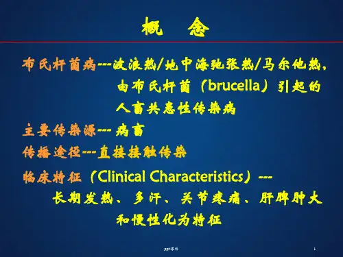
布氏杆菌病布氏杆菌病又称波浪热,是由布氏杆菌引起的人畜共患的传染病。
以长期发热、多汗、关节疼痛、肝脾肿大这主要临床特征。
本病易复发,容易转为慢性。
【流行病学】1、传染源:主要为病畜,包括:羊、猪,其中羊为主要传染源。
其他动物如狗、鹿、马、骆驼等亦可为传染源。
病原存在于病畜的皮毛、胎盘、羊水、尿液中,其中乳汁排菌可达数月至数年。
人传人虽有可能,但极少发生。
2、传播途径:接触传播如(接产、挤奶、屠宰等);实验室工作人员接触染菌标本;加工畜产品等通过粘膜的接触感染人体。
消化道传播如进食含布氏菌的生奶、奶制品或被污染的水和肉类而感染。
其他还可经呼吸道粘膜、眼结膜、性器官粘膜进入人体。
3、人群易感性:人群普遍易感,病后有一定的免疫力,各型间有交叉免疫。
【发病机制】侵入人体的布氏菌经淋巴管进入局部淋巴结,在此大量繁殖成为原发病灶,进入血液循环形成菌血症。
细菌释放内毒素和其他物质,导致毒血症的出现。
细菌随血流播散全身各部位(主要是肝、脾、骨髓和肾等处)进一步繁殖,引起组织细胞的变性、坏死。
布氏杆菌寄生于单核-巨噬胞内,不易被清除导致病毒反复发作和难以根治。
【临床表现】潜伏期1~3周或数月(3日至数月)。
临床上分为急性期和慢性期。
(一)、急性期大多缓慢起病,少数突然发病。
1、发热热型不一,以不规则多见,典型的波浪热已不多见。
2、多汗是本病主要症状之一,无论患者发热与否,常有明显多汗,大量出汗后可发生虚脱。
3、关节疼痛为关节炎所致,常在发病之初出现,亦有发病后1个月才出现者。
4、神经系统症状以神经痛多见,常有坐骨神经痛和腰骶神经痛。
5、泌尿、生殖系统症状可发生睾丸炎、附睾炎、前列腺炎、卵巢炎等。
6、肝、脾及淋巴结肿大大约半数患者可出现肝肿大和肝区疼痛。
急性期布氏杆菌病患者经抗菌治疗后,约有10%以上复发。
(二)、慢性期病程长于一年者为慢性期。
主要表现为疲乏无力,有固定的或反复发作的关节和肌肉疼痛可存在骨和关节的器质性损害。