小细胞肺癌
- 格式:pdf
- 大小:1.07 MB
- 文档页数:9
小细胞肺癌确定标准
小细胞肺癌(SCLC)的确诊通常基于以下几个方面:
1. 病史和体格检查:医生会询问患者的病史,包括吸烟史、家族史等,并进行体格检查,寻找可能的肺部异常。
2. 影像学检查:胸部X光或CT扫描可以显示肺部的异常结构,如肿瘤、肺门淋巴结肿大等。
PET-CT扫描可以帮助确定肿瘤的范围和是否有远处转移。
3. 病理学检查:通过支气管镜检查或经皮肺穿刺活检获取组织样本,然后在显微镜下观察细胞形态和结构。
如果细胞具有典型的小细胞特征,如小的、圆形的细胞核和较高的核质比,就可以确诊为小细胞肺癌。
4. 免疫组化染色:通过免疫组化染色检测肿瘤细胞中的特定蛋白,如神经内分泌肿瘤标志物(如CD56、Syn和CgA),有助于确诊小细胞肺癌。
5. 分子生物学检测:通过检测肿瘤细胞中的基因突变和融合,可以为治疗选择提供更精确的信息。
例如,ALK融合基因阳性的患者可能对ALK抑制剂有较好的反应。
6. 分期:根据肿瘤的大小、淋巴结转移情况和远处转移情况,对小细胞肺癌进行分期,以指导治疗方案的选择。
综上所述,小细胞肺癌的确诊需要结合多种检查方法和临床表现,由专业医生进行综合判断。
肺癌小细胞癌治疗方案引言肺癌是一种常见的恶性肿瘤,其中小细胞癌为其中一种类型。
小细胞肺癌具有高度侵袭性和快速生长的特点,早期诊断和治疗对于患者的生存率至关重要。
本文将介绍肺癌小细胞癌的常见治疗方案,包括手术治疗、放疗和化疗。
手术治疗手术治疗在小细胞肺癌患者中并不常见,通常仅适用于早期病变并且没有远处转移的患者。
手术治疗的主要目的是完全切除肿瘤组织,并减少术后复发的风险。
根据患者的具体情况,常见的手术治疗方法包括肺叶切除、楔形切除和肺组织切除。
手术治疗通常需要在全身麻醉下进行,并需要在术后进行一段时间的恢复。
然而,有一些局限性限制了手术治疗在小细胞肺癌中的广泛应用。
首先,小细胞癌具有高度侵袭性,手术治疗只能切除肿瘤的局部部分,无法消除微小转移病灶。
其次,手术治疗对于晚期患者来说,不再是一个合适的选择,因为此时已经出现远处转移。
放疗放疗是小细胞肺癌的常规治疗方案之一,可以用于早期和晚期患者。
放疗的目标是通过高能射线破坏肿瘤细胞的DNA,达到抑制肿瘤生长和扩散的效果。
放疗可以根据患者的具体情况进行定制,包括放疗方式、剂量和持续时间。
常见的放疗方式包括外部放疗和内部放疗。
外部放疗是通过将放射线束照射到肿瘤区域来进行治疗,而内部放疗则是通过在肿瘤附近放置放射性物质来进行治疗。
放疗的副作用可能会引起一系列不适,如恶心、呕吐、疲劳和皮肤炎症。
然而,这些副作用大多数都是暂时的,可以通过药物和其他支持治疗进行缓解。
化疗化疗是小细胞肺癌的关键治疗方案之一,它可以通过使用抗癌药物来杀死肿瘤细胞并抑制其生长。
化疗可以用于早期和晚期患者,并可以单独使用或与其他治疗方法(如手术治疗或放疗)联合使用。
常见的化疗药物包括顺铂、卡铂、依托泊苷和培美曲塞。
这些药物可以通过静脉注射或口服的方式给予患者。
化疗的副作用是普遍存在的,包括恶心、呕吐、脱发、免疫功能下降等。
然而,现代化疗药物的不断改进已经大大减轻了这些副作用。
综合治疗方案对于小细胞肺癌患者来说,综合治疗方案往往是最有效的治疗方法。
小细胞肺癌名词解释小细胞肺癌是一种危及人类生命的癌症。
小细胞肺癌是肺癌的一种,也是最常见的肺癌类型之一。
肺癌:肺癌是一种疾病,是指肺细胞突变开始失去控制,肿瘤由此而形成,肿瘤可以进一步发展,滋生到肺部腔隙,并慢慢地蔓延到其它器官,形成恶性肿瘤。
它是一种恶性肿瘤,是由已知或未知原因引起肺细胞增殖和发展导致的,最终形成肿瘤。
小细胞肺癌:小细胞肺癌(SCLC)是一种非常恶劣的恶性肿瘤,具有高度恶性的特性,更容易转移到周围器官和其它部位,比起其他类型的肺癌,小细胞肺癌更容易迅速增长和蔓延,也更不易被发现,治疗起来十分困难。
症状:但是,小细胞肺癌早期症状是几乎没有的,因此会导致病情发展到一定阶段才被发现,这使得治疗的效果大打折扣。
一般而言,小细胞肺癌的症状主要有以下几种:咳嗽、喘息、咯血、气促等症状,同时还可能出现贫血、体重明显下降等。
诊断:对于小细胞肺癌的诊断,依赖于多项检查,其中常用的包括血液检查、影像学检查(包括X射线、CT、MRI)等,还有组织活检(也就是把细胞取下来进行检查)等。
这样的检查既能发现病灶的位置,也可以了解有关细胞的情况,从而对病情作出准确的诊断。
治疗:对于小细胞肺癌的治疗,现在主要有两个方法:手术和放疗。
如果是早期的小细胞肺癌,手术可能是最有效的,而放疗的效果则不佳,因为小细胞肺癌的细胞更容易抗放疗,而且肿瘤容易在术后再次复发。
另外,对于小细胞肺癌的治疗还有其他的方法,比如药物治疗,这里面有三类药物,分别是抗癌药物、激素药物和生物疗法,这些药物可以有效地抑制肿瘤的生长,延缓病情发展,从而增加治疗的机会。
也许小细胞肺癌是一场及其痛苦的疾病,但是,如果及时进行检查和治疗,仍然有可能改善或治愈病情。
毕竟,小细胞肺癌的治疗还处于并不完善的状态,但随着药物抗癌和数字技术的发展,科学家们努力地研究出更有效的治疗方法,期盼能清除这种癌症,让小细胞肺癌的病人可以尽快重获康复。
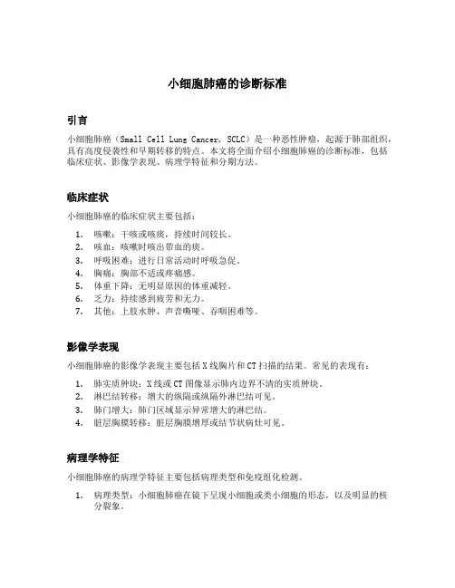
小细胞肺癌的诊断标准引言小细胞肺癌(Small Cell Lung Cancer, SCLC)是一种恶性肿瘤,起源于肺部组织,具有高度侵袭性和早期转移的特点。
本文将全面介绍小细胞肺癌的诊断标准,包括临床症状、影像学表现、病理学特征和分期方法。
临床症状小细胞肺癌的临床症状主要包括:1.咳嗽:干咳或咳痰,持续时间较长。
2.咳血:咳嗽时咳出带血的痰。
3.呼吸困难:进行日常活动时呼吸急促。
4.胸痛:胸部不适或疼痛感。
5.体重下降:无明显原因的体重减轻。
6.乏力:持续感到疲劳和无力。
7.其他:上肢水肿、声音嘶哑、吞咽困难等。
影像学表现小细胞肺癌的影像学表现主要包括X线胸片和CT扫描的结果。
常见的表现有:1.肺实质肿块:X线或CT图像显示肺内边界不清的实质肿块。
2.淋巴结转移:增大的纵隔或纵隔外淋巴结可见。
3.肺门增大:肺门区域显示异常增大的淋巴结。
4.脏层胸膜转移:脏层胸膜增厚或结节状病灶可见。
病理学特征小细胞肺癌的病理学特征主要包括病理类型和免疫组化检测。
1.病理类型:小细胞肺癌在镜下呈现小细胞或类小细胞的形态,以及明显的核分裂象。
2.免疫组化检测:免疫组化检测常用的标记物包括神经内分泌标记物(如CD56、Synaptophysin)和细胞角蛋白标记物(如CAM5.2、AE1/AE3等)。
分期方法小细胞肺癌的分期方法主要采用国际上广泛应用的TNM分期系统。
根据肿瘤的病理特点和淋巴结转移情况,分期分为以下几个阶段:1.分期0:原位癌,肿瘤仅局限于上皮层内,未侵犯基底膜。
2.分期I:肿瘤侵犯肺组织,但未侵犯纵隔淋巴结。
3.分期II:肿瘤侵犯纵隔淋巴结或肺门淋巴结。
4.分期III:肿瘤侵犯对侧肺和纵隔淋巴结。
5.分期IV:肺癌晚期,肿瘤转移到体内其他部位。
结论小细胞肺癌的诊断标准涵盖临床症状、影像学表现、病理学特征和分期方法。
临床医生应根据病人的症状、影像学结果和病理学特征进行综合判断,以确定诊断和分期,并制定相应的治疗方案。

肺癌小细胞癌化疗方案肺癌是全球范围内最常见的癌症之一,而小细胞肺癌是肺癌的一种常见类型。
小细胞肺癌的特点是恶性程度高,生长迅速,并且易于扩散到其他器官。
化疗是小细胞肺癌的主要治疗方式之一,本文将介绍肺癌小细胞癌化疗方案方面的内容。
1. 化疗在小细胞肺癌治疗中的地位小细胞肺癌对放射治疗和手术治疗的敏感性相对较低,因此化疗一直被认为是小细胞肺癌的首选治疗方式。
化疗可以通过药物的系统性作用,杀死癌细胞及其在身体中的转移细胞,以达到控制肿瘤生长及扩散的目的。
2. 典型的化疗方案典型的小细胞肺癌化疗方案是使用多种药物组合,通常包括铂类化合物(如顺铂)和培美曲塞(etoposide)等。
这种方案被称为EP方案(Etoposide and Platinum),已被广泛用于小细胞肺癌的治疗。
EP方案在一般情况下,每21天为一个疗程,连续进行4-6个疗程。
3. 化疗方案的个体化虽然EP方案是目前最常用的小细胞肺癌化疗方案,但医生在制定化疗方案时会根据患者的具体情况进行个体化的调整。
例如,对于某些患有心脏疾病或者肾脏功能不全的患者,医生可能会减少药物剂量或者选择其他药物组合进行治疗,以降低治疗对患者的不良影响。
4. 化疗方案的持续性小细胞肺癌的化疗通常需要持续进行,这是因为小细胞肺癌的细胞具有较高的反应性和变异性。
即使在看似完全消失的情况下,残留的癌细胞也可能随时复发。
因此,大多数患者需要进行较长时间的化疗,以确保肿瘤的完全消除。
5. 化疗的副作用与处理化疗药物不仅对癌细胞有毒性作用,也对正常细胞造成一定程度的损害,因此会伴随一系列副作用。
常见的化疗副作用包括恶心、呕吐、脱发、食欲不振等。
针对这些副作用,医生会采取相应的措施进行处理,如使用抗恶心药物、营养支持等。
6. 其他治疗方式的结合在一些情况下,化疗可能与其他治疗方式结合使用,以期达到更好的治疗效果。
常见的联合治疗方式包括放疗、靶向治疗和免疫治疗等。
这些治疗方式的选择和顺序,需要根据患者的具体情况和肿瘤的特征来决定。

小细胞癌最佳治疗方案小细胞肺癌是一种具有高度侵袭性和早期转移倾向的恶性肿瘤。
由于其发展迅速且容易在早期就扩散到其他部位,小细胞肺癌的治疗方案具有特殊的挑战性。
本文将介绍并讨论小细胞肺癌的最佳治疗方案。
一、手术治疗对于小细胞肺癌,手术治疗的作用相对有限。
由于该癌症常在早期就已转移扩散,手术切除的机会较少。
然而,在临床上,对于早期小细胞肺癌患者,当病变局限于肺部且没有明显转移时,手术治疗可能仍然是一种有效的治疗选择。
二、化疗化疗是目前治疗小细胞肺癌的主要方法。
通常采用的方案是将顺铂与依托泊苷联合使用。
这一化疗方案已被广泛认可,并取得了良好的治疗效果。
顺铂与依托泊苷的联合应用可以有效地抑制癌细胞的增殖,并减少转移的风险。
对于无手术切除可能性的晚期小细胞肺癌患者,化疗是常用的治疗方式。
三、放疗放疗在小细胞肺癌治疗中具有重要的地位。
放疗可以用来控制原发肿瘤的生长,减少症状和缓解疼痛。
对于局限期患者,放疗结合化疗可以有效提高生存率和缓解症状。
对于广泛期患者,放疗可以用来缓解症状,改善生活质量。
四、免疫治疗近年来,免疫治疗在肿瘤治疗中取得了显著的进展,也为小细胞肺癌患者带来了新的希望。
免疫检查点抑制剂如PD-1抑制剂和PD-L1抑制剂已被证明在小细胞肺癌的治疗中表现出良好的疗效。
这些药物可以激活患者自身的免疫系统,增强对癌细胞的免疫攻击能力。
免疫治疗相对较新,但正在不断发展,并在许多临床试验中取得了积极的结果。
五、靶向治疗靶向治疗是根据肿瘤的特定突变或异常基因表达来选择特定的药物进行治疗。
目前已发现在小细胞肺癌中存在着一些靶向可治疗的突变,如EGFR、ALK等。
对于存在这些突变的患者,靶向药物可以显著提高治疗效果并减少毒副作用。
综上所述,小细胞肺癌的最佳治疗方案通常是综合治疗。
对于早期患者而言,手术切除可能会是有效的治疗选择。
对于晚期患者,化疗联合放疗可以带来良好的治疗效果。
此外,免疫治疗和靶向治疗也为小细胞肺癌患者带来了新的治疗机会。
小细胞肺癌确诊标准1. 引言1.1 小细胞肺癌的定义小细胞肺癌是肺癌的一种亚型,通常被认为是一种速度较快且具有高度侵袭性的肿瘤。
它通常起源于肺部的支气管上皮细胞,具有小细胞形态特征,细胞核较大、核浆比较大以及核质较少,常见的细胞器也较少。
小细胞肺癌与非小细胞肺癌不同,其细胞形态较小细胞肺癌更为小型,故得名小细胞肺癌。
小细胞肺癌的发病率逐年上升,尤其在吸烟人群中更为常见。
据统计,在全球范围内,小细胞肺癌大约占据了所有肺癌病例的10%至15%左右。
由于其生长速度快,常常在早期便已经转移至其他部位,使得小细胞肺癌的治疗难度增加,预后相对较差。
早期确诊和治疗对于小细胞肺癌患者的生存率至关重要。
1.2 小细胞肺癌的发病情况小细胞肺癌是一种高度侵袭性的肺癌类型,通常起源于支气管上皮细胞。
根据统计数据显示,小细胞肺癌在所有肺癌病例中占比约10%-15%。
其在吸烟者中的发病率更高,而且几乎所有小细胞肺癌患者都有长期吸烟史。
男性患者的患病率明显高于女性。
尽管小细胞肺癌的患病率并不高,但其侵袭性极强,病情发展迅速,预后较差。
小细胞肺癌的高度恶性特性使其成为一种严重威胁人类健康的疾病。
对于小细胞肺癌的早期诊断至关重要,只有及早发现和及时治疗,才能提高患者的生存率和生活质量。
了解小细胞肺癌的发病情况及其诊断标准对于临床医生和患者都具有重要意义。
2. 正文2.1 临床症状小细胞肺癌在临床上通常表现出以下症状:咳嗽、咳痰、胸痛、呼吸困难、咳血、声音嘶哑等。
患者还可能出现全身性症状如消瘦、乏力、食欲减退等。
部分患者可能伴有肝脾肿大、淋巴结肿大等特征性体征。
由于小细胞肺癌生长迅速,病情进展快,患者往往在早期就出现明显的症状,这使得早期诊断和治疗十分关键。
在日常临床实践中,医生需要综合患者的症状表现和体征,结合影像学检查和实验室检查结果,才能做出正确的诊断。
由于小细胞肺癌的症状非特异性,容易与其他类型的肺癌或呼吸系统其他疾病混淆,因此对于可能患有小细胞肺癌的患者,及时就医、全面检查至关重要。
摘要小细胞肺癌(Small Cell Lung Cancer,SCLC)是一种高度恶性的肺癌,具有快速生长和广泛转移的特点。
化疗是小细胞肺癌治疗的主要手段之一。
本文旨在介绍小细胞肺癌的化疗方案,包括化疗药物的选择、剂量、疗程以及可能的不良反应和应对措施。
一、小细胞肺癌化疗药物的选择1. 紫杉类紫杉类药物是治疗小细胞肺癌的一线化疗药物,包括紫杉醇和多西紫杉醇。
它们通过抑制微管蛋白的聚合,导致肿瘤细胞的有丝分裂停滞。
2. 阿霉素类阿霉素类药物是治疗小细胞肺癌的另一类重要化疗药物,包括阿霉素和表阿霉素。
它们通过干扰DNA复制和转录,抑制肿瘤细胞的增殖。
3. 拉司汀类药物拉司汀类药物是治疗小细胞肺癌的第三代铂类药物,包括顺铂和卡铂。
它们通过与DNA结合,干扰DNA复制和转录,从而抑制肿瘤细胞的生长。
4. 紫杉醇类药物紫杉醇类药物是治疗小细胞肺癌的一线化疗药物,包括紫杉醇和多西紫杉醇。
它们通过抑制微管蛋白的聚合,导致肿瘤细胞的有丝分裂停滞。
二、化疗剂量和疗程1. 剂量化疗药物的剂量取决于患者的体重、肝肾功能、病情严重程度等因素。
以下为常用化疗药物的剂量:(1)紫杉醇:135-175mg/m²,静脉滴注,每3周一次。
(2)阿霉素:45-50mg/m²,静脉滴注,每3周一次。
(3)顺铂:25-75mg/m²,静脉滴注,每3周一次。
(4)卡铂:AUC=5-7,静脉滴注,每3周一次。
2. 疗程小细胞肺癌的化疗疗程一般为4-6个周期。
在化疗过程中,需根据患者的病情、疗效和耐受情况调整治疗方案。
三、化疗方案1. 单药化疗对于初治的小细胞肺癌患者,可先进行单药化疗,如紫杉醇、阿霉素、顺铂等。
单药化疗适用于病情较轻、体质较弱的患者。
2. 联合化疗对于病情较重、转移范围较广的患者,可进行联合化疗。
以下为常见的联合化疗方案:(1)EP方案:阿霉素+顺铂。
(2)CP方案:顺铂+卡铂。
(3)TP方案:紫杉醇+顺铂。
小细胞肺癌分期标准小细胞肺癌(SCLC)是一种高度侵袭性的肺癌类型,通常在诊断时已经扩散到其他部位。
由于其快速生长和早期转移的特点,小细胞肺癌的分期对于治疗和预后具有重要意义。
目前,小细胞肺癌的分期主要采用国际抗癌联盟(UICC)和美国癌症学会(AJCC)颁布的TNM分期系统。
根据肿瘤的大小(T)、淋巴结的受累情况(N)和是否远处转移(M)来确定肺癌的分期,从而指导临床治疗方案的选择。
T分期是指原发肿瘤的大小和范围,根据肿瘤的直径和侵袭性分为T1、T2、T3和T4四个阶段。
T1期肿瘤直径小于3厘米,T2期肿瘤直径在3-7厘米之间,T3期肿瘤直径大于7厘米或侵犯邻近结构,T4期肿瘤侵犯了纵隔器官或胸膜,或者出现了双侧肺内转移。
N分期是指淋巴结的受累情况,根据受累淋巴结的数量和位置分为N0、N1、N2和N3四个阶段。
M分期是指是否有远处转移,分为M0和M1两个阶段。
根据TNM分期系统,小细胞肺癌的分期分为四个阶段,Ⅰ期、Ⅱ期、Ⅲ期和Ⅳ期。
Ⅰ期小细胞肺癌指肿瘤局限于一个肺叶或一个肺段,没有淋巴结转移或远处转移。
Ⅱ期小细胞肺癌指肿瘤扩散到同一侧的另一肺叶、肺段或同侧纵隔淋巴结,但没有远处转移。
Ⅲ期小细胞肺癌分为ⅢA和ⅢB两个亚型,ⅢA期指肿瘤扩散到对侧肺叶或肺段、同侧或对侧纵隔淋巴结,ⅢB期指肿瘤侵犯纵隔器官或胸膜,或者出现了双侧肺内转移。
Ⅳ期小细胞肺癌指肿瘤远处转移,可以是对侧肺叶、纵隔器官、胸膜、脏层胸膜、心包、脊柱、颅骨或其他器官。
对于小细胞肺癌患者的治疗方案选择和预后评估,分期是至关重要的。
早期诊断的小细胞肺癌患者通常可以接受手术切除肿瘤或放疗,预后相对较好。
而晚期诊断的小细胞肺癌患者通常需要接受化疗或靶向治疗,预后较差。
因此,对小细胞肺癌的准确分期可以帮助医生选择最合适的治疗方案,提高患者的生存率和生活质量。
总之,小细胞肺癌的分期对于治疗和预后具有重要意义,目前主要采用TNM 分期系统。
通过对肿瘤的大小、淋巴结受累情况和是否远处转移的评估,可以确定小细胞肺癌的分期,从而指导临床治疗方案的选择。
小细胞肺癌小细胞肺癌是一种恶性肿瘤,起源于肺部的细胞,尤其是呼吸道的上皮细胞。
它是肺癌中最常见的类型之一,通常发生在吸烟者身上。
小细胞肺癌非常侵袭性,扩散速度快,所以早期发现和诊断对于治疗至关重要。
本文将讨论小细胞肺癌的原因、症状、诊断和治疗。
小细胞肺癌的主要原因是长期吸烟。
吸烟是导致肺癌的主要危险因素,其中小细胞肺癌更多发生在烟龄较长且吸烟量较大的人群中。
此外,职业暴露于某些致癌物质(如石棉、镍、镉等)也会增加患小细胞肺癌的风险。
遗传因素也可能在小细胞肺癌的发生中起到一定作用,有些人患有特定的基因突变,增加了患病的风险。
小细胞肺癌早期往往没有明显的症状,使得早期发现和治疗成为一项巨大的挑战。
随着肿瘤的生长和扩散,症状才会逐渐出现。
常见的症状包括咳嗽、咳痰、咳血、呼吸困难、胸痛等。
一些患者还可能出现乏力、食欲减退、体重下降等非特异性症状。
肺癌也可能反应到其他部位,导致骨科症状、神经系统症状等。
诊断小细胞肺癌通常包括病史询问、体格检查、影像学检查和组织学检查。
病史询问主要了解症状、家族史、吸烟史等。
体格检查可以发现一些肺部或其他器官的异常体征。
影像学检查如X线、CT、MRI等可帮助查明肿瘤的大小、位置、扩散情况。
对怀疑为小细胞肺癌的患者,需要进行组织学检查以确诊,通常通过支气管镜检查、穿刺活检等方法采集肿瘤组织进行病理学分析。
小细胞肺癌的治疗一般采用综合治疗的策略,包括手术、放疗和化疗。
然而,由于小细胞肺癌生长扩散较快,大部分患者在诊断时已处于晚期,无法进行手术治疗。
对于早期小细胞肺癌患者,手术切除可带来较好的治疗效果。
放疗主要针对肺部的肿瘤进行,可以通过杀死肿瘤细胞来控制病情。
化疗则是使用一系列抗癌药物,通过杀死肿瘤细胞来控制病情。
化疗方案通常是联合用药,根据患者的具体情况选择药物和治疗方案。
小细胞肺癌是一种非常严重的疾病,预后通常较差。
这主要是因为大部分患者在诊断时已处于晚期,治疗效果不佳。
尽管如此,通过积极的治疗,仍然有一些患者可以取得较长时间的生存。
小细胞肺癌能活多久小细胞肺癌是一种高度恶性的肺癌,因其生长迅速和易于转移的特点而令人忧心。
对于患有小细胞肺癌的患者来说,了解其能够活多久对于治疗和生活规划非常重要。
然而,要回答这个问题并不容易,因为每个患者的情况都是独特的。
在本文中,我们将探讨小细胞肺癌的预后以及提高生存率的方法。
首先,了解小细胞肺癌的预后需要考虑多个因素。
其中最重要的是疾病的分期。
根据不同的分期,患者的生存率也会有所不同。
根据美国癌症协会的数据,小细胞肺癌的五年生存率在早期阶段(仅限于已局限于一侧肺部且无淋巴结侵犯的患者)可能高达30%至40%,但在晚期阶段(癌细胞已经扩散到其他部位)通常不到5%。
其次,治疗方案也对小细胞肺癌的生存率具有重要影响。
小细胞肺癌通常是通过化疗来管理的,因为它对放疗和手术不太敏感。
标准的治疗方案包括多药化疗,如顺铂和依托泊苷。
这种化疗方案在大多数情况下可以显著降低肿瘤负荷,并延长生存期。
然而,小细胞肺癌的复发率非常高,而且通常在化疗后很快复发。
因此,医生们经常会建议进行二线治疗,如放疗或其他新型抗癌药物治疗。
个体化的治疗方案和临床试验也可以提供额外的机会。
此外,患者的身体健康状况和个人因素也会影响小细胞肺癌的预后。
有些患者可能同时患有其他疾病,如心脏病或糖尿病,这可能会影响其治疗方案和生存期。
年龄也是一个重要的因素,老年患者通常有较差的预后。
此外,吸烟也会显著增加小细胞肺癌患者的死亡风险。
戒烟对改善预后非常重要,因此患者应该积极采取措施戒烟或减少烟草消耗。
除了医疗手段,生活方式和心理因素也对患有小细胞肺癌的患者的生活质量和预后有着重要影响。
保持积极的心态和良好的营养是至关重要的。
这些可以帮助患者提高抵抗力,并更好地应对治疗的不良反应和副作用。
此外,身体活动和定期锻炼对于维持健康状态和减少治疗相关的疲劳和身体不适也非常重要。
尽管小细胞肺癌的预后相对较差,但新的治疗和护理方法的出现给患者带来了希望。
免疫疗法和靶向治疗等新型治疗手段正在不断发展,并显示出一定的疗效。
小细胞癌影像学特点
小细胞肺癌(Small Cell Lung Cancer,SCLC)是一种高度侵袭性的肺癌类型,其特点在于细胞体积小,核与胞质之间的比例大,细胞核的形态多变,并且有明显的核的细胞质不均等现象。
在影像学方面,小细胞肺癌的表现有一定的特点。
在胸部X线片上,通常可见到类似于肺实质的阴影,与周围正常肺组织的边界不清晰,可能有不规则的边界和模糊的轮廓。
这种阴影可呈现单发或多发的形式,并且在周围肺野中也可能伴有多发肺转移灶。
胸部CT扫描是诊断和评估小细胞肺癌的主要方法。
CT扫描可以提供更为详细的肿瘤形态特点与定位信息。
小细胞肺癌在CT扫描上通常呈现为较大的肺实质肿块,形态不规则,边缘模糊,有时可以看到肺门的淋巴结肿大以及胸腔积液。
此外,CT扫描还可以评估是否存在远处的淋巴结转移、肺外转移以及是否有其他病灶存在。
对于可能伴有脑转移的小细胞肺癌患者,进行脑部影像学检查也是必需的。
脑
部MRI可以检测到小细胞肺癌在脑内的转移病灶。
这些病灶通常呈现为多发的结
节状或节段状影像,有时可见囊性变或出血。
综上所述,小细胞肺癌在影像学上的特点是肺部阴影呈现为不规则的肺实质肿块,边缘模糊,常伴有淋巴结肿大和胸腔积液。
此外,脑转移常见,可通过脑部影像学检查来评估。
这些影像学特点对于小细胞肺癌的诊断和治疗具有重要的参考价值。
小细胞肺癌二线方案简介小细胞肺癌(small cell lung cancer,SCLC)是一种恶性程度较高的肺癌类型,常见于吸烟人群。
尽管在早期诊断和治疗取得了一些进展,但其预后仍然较差。
目前,一线治疗方案中的放化疗已成为标准治疗。
然而,在一线治疗失败或复发的患者中,需要选择适合的二线方案来延长患者的生存期和提高生活质量。
本文将介绍小细胞肺癌二线方案的选择和管理,以帮助医生更好地处理这一挑战性的临床情况。
选择二线方案的考虑因素在选择适当的二线治疗方案之前,需要综合考虑以下几个因素:1. 患者特征包括年龄、性别、身体状况、合并症等。
2. 前一线治疗的反应和耐药性评估患者对前一线治疗的反应及可能存在的耐药性。
3. 肿瘤特征包括肿瘤分期、组织学类型、基因表达等。
4. 治疗目标确定治疗的目标,是延长患者的生存期还是缓解症状。
5. 患者意愿和选择需要与患者进行充分沟通,了解患者的意愿和选择。
常用的二线治疗方案在小细胞肺癌的二线治疗中,常用的方案包括以下几种:1. 二线化疗方案常用的二线化疗药物包括卡铂、依托泊苷、顺铂等。
通常采用多药联合化疗,通过不同机制的药物联合起来,提高治疗的效果。
2. 靶向治疗针对小细胞肺癌的靶点,使用相应的靶向药物进行治疗。
例如,针对抑制DNA 合成和修复的PARP酶的靶向药物奥拉帕尼布(olaparib)可以用于维持治疗。
3. 免疫治疗针对小细胞肺癌的免疫检查点,通过激活免疫系统来抑制肿瘤生长。
PD-1抑制剂(如帕博利珠单抗)和PD-L1抑制剂(如仰沙珠单抗)是目前应用较广泛的免疫治疗药物。
4. 支持性治疗针对患者的症状进行缓解,包括镇痛治疗、抗恶心药物、营养支持等。
这些治疗可以改善患者的生活质量。
管理二线治疗的注意事项在使用二线治疗方案时,需要注意以下几个方面:1. 严密监测治疗效果定期进行影像学检查和临床评估,及时评估治疗的效果。
2. 处理副作用和不良反应及时发现和处理治疗引起的副作用和不良反应,保证患者的生活质量。
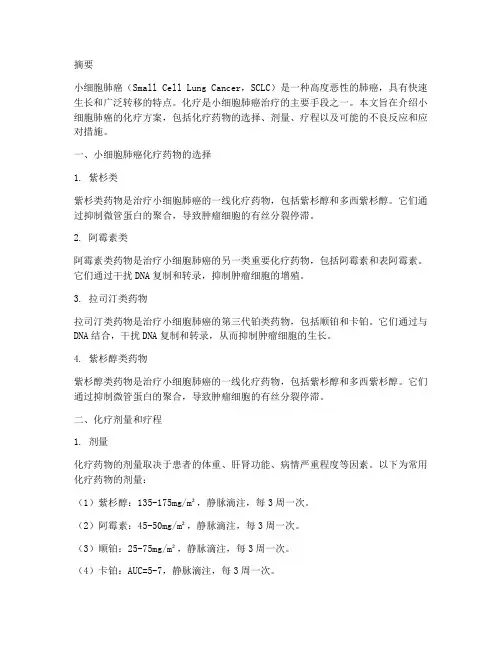
Tumorigenesis and Neoplastic ProgressionEvaluation of Circulating Tumor Cells and Serological Cell Death Biomarkers in Small Cell Lung Cancer Patients Undergoing ChemotherapyJian-Mei Hou,*Alastair Greystoke,*Lee Lancashire,*Jeff Cummings,*Tim Ward,* Ruth Board,*Eitan Amir,†Sarah Hughes,* Matthew Krebs,*Andrew Hughes,‡Malcolm Ranson,*†‡Paul Lorigan,†‡Caroline Dive,*‡and Fiona H.Blackhall*†‡From the Clinical and Experimental Pharmacology Group,* Paterson Institute for Cancer Research,University of Manchester, Manchester;the Christie Foundation Trust,†Manchester;and the School of Cancer and Imaging Sciences,‡University of Manchester,Manchester,United KingdomSerological cell death biomarkers and circulating tu-mor cells(CTCs)have potential uses as tools for phar-macodynamic blood-based assays and their subse-quent application to early clinical trials.In this study, we evaluated both the expression and clinical signif-icance of CTCs and serological cell death biomarkers in patients with small cell lung cancer.Blood samples from88patients were assayed using enzyme-linked immunosorbent assays for various cytokeratin18 products(eg,M65,cell death,M30,and apoptosis)as well as nucleosomal DNA.CTCs(per7.5ml of blood) were quantified using Veridex CellSearch technology. Before therapeutic treatment,cell death biomarkers were elevated in patients compared with controls. CTCs were detected in86%of patients;additionally, CD56was detectable in CTCs,confirming their neo-plastic origin.M30levels correlated with the per-centage of apoptotic CTCs.M30,M65,lactate dehy-drogenase,and CTC number were prognostic for patient survival as determined by univariate ing multivariate analysis,both lactate dehy-drogenase and M65levels remained significant.CTC number fell following chemotherapy,whereas lev-els of serological cell death biomarkers peaked at48 hours and fell by day22,mirroring the tumor re-sponse.A48-hour rise in nucleosomal DNA and M30levels was associated with early response and severe toxicity,respectively.Our results provide a rationale to include the use of serological biomark-ers and CTCs in early clinical trials of new agents for small cell lung cancer.(Am J Pathol2009,175:808–816; DOI:10.2353/ajpath.2009.090078)Small cell lung cancer(SCLC)is initially chemosensitive but invariably relapses with a chemoresistant pheno-type.1A number of molecularly targeted therapies have been evaluated attempting to improve outcome,but none have succeeded to date.2Ideally,early clinical trials should incorporate validated pharmacodynamic biomar-kers,conducted to good clinical laboratory practice,that demonstrate both proof of mechanism(drug hits target) and proof of concept(tumor responds to drug).3Al-though possible,serial biopsies are rare in SCLC,and the tissue obtained often insufficient for extensive molecular profiling.Thus,there is a pressing need for blood-based biomarkers that report therapeutic response.Assays of drug-induced cell death are potential proof of concept biomarkers for multiple therapeutics.4The M30Apoptosense and M65assays(Peviva,Bromma, Sweden)detect cytokeratin(CK)18,expressed in epi-thelial but not hematopoietic cells,and released into the blood following cytoskeletal disassembly and degrada-tion during apoptotic and/or necrotic cell death.5The M30antibody recognizes a caspase-cleaved neoepitope of CK18that is only revealed during apoptosis,whereas the M65assay detects full length and cleaved forms of CK18reporting apoptosis and necrosis.6Nucleosomal DNA(nDNA)results from cleavage of chromatin by apo-ptotic endonucleases into membrane bound DNA frag-ments that are phagocytosed by macrophages and sub-Supported by Cancer Research UK grant C147/A6058and European Union CHEMORES FP6contract LSHC-CT-2007-037665;Cancer Research UK China fellowship C480/A7421(to J.-M.H.);and a clinical pharmacology fel-lowship from Cancer Research UK and AstraZeneca Ltd.(to A.G.).J.-M.H.and A.G.contributed equally to this work.Accepted for publication April15,2009.Address reprint requests to F.Blackhall or Caroline Dive,Clinical and Experimental Pharmacology,Paterson Institute for Cancer Research, Wilmslow Road,Manchester,M204BX,UK.E-mail:fiona.blackhall@christie or cdive@.The American Journal of Pathology,Vol.175,No.2,August2009 Copyright©American Society for Investigative PathologyDOI:10.2353/ajpath.2009.090078808sequently released into the blood.7nDNA release is also detected when levels of apoptosis overwhelm macro-phage capacity for phagocytosis and a more necrotic cell fate ensues.7We have previously validated these cell death biomarkers8,9and optimized them for application to a busy, clinical setting.6Here we report on the behavior and clinical utility of these assays in patients with SCLC.Importantly,cell death assays may report host toxicity in addition to tumor response.However,circulating tumor cell(CTC)numbers can,in theory,be used to directly evaluate drug effect on malignant cells.10The cytometric approach using CellSearch technology(Veridex Inc., Huntingdon Valley,PA)is now approved by the Food and Drug Administration for clinical decision-making in pa-tients with metastatic breast,colorectal and prostate cancers.11–13This is the first report on the use of this technology platform for CTC enumeration in patients with SCLC and the first direct comparison of serological bi-omarkers of cell death and cell death in CTCs.This study was conducted to evaluate cell death as-says(M30,M65,and nDNA)and CTC profiles in patients with SCLC undergoing standard chemotherapy,as a pre-lude to their incorporation as biomarkers in early clinical trials.The hypothesis tested was that increases in cell death biomarkers immediately following therapy would predict outcome and that given the metastatic potential of SCLC high numbers of CTCs would be detectable.The results from this study are most encouraging for the de-velopment of CTCs as pharmacodynamic biomarkers in SCLC,provide novel insights into the clinical significance of serological cell death assays,and demonstrate agree-ment between measures of cell death at the molecular and cellular level in this disease.Materials and MethodsPatientsBlood samples were collected from SCLC patients under-taking chemotherapy at the Christie Hospital,Manchester, UK,and from healthy volunteers according to ethically ap-proved protocols.Eligible patients had pathologically con-firmed chemo-naive SCLC,staged and managed using standard protocols.Patients received platinum-based che-motherapy,given in combination with etoposide where age, performance status,and comorbidities allowed.One pa-tient received non-platinum-based combination therapy due to disease related thrombocytopenia at baseline.Rad-ical radiotherapy and prophylactic cranial irradiation were administered either concurrently with chemotherapy(start-ing with cycle2)or sequentially following cycle6in patients with limited disease depending on clinical status and extent of disease.Data were collected on World Health Organization per-formance status(PS),complete blood count,alkaline phosphatase,lactate dehydrogenase(LDH),sodium(Na), radiology,treatment received,toxicity,response,and survival.Early response was assessed by an indepen-dent radiologist on chest radiographs taken on days1 and15of the first cycle of treatment.Blood Sampling,Processing,and Serological Cell Death Enzyme-Linked Immunosorbent AssaysBlood samples(15ml)were collected using the Vacu-tainer system(BD,Franklin Lakes,NJ)within24hours before the first treatment then48hours and22days after therapy.The first5ml of blood was discarded to minimize the risk of skin contamination.Additional samples were obtained from a subgroup of patients(nϭ12)after24 hours,on day8,and on day15to explore the optimal time points for the assays.Plasma samples were col-lected in lithium-heparin tubes,refrigerated at4°C for a maximum of2hours before centrifugation at1000ϫg for 10minutes,and transferred immediately toϪ80°C.Se-rum samples were collected in silica tubes,clotted at room temperature for30minutes to2hours,before cen-trifugation at2000ϫg for10minutes and transferred immediately toϪ80°C.6Plasma samples were analyzed for M30and M65(Peviva)using our previously described assays validated to good clinical laboratory practice.8,9 Serum samples were analyzed for nDNA[Cell Death Detection ELISA(Roche,Basel,Switzerland)]as previ-ously described.14CTC EnumerationBlood samples(7.5ml)were collected into CellSave tubes(Veridex),containing EDTA and a cellular preser-vative.Samples were maintained at room temperature and processed within72hours using the CellSearch platform(Veridex)as previously described.15In brief, blood was diluted,centrifuged and incubated with fer-rofluid particles coated with anti-EpCAM antibodies.After immunomagnetic enrichment,ferrofluid-labeled cells were permeabilized and fluorescently labeled using phy-coerythrin-conjugated anti-CK antibodies(pan-keratin antibody C-11that recognizes keratins4,5,6,8,10,13, and18)to identify epithelial cells and allophycocyanin-conjugated anti-CD45antibody to identify and discount white blood cells.4Ј6-Diamidino-2-phenylindole(DAPI) was incorporated to identify cell nuclei and to reveal mitotic or apoptotic nuclear morphologies.Analysis of CD56expression on CTCs was performed using Alexa Fluor488-conjugated mouse anti-human CD56antibody (BD PharMingen,San Diego,CA)analyzed in the fourth channel of the CellSearch system.The antibody concen-tration was12g/ml and the integration time for CellSearch Analyzer II was configured to0.4second.The positivity of CD56in SCLC CTCs was obtained using the research mode of CellSearch Analyzer II.After repeated magnetic separation,the fluorescently labeled cells were oriented to the cartridge surface for analysis using the CellTracks Analyzer II,a semiautomated fluorescent mi-croscope.Captured images,processed by CellTracks software,were analyzed blinded without knowledge of patient data.CTCs were defined as nucleated cells stain-ing positively for cytokeratin and negatively for CD45and reported as CTC number per7.5ml of blood.Based on the nuclear and CK morphology,apoptotic and mitoticCirculating Biomarkers in SCLC809AJP August2009,Vol.175,No.2CTCs were enumerated in cases where CTC number was Ͼ100.The reproducibility,intra-and interassay variabil-ity,cell stability,and recovery precision of the CellSearch system have been validated by the manufacturer.Statistical AnalysisStudy design and statistical analysis was performed us-ing SPSS for Windows (release 13.0.2004,SPSS Inc.,Chicago,IL)where P values of Յ0.05were considered significant.A minimum sample size of 36cases was required for enzyme-linked immunosorbent assay and CTC analyses to detect with 80%power a difference in survival between good and poor prognosis groups of 100days,with an accrual and median follow-up of 365days.In addition,data from Phase 1trials have shown that progressive disease is associated with a 12.5%(SD 28%)increase in M65at day 22.16However,in patients re-sponding to cytotoxic chemotherapy in other solid tu-mors,a decrease of 20%(SD 44%)has been seen.17To detect this difference with 80%power (P ϭ0.05)assum-ing a 70%response rate 34patients were required.Variables were positively skewed and were log (base 2)transformed before analysis to stabilize the sample vari-ance,and nonparametric tests were used to satisfy the assumptions of variance between sample groups.Cell death biomarker levels were analyzed as continuous vari-ables.Differences between groups were tested using the Mann-Whitney U Test for continuous variables and the 2test for categorical variables.Relationships were examined using the non-parametric Spearman’s rho bivariate correla-tion with a two-tailed test for significance.Univariate survival analysis was performed using the Kaplan-Meier method,with data categorized into three groups based on upper (75th),middle (median),and lower (25th)quartiles,which were compared using the log rank (Mantel-Cox)test.Mul-tivariate analysis,using a forward stepwise Cox proportional hazards regression model,tested for associations between the variables and survival (defined as time from consent to death).Separate multivariate analysis was performed for M30and M65due to the strong correlation between these variables.Subjects with missing data were removed before this analysis.This did not have an adverse effect on the power of the study.The REMARK guidelines for reporting results from prognostic biomarkers statistical analysis were followed.18ResultsClinical Characteristics of PatientsA total of 88patients were enrolled prospectively be-tween October 2006and December 2008;78patients were assessable for cell death biomarkers and 50pa-tients were assessed for CTCs.Insufficient blood volume prevented analysis of all biomarkers in all patients.Clin-ical characteristics are summarized in Table 1,and there were no significant differences between the total cohort,the cell death biomarker cohort,and the CTC cohort.Cell Death Biomarkers and CTCs before ChemotherapyBefore treatment all three cell death biomarkers were higher in 78patients with SCLC compared with 85healthy controls (median values for M30were 268versus 198U/L P ϭ0.02,for M65were 609versus 245U/L P Ͻ0.0001,and for nDNA were 1.40versus 0.30,respec-tively,P Ͻ0.0001).CTCs were detected in 43of 50(86%)patients analyzed with median CTC number of 28(range,0–44,896,mean ϮSD ϭ2915Ϯ8115).Table 1.Clinical Characteristics of SCLC PatientsCharacteristicTotal cohort N ϭ88Cell death biomarkercohort N ϭ78CTC cohort N ϭ50Age,median years (range)67(27–86)67(27–86)67(28–84)Sex,n (%)Male 44(50)39(50)27(54)Female 44(50)39(50)23(46)Stage,n (%)Limited stage 35(40)33(42)20(40)Extensive stage53(60)45(58)30(60)Liver metastases (%of extensive stage)32(60)29(64)19(63)Performance status,n (%)07(9)6(8)4(8)148(55)42(54)27(54)230(34)28(36)17(34)32(2)2(3)2(4)Treatment,n (%)Cisplatin ϩetoposide 19(22)17(22)10(20)Carboplatin ϩetoposide 55(62)50(64)29(58)Carboplatin13(15)10(13)10(20)Vincristine,adriamycin,cyclophosphamide 1(1)1(1)1(2)Baseline laboratory values Na,median (range)137(113–149)137(114–149)137(114–145)Na Ͻ132,n (%)15(17)15(19)7(14)LDH,median (range)548(240–6183)562(294–6183)583(240–6010)LDH Ͼ450,n (%)56(64)50(64)31(62)810Hou et alAJP August 2009,Vol.175,No.2M65and M30correlated with each other,consistent with detection of all forms of CK18by M65,and the apoptosis cleaved fragment by M30(P Ͻ0.001).In contrast,nDNA did not correlate with M65or M30.Pretreatment CTC num-ber correlated significantly with M30and M65(P Ͻ0.001)but not with nDNA in 40patients assessable for all biomar-kers.Morphological assessment for apoptotic and mitotic CTCs was performed in 14samples from 12patients con-taining a minimum CTC number of 100.Typical examples of cellular morphology are shown in Figure 1A.Apoptotic cells were observed in 13of 14evaluable patient samples (range,0–5.9%)and mitotic cells were observed in 12of 14patient samples (range,0–12.7%).A positive correlation was observed between apoptotic CTC number and serological levels of the apoptotic product M30(P Ͻ0.05).CD56expression profiling was performed on 15CTC samples in which the CTC number varied from 0to 5884.In all samples with CTCs there were CD56positive cells,in keeping with the IHC findings from matched tumor biopsies (Figure 2,A–C).This confirmed the dual epithelial-neuroendocrine nature and neoplastic origin of CTCs.Associations between Pretreatment Cell Death Biomarkers,CTCs,and Clinical FactorsM30,M65,and CTC number correlated with stage and PS.Patients with extensive stage disease had higher levels of M65compared with patients with limited stage disease (P Ͻ0.0001by Mann-Whitney test).The median M30,M65,and CTC number for patients with extensive stage disease and limited stage disease were 379U/L (range,82–1331U/L)versus 185U/L (range,68–466U/L)for M30,956U/L (range,185–4792U/L)versus 390U/L (range,162–1370U/L)for M65,and 237(range,1–44896)versus 2(range,0–91)for CTC number,re-spectively.Seven patients with no detectable CTCs all had limited stage disease SCLC.M30,M65,and CTC number also correlated with the presence of liver metastases (P Ͻ0.0001by Mann-Whitney test)with median 424U/L (range,98–1331U/L)versus 209U/L (range,68–725U/L)for M30,1363U/L (range,202–4792U/L)versus 449U/L (range,162-2808U/L)for M65,and 1197(range,2–44896)versus 4(range,0–487)for CTC number,respectively.M30,M65,and CTC number correlated with alkaline phosphatase,LDH (P Ͻ0.001);and inversely with Na (P Ͻ0.01).nDNA did not correlate with any clinical factors but correlated with LDH and alkaline phosphatase (P Ͻ0.01).Prognostic Significance AnalysisAt the time of the analysis 60patients in the total cohort had died and the median follow-up for surviving patients was 289days (14–707days).Of the CTC cohort of 50patients,32patients had died with a median follow-up of 210days.Of the 78patient cohort with cell death biomar-kers,57had died with a median follow-up of 336days.In univariate analysis,significant clinical and biochem-ical factors that adversely impacted on survival were stage,liver metastases,PS,Na,alkaline phosphatase,and LDH.Higher values of M30,M65,CTC number but not nDNA,were associated with shorter survival (Table 2).In the multivariable model,PS was the strongest factor for survival among standard clinical and biochemical factors.When cell death biomarkers were included in the model M65(HR ϭ1.68(95%confidence interval 1.13–2.51)P ϭ0.011)and LDH emerged as independent prognostic factors (HR 1.56(95%confidence interval 1.06–2.29)P ϭ0.023),(Table 2).The median survival for patients with CTC number Ͼ300(highest quartile)was 134days compared with 443days for patients with CTC number Ͻ2(lowest quartile)(P Ͻ0.005).The median survival for patients with M65Ͼ1061U/L (highest quar-tile)was 151days compared with 388days for patients with M65Ͻ309U/L (lowest quartile)(Figure 3)(P Ͻ0.0001).Figure 1.SCLC CTC morphology and effect of chemotherapy on CTC number.Based on CK and DAPI staining profile,CTCs represent as a heterogeneous population among which there are mitotic,apoptotic,and aggregated CTCs (A ).A majority of patients have decreased CTC number on day 22after the first cycle of chemotherapy (B ).Circulating Biomarkers in SCLC 811AJP August 2009,Vol.175,No.2Serial Assay of Cell Death Biomarkers and CTCs during ChemotherapyA pilot study to establish the optimal sampling times post therapy for the cell death assays in 12patients demon-strated similar patterns with a rise in M30,M65(Figure 4A),and nDNA (Figure 4B)24to 48hours after chemo-therapy,falling by day 22.Therefore the 48hours and day 22time points were selected for exploration in the larger cohort.Samples were evaluable for cell death biomarkers in 35patients at 48hours and 45patients at day 22(summarized in Figure 4C).M30and M65were higher at 48hours compared with baseline [mean increase 50%,(95%confidence interval 19to 81%)and 45%,(95%confidence interval 16to 74%)],respectively,P Ͻ0.001)(Figure 4C).By day 22after treatment M30values were similar to baseline,whereas M65and nDNA were lower,but still higher than healthy controls (medians 363U/L and 0.87P Ͻ0.001)(Figure 4C).Samples were evaluable for CTCs in 24patients at day 22.The CTC number also decreased on day 22(median CTC number ϭ1,range,0–2960,mean ϮSD ϭ210Ϯ673P Ͻ0.05)(Figure 1B),and CTCs were detectable in only 60%of patients compared with 86%at baseline.Relationship between Serial Assay of Cell Death Biomarkers with Survival,Chemotherapy Response,and ToxicityPersistently elevated M30(HR ϭ1.54,95%confidence interval 1.10–2.14,P ϭ0.01)and CTC number (HR ϭ1.43,95%confidence interval 1.09–1.86,P ϭ0.01)atFigure 2.CD56in SCLC CTC and tumor biopsies.The CD56staining profile is consistent in the matched tumor biopsies and CTC samples.A and B:Tumor biopsies from primary lung lesions and isolated CTCs from the same patients.C:Tumor biopsy from liver metastasis and paired CTCs.The asterisk shows a SCLC CTC with dual staining for CK and CD56that can be seen next to a white blood cell (DAPI positive only).812Hou et alAJP August 2009,Vol.175,No.2day 22were adverse prognostic factors in univariate analysis.There was a significant association between an increase in nDNA at 48hours and response (n ϭ22)compared with stable disease (n ϭ10)(mean change ϩ58%versus Ϫ41%,P Ͻ0.05).Patients who developed toxicity requiring hospitalization had higher baseline lev-els of cell death biomarkers,potentially reflecting a higher disease burden and poorer performance status before therapy.The peak in M30at 48hours was signif-icantly higher in patients requiring admission for toxicity (mean change 63%versus 15%,P Ͻ0.05).DiscussionHere we report for the first time the behavior of an inte-grated panel of cell death biomarkers,M30,M65,and nDNA,in patients undergoing standard chemotherapytreatment for SCLC together with enumeration and cell fate of CTCs.We demonstrate the large numbers of CTCs detectable in SCLC compared with other cancer types 19and their potential as a pharmacodynamic biomarker.While biomarker research is now an adjunct to most oncology drug trials,the data generated are usually ex-ploratory,limited by the use of unvalidated assays and lacking sufficient robustness to inform on future clinical drug development.In our previous work these assays were validated and optimized to good clinical laboratory practice standard for clinical trials 8,9and the CellSearch technology for CTCs currently dominates the technology platforms for CTC enumeration with respect to reproduc-ibility.15Our current results will inform the application and interpretation of these bioassays when incorporated into upcoming trials of novel therapies for SCLC,not least those agents targeted to apoptosis regulatory compo-nents such as Bcl-2family antagonists and inhibitors of the IAP family of proteins.20Interest in serological biomarkers for SCLC has fo-cused mainly on disease-related biomarkers rather than the mechanism-related biomarkers evaluated in the present study.Our data demonstrate the confounding influence of tumor burden and the prognostic importance of biomarkers related to the critical pathways of cell sur-vival.Both M30and M65correlated with known clinical and biochemical prognostic factors in this study includ-ing stage and LDH.The latter,together with neuron spe-cific enolase (NSE)and the precursor of gastrin-releasing peptide (proGRP)are among the best characterized prognostic markers of SCLC.Both serum NSE and pro-GRP are elevated in 60to 90%of patients,correlate with stage and may predict for response to treatment 21and for survival.22LDH is the most commonly used prognos-tic factor for SCLC in clinical trials although NSE may be superior.23The results from this study suggest additional prognostic information from the measurement of M65over LDH alone.Table 2.Prognostic Significance of Clinical Factors and BiomarkersCovariateUnivariate P valueUnivariate HR95%Confidence intervalRisk groupClinical factors Age 0.091 1.021–1.05PSϽ0.001 2.63 1.8–4.1Stage (limited versus extensive)Ͻ0.0010.380.22–0.66Extensive Ͼlimited Liver metastases (Ab versus Pres)Ͻ0.001 2.61 1.55–4.38Pres ϾAb Pre-therapy biomarkers LDH Ͻ0.001 1.53 1.19–1.95High values M30Ͻ0.001 1.74 1.3–2.33High values M65Ͻ0.001 1.82 1.4–2.36High values CTC no.0.015 1.1 1.02–1.18HighvaluesDay 22biomarkers LDH 0.005 2.09 1.24–3.52High values M300.011 1.54 1.1–2.14High values M650.26 1.210.87–1.67CTC no.0.011.43 1.09–1.86High values Covariate Adjusted P value*HR 95%Confidence intervalRisk group LDH 0.023 1.56 1.06–2.29High values M650.0111.681.13–2.51High values*Multivariable analysis.Figure 3.Kaplan-Meier curve demonstrating overall survival according to M65determined before therapy.Circulating Biomarkers in SCLC 813AJP August 2009,Vol.175,No.2Although potentially counterintuitive,it is well recog-nized that increased tumor apoptosis and necrosis are associated with increased proliferation and higher grade and may be regarded as adverse biological factors.24The disproportionately high pretherapeutic levels of M65and nDNA with relatively normal M30in this study are consistent with the large areas of necrosis commonly seen in SCLC at biopsy.There are conflicting reports for the clinical significance of pretherapeutic M30in other types of cancer with one small series failing to confirm any associations 25but others noting correlation with stage.26A recent study in lung cancer,biased to non-small cell lung cancer,but with 7SCLC patients included,reported elevated baseline M30as a poor prognostic factor consistent with our data.27No data have yet been published on the prognostic significance of M65;how-ever,tissue polypeptide antigen,which recognizes com-plexes of CKs 8,18,and 19,has previously been re-ported as a prognostic factor in SCLC.28The pattern of release of the cell death biomarkers into blood following chemotherapy treatment is consistent with pharmacodynamic relevance.Elevated levels of cir-culating free DNA are detectable in cancer patients in-cluding those with SCLC 29and several studies have sup-ported a role for nDNA in monitoring and predicting clinical outcome.30In a recent study of 128patients with SCLC the utility of nDNA measured immediately before the first three cycles of therapy was compared with a panel of other putative SCLC biomarkers (CYFRA21-1,CEA,NSE,and proGRP).They found that falling levels of all biomarkers,including nDNA,at the start of cycle 2could be used with reasonable sensitivity and specificity to predict early response to therapy.Interestingly in a multivariate model only PS and the value of nDNA immediately before the second cycle predicted re-sponse to therapy.In our study we also observed lower baseline nDNA levels in patients with early response to therapy,but did not see any prognostic significance of the level immediately before the second cycle.31In our study we show for the first time a surge in nDNA at 48hours following therapy in keeping with increased cell death,predicted for early response to chemotherapy treatment.Several studies have reported increases in M30after chemotherapy,although the ability of this peak to predict tumor response has varied between reports,disease type,and chemotherapy administered.27,32,33We did not confirm a statistically significant relationship between ra-diological response and M30or M65dynamics in this study.This may be due to the high response and disease stabilization rate compared with the few non-responders in the cohort.In this series,a rise in M30at 48hours after chemo-therapy was found to correlate with severe toxicity requir-ing hospital treatment.Kramer et al previously attributed a rise in M65to toxicity from estramustine among patients with prostate cancer on the basis of a lack of a corre-sponding rise in M30and fall in prostate specific antigen but did not demonstrate any relationship to clinical tox-icity.32In this study both M30and M65rose in response to treatment;however,patients experiencing serious tox-icity had a disproportionate rise in M30at 48hours,suggesting that M30reports apoptosis of normal epithe-lium,in addition to tumor cell death.Indeed,recent re-ports have suggested utility of M30in the assessment of hepatitis and sepsis.34,35SCLC tumor markers proGRP and NSE may be complementary to M30and M65in future studies to determine the dominant process leading to CK18release.The choice of sample collection time points is vital in the application of these assays to early clinical trials.Consistent with our results,other studies have observed peak M30and nDNA levels at 24to 48hours after che-motherapy.27,31,32However,this may not hold true for novel agents 36where it would be prudent to perform pilot studies to select optimal time points.Our findings illustrate several caveats to the interpre-tation of cell death biomarkers in early clinical trials due to confounding influences of tumor burden,prognostic sig-nificance and lack of specificity for cancer cells.In the absence of tumor biopsies,CTCs hold great promise as a surrogate tissue.Although SCLC is often referred to as a neuroendocrine tumor,it is an epithelial tumor that co-expresses neural,endocrine,and epithelial markers 37and so as we show,the latter can be exploited for detec-100110120130B i o m a r k e r (% o f h e a l t h yc o n t r o l s )-40-200204060%C h a n g e i n B i o m a r k e r-100-50050100150Day of Chemotherapy%C h a n g e i n B i o m a r k e rABCFigure 4.Serial assays of serological cell death biomarkers during chemo-therapy.A:Determination of optimal time points for sampling M30/M65(n ϭ12).B:Determination of optimal time points for sampling nDNA (n ϭ12).C:Comparison of patient values (n ϭ39)with controls (n ϭ85).814Hou et alAJP August 2009,Vol.175,No.2。