医学影像-胃淋巴瘤1例-影像学简介
- 格式:pdf
- 大小:1.27 MB
- 文档页数:41

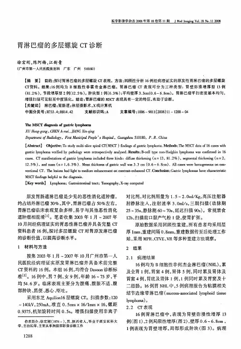
胃肠道恶性淋巴瘤的CT/MRI诊断王守安,吴晶涛,陈文新,孙 骏,董 颖(扬州大学临床医学院,江苏省苏北人民医院影像科 江苏 扬州 225001) 【摘 要】 目的 提高胃肠道恶性淋巴瘤的CT/MRI诊断水平。
方法 回顾性分析经手术病理证实的16例胃肠道恶性淋巴瘤的CT/MRI平扫+增强表现。
结果 16例经手术病理证实的淋巴瘤均为非霍杰金淋巴瘤,12例为B细胞来源,3例为小淋巴细胞型,1例为T细胞型。
10例为胃壁肿瘤,5例为小肠肿瘤,1例为结肠肿瘤。
13例表现为弥漫增厚型,3例表现为溃疡型。
增强扫描后15例明显均匀强化,1例不均匀强化。
结论 CT/MRI对胃肠道恶性淋巴瘤具有较高诊断价值。
【关键词】 胃肠道;恶性淋巴瘤;体曾摄影术,X线计算机;磁共振成像中图分类号:R735.2;R814.42;R 445.2 文献标识码:A 文章编号:1006-9011(2012)07-1110-03CT/MR diagnosisi of primary gastriointerstinal malignant lymphomaWANG Shou-an,WU Jing-tao,CHEN Wen-xin,SUN Jun,DONG YinDepartment of Medical Imaging,Clinical Medical College of Yangzhou University,Subei Peoples Hosipital of Jiang-su Province,Yangzhou 225001,P.R.China【Abstract】 Objective To improve the CT/MR diagnostic level of primary gastrointerstinal malignant lymphoma.Meth-ods Imaging data and pathology or opertative results of 16patients are erviewed retrospectively.Results All patientswere non-hodgkin lymphoma,of whom 12were B cell,3were small cell,1was T cell.10located in stomch,5in intersti-nal,and 1in colon.13patients were well thickening type,and 3were ulceration type.15patients were homogeneously en-hanced after intervenous injection of contrast agent,1patient was heterogensis enhanced.Conclusion CT/MR have highdiagnotic value to the gastrointestinal malignant lymphoma.【Key words】 Gastrointestinal;Malignant lymphoma;CT;MR 胃肠道恶性淋巴瘤是原发于胃肠道肠壁内淋巴细胞的恶性肿瘤,是常见的结外淋巴瘤,约占结外淋巴瘤的30%~45%[1],占胃肠道恶性肿瘤的1%~5%,临床表现与胃肠道恶性肿瘤类似,但预后却明显优于后者,与节内淋巴瘤相比,预后也比较好。
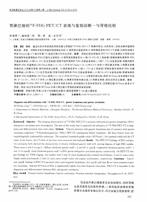

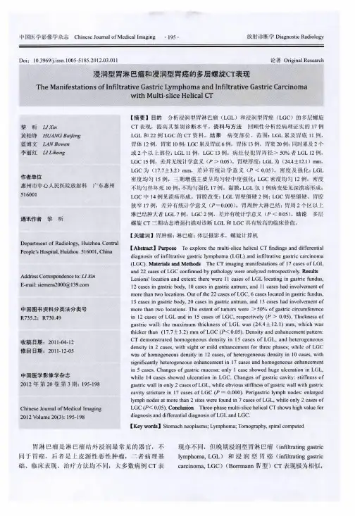
胃淋巴瘤的影像诊断分析胃淋巴瘤是胃肠道最常见的淋巴瘤,也是胃癌的一种。
由于临床表现不典型,因此影像诊断非常重要。
本文主要从两个方面进行分析,一是影像表现,二是影像诊断。
一、影像表现1、CT表现:CT检查是胃淋巴瘤的重要检查方法。
胃淋巴瘤CT表现多种多样,其中以胃肿瘤为主要表现。
胃淋巴瘤一般表现为胃壁局限性增厚,伴有胃腔狭窄,边界不清,部分病灶边缘可呈现溃疡样改变。
肿瘤密度不均匀,可以出现坏死、囊变。
少数表现为胃腔外肿块,多数伴随胃壁广泛浸润、肠壁受压以及节点转移等表现。
2、MRI表现:MRI检查可评估胃淋巴瘤的扩散情况和周围组织的受累程度。
表现为T1WI上信号略低,T2WI上信号略高,增强后呈不均匀强化。
3、PET-CT检查:PET-CT检查对定期评估胃淋巴瘤的疗效和复发具有较高的敏感性和特异性。
胃淋巴瘤在PET图像上显示为代谢亢进的病灶,一般表现为SUV值较高、分布不均。
二、影像诊断1、胃淋巴瘤的定性诊断:当胃肠道内检查触及到可疑病变时,常用CT或MRI进一步明确诊断。
在鉴别胃癌和胃淋巴瘤时,胃癌多见于胃幽门部,肿瘤边缘鳞状上皮变化明显,与胃壁分界清晰;而胃淋巴瘤多见于胃中段,肿瘤边缘不清,呈现淡漠性胃壁增厚。
2、胃淋巴瘤的定量诊断:通过CT图像计算肿瘤大小、SUV值等进行定量诊断,可为临床提供更多的信息。
例如,胃淋巴瘤病灶直径>10mm积极处理,SUV>8为较高代表瘤细胞密度的指标。
综上所述,胃淋巴瘤的影像诊断是胃淋巴瘤诊断过程中一个必要且重要的环节,合理选择影像检查方法和准确解读影像结果可以为胃淋巴瘤的诊治提供重要的参考。