肉瘤样癌
- 格式:docx
- 大小:13.42 KB
- 文档页数:1

肺肉瘤样癌CT表现与病理分析【摘要】目的:通过12例肺肉瘤样癌患者的临床表现,CT征象及手术后病理分型来评估CT对肺癌肉瘤的诊断价值。
方法:将手术证实的12例肺癌肉瘤的临床表现、病理类型与CT特征对照分析。
结果:肺肉瘤样癌的CT表现显示具有肺癌和肉瘤的双重特征。
既具有癌块的边缘征象,如分叶、毛刺等周边浸润征象,增强扫描又具有肉瘤的强化表现,如较厚的环状强化、肿块中央常为不规则的密度减低区。
结论:肺内圆形或椭圆形较大肿块,增强扫描肿块呈不规则环状强化,中央显示不规则密度减低区,酷似肉瘤的CT表现,而其边界不甚规则,边缘不光整,有分叶、毛刺等浸润表现时应考虑为肺肉瘤样癌之可能。
胸部CT 增强扫描对肺肉瘤样癌的初步诊断具有一定的临床价值。
【关键词】肺肉瘤样癌CT 病理CT Imaging Analysis of Lung Sarcomatoid Carcimoma Correlated with Pathological FindingsWANG Jian-hua,ZHAO Zhi-qing,LUO Di-lin,HOU Yu,YANG Mao-hong,ZHUANQing-chun(Shenzhen Shajing Hospital Affliated to Guangdong Medical College,Shenzhen,Guangdong 518104,China)Abstract:Objective To investigate the diagnostic values of CT by studying the imaging and clincopathologic features of primary pulmonary sarcomatoid carcinoma (PSC). Methods The CT findings and clinicopathologic manifestations of PSC proved by operation and pathology in 12 cases were reviewed retrospectively. Results The CT findings of PSC had both lung cancer and sarcomatous features.The features including the marginal sign of cancer, such as lobular sign and spiculated sign, and thecontrast-enhanced CT findings of sarcoma, such as rim enhancement and irregular low hypodense areas in the center.Conclusion The PSC was a rounded or elliptic mass in the lung. It had rim enhancement and irregular low hypodense areas in the center after contrast-enhanced CT, lobular sign and spiculated sign. The Contrast-Enhanced CT had some clinic values in preliminary diagnostic PSC.Key words: Lung;Sarcomatoid carcinoma;CT; Pathology肉瘤样癌(Sarcomatoid Carcinoma,SC )是一种较少见的癌和肉瘤样形态混合于一个瘤体内的恶性肿瘤,可以发生在全身许多部位[1~5],但以上呼吸道、肺、乳腺和肾常见。


肉瘤样肝细胞癌1例赵丹红虞晓丽经风华王永仁义乌市中医院322000病例男性,55岁,因“右上腹胀痛半月余”入院。
既往发现“慢性乙型病毒性肝炎”病史10年,一直予正规口服“拉米夫定抗病毒治疗。
入院时查体:腹部平软,右上腹轻微压痛,无反跳痛,肝脾肋下未及。
辅助检查:AFP1.6ng/mL;上腹部CT增强:肝右叶直径约径约5.3cm的不规则形低密度区,边界不清,密度不均,增强扫描动脉期轻度强化,门脉期、延时期病灶边缘部分进一步强化,提示炎性病变、肝转移性肿瘤均有可能;进一步MR检查提示:右肝占位,考虑不典型增生结节,不排除外。
术中所见:轻度肝硬化改变,肝IV、V、VIII段与下腔静脉间探及一直径约5cm×6cm质硬肿块,行右半肝切除术。
术后病理:右半肝肉瘤样细胞癌。
免疫组化:CK、CK19、EMA、Vim(+);Ki67(30%阳性);AFP、Hep-par-1、CEA、SMA(‐)。
术后三个月患者死亡。
图2:梭形瘤细胞,异型明显H&E×100Figure2:The spindle cells had obvious abnormity(H&E×100)图1:(a):CT平扫示肝右叶低密度占位,约5.0*5.3cm,边界不清,病灶中央见片状囊性密度区(b):CT增强扫描动脉期,病灶边缘实性成分轻度强化(c、d):门脉期及延时期显示病灶边缘实性成分持续强化Figure1:(a):Axial NECT image shows a 5.0*5.3cm,ill—defined hypodense lesion in the right lobe of liver,with central areas of more hypodense(b):arterial phase ,the lesion shows mild enhancement of the peripheral solid part(c、d):portal venous and delayed phase, the peripheral solid part shows continued enhancement讨论肝肉瘤样癌是指原发于肝的恶性上皮样肿瘤,恶性程度高,十分少见,约占外科手术切除肝癌的1.8%[1]。
38例肺肉瘤样癌临床特点及预后的回溯性分析李园园;张丽丽;蒋娟;杨华平;曹立明;顾其华;李敏;胡成平【摘要】Background and objective Pulmonary sarcomatoid carcinoma is a rare histologic subtype of non-small cell lung cancer. hTe effective treatment for this disease has not well deifned due to its extremely low morbidity. hTis study explores the clinicopathological characteristics and prognosis of 38 patients with PSC, so as to provide some clues for its diagnosis and treatment. Methods hTe study enrolled 38 patients with PSC that were diagnosed with histology or cytology in our hospital between January 2000 and December 2013. We retrospectively analyzed general clinical characteristics, smoking history, tumor size, TNM staging, pathology, immunohistochemistry, diagnostic method, treatment and prognosis. We used SPSS 19.0 statistical sotfware and Kaplan-Meier method to analyze our data. Results Patients in this study were aged from 26 to 76 years old (the median age was 57.5 years old). Among all of them, the male to female ratio was 4:1, and 81.6%of patients had smoking history. Cough and hemoptysis were the most common primary symptoms. hTe median survival was 21 months, while one-year survival rate, three-year survival rate and five-year survival rate were 68.4%, 31.6%and18.4%respectively. Tumor size, TNM staging, distant metastasis and surgery therapy were associated with the prognosis of patients. Conclusion Patients with PSC present with no special symptoms generally. According to our study, factors that affect patients’ prognosis include tumor s ize,TNM staging, distant metastasis and surgery. Complete resection is the key treatment for PSC patients, but comprehensive chemoradiotherapy needs further exploration in evidence-based medicine. Biological target therapy may give new insight into treatment for PSC.%背景与目的肺肉瘤样癌(pulmonary sarcomatoid carcinoma, PSC)是一种罕见的肺部恶性肿瘤,属于非小细胞癌。
PD-1肉瘤样癌完全缓解
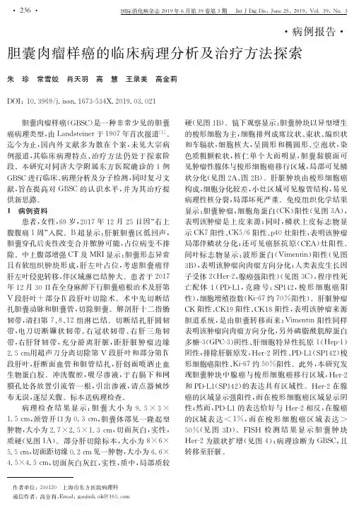
胆管肉瘤样癌属于一种特殊类型的胆管癌,其非肉瘤或癌肉瘤,又称为梭形细胞癌或化生性癌[1]。
肉瘤样癌的肉瘤样成分是癌的化生,此特点区别于单纯的肉瘤及同时有上皮与间质来源的癌肉瘤。
部分病例还可见巨细胞[2-3]。
肉瘤样癌可见于多个器官,文献报道的有上消化道、乳腺、皮肤、女性生殖道、泌尿道和肺等[3]。
肝内胆道系统的肉瘤样癌非常罕见,仅占肝内原发性恶性肿瘤的0.2%[
Rh阴性血
患者因胆囊占位于2019.05.19全麻下行胆囊癌根治术,术后病理示:胆囊管与胆总管结合部浸润型肉瘤样癌,大小:5.0×4.0×3.5cm,间质伴少量淋巴细胞浸润,中央伴片状肿瘤性坏死;肿瘤侵透胆管壁深肌层达外膜,肿瘤广泛侵犯神经,未见脉管内癌栓;胆管下切缘、左侧胆管上切缘及右侧胆管上切缘均未查见癌。
慢性胆囊炎;淋巴结未查见转移性癌(0/7)(1)(胆囊颈周围淋巴结)0/2(2)(第12P组淋巴结)0/5 免疫组化结果示:CK广谱(+),Vimentin(3+),EMA(-),S-100(-),CK7(-)CK8/18(2+),CK19(-),Bcl-2(-),Desmin (2+),SMA(+),CD34(-),CR(-)β-cantenin(-),CD117(-),Dog-1(-),CD99(-),Actin(-),Lysozyme(+),CD68(3+),CD163(+),CD31(-),NSE(-),NF(-),FN(3+),CD10(3+),P53(3+),Ki-67(+,45%-50%)。
肿瘤病理分期:pT2N0。
术后恢复较顺利,2020.01.06于我院门诊行CT示胆囊管癌术后,肝门区病变较前新发,考虑复发可能并肝左叶胆管扩张;左肾下极小囊肿;右肺下叶支气管扩张;双肺多发纤维灶。
2020.01.11行MR示:胆囊术后,肝门区肿物并肝左叶胆管梗阻,考虑复发。
患者胆囊管肉瘤样癌复发,放化疗不敏感,
与患者及家属沟通后2020.01.12行靶向及免疫治疗,今为行进一步治疗,门诊以“胆囊癌术后”收入院。
患者自发病以来,神志清,精神可,食欲可,体重无明显减轻。