肝腺瘤的CT诊断与鉴别
- 格式:pdf
- 大小:163.12 KB
- 文档页数:2


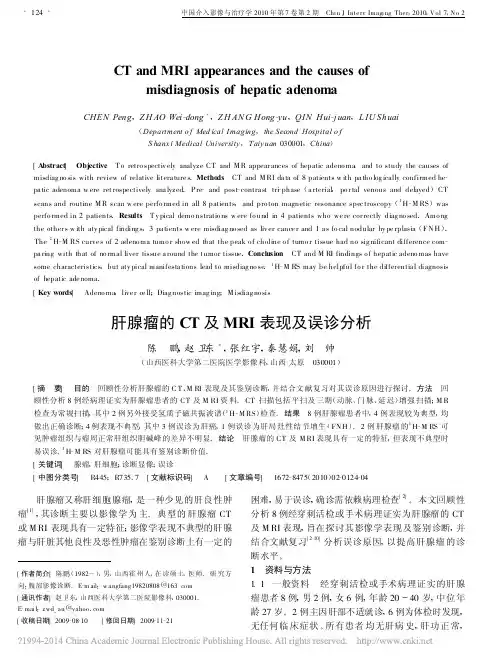
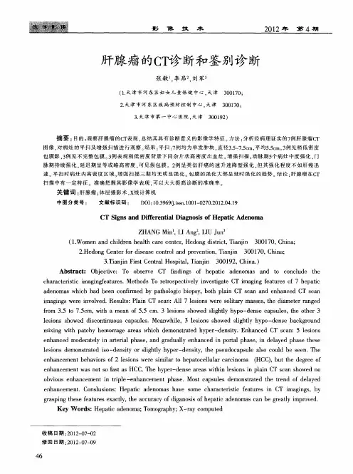
CT and MRI appearances and the causes ofmisdiagnosis of hepatic adenomaCHEN Peng ,Z H AO Wei -dong *,Z H AN G Hong -yu ,QIN Hui -j uan ,L IU Shuai(Department o f Med ical I maging ,the Second Hospital o f S hanx i Medical University ,T aiy uan 030001,China )[A bstract ] Objective T o retro spectiv ely analyze CT and M R appear ances of hepatic adeno ma ,and to study the causes of misdiag no sis with review of relative litera ture s .Metho ds CT and M RI da ta of 8patients w ith pa tho log ically confirmed he -pa tic adenoma w ere ret rospectively ana lyzed .P re -and post -contra st tri -phase (a rterial ,po r tal venous and delayed )CTscans and routine M R scan w ere perfo rmed in all 8patients ,and pro ton magnetic resonance spec troscopy (1H -M RS )wasperfo rmed in 2patients .Results T y pical demo nstratio ns w ere fo und in 4patients who we re cor rectly diag no sed .Amo ng the other s w ith aty pical finding s ,3pa tients w ere misdiag no sed as liv er cance r and 1a s fo cal nodular hy pe rplasia (F N H ).The 1H -M RS curve s of 2adeno ma tumo r show ed that the peak o f choline o f tumo r tissue had no sig nificant difference com -pa ring with that of no rmal liver tissue a round the tumor tissue .Conclusion CT and M RI findings o f hepatic adeno mas have some characteristics ,but aty pical manifesta tions lead to misdiag no se .1H -M RS may be helpful fo r the differentia l diagnosis of hepatic ade noma .[Key words ] Adeno ma ,live r ce ll ;Diag nostic imag ing ;M isdiag no sis肝腺瘤的CT 及MRI 表现及误诊分析陈 鹏,赵卫东*,张红宇,秦慧娟,刘 帅(山西医科大学第二医院医学影像科,山西太原 030001)[摘 要] 目的 回顾性分析肝腺瘤的C T 、M RI 表现及其鉴别诊断,并结合文献复习对其误诊原因进行探讨。
肝腺瘤ct报告模板
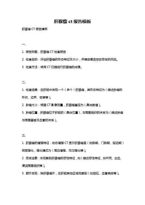
肝腺瘤CT报告模板
一、
1. 报告标题:肝腺瘤CT检查报告
2. 检查目的:评估肝腺瘤的形态特征及大小,并确定是否存在恶变的风险。
3. 检查方法:使用CT扫描进行肝腺瘤的成像。
二、
1. 检查结果:在肝脏中发现一个(多个)肝腺瘤,其形态特征为(描述肿瘤的形状、边界、密度等)。
2. 肿瘤大小:根据CT影像测量,肝腺瘤直径为(具体数值)。
3. 肿瘤位置:肝腺瘤位于肝脏的(具体位置),与周围组织的关系为(描述肿瘤与周围器官及血管的关系)。
三、
1. 肝腺瘤的增强特征:动态增强CT显示肝腺瘤呈(动脉期、门脉期、延迟期)明显强化,强化模式为(周边增强、均匀强化等)。
2. 恶变迹象:未观察到肝腺瘤的恶性特征,如(描述恶性特征,如坏死、出血、浸润周围组织等)。
3. 额外发现:除肝腺瘤外,在肝脏其他区域观察到(如结石、血管病变等)。
四、
1. 诊断建议:综合CT结果,初步诊断为良性肝腺瘤,但需结合临床资料,进一步评估肿瘤的性质和风险。
2. 随访建议:对于较大或存在不确定性的肝腺瘤,建议定期复查CT或进行其他相关检查,以评估肿瘤的生长情况及可能的恶变迹象。
3. 注意事项:如有不适或症状加重,请及时就医,并告知医生有关CT检查的结果。
希望以上模板可以帮助您完成相应的文章写作。
使用时请根据具体情况进行适当修改。

FNH和肝腺瘤的影像诊断与鉴别诊断Focal Nodular Hyperplasi FNH a1•肝脏局灶性结节性增生(Focal nodular hyperplasia,FNH)是良性增生性疾病,由异常排列的肝细胞结节构成,组织学正常或结节正常。
•是仅次于海绵状血管瘤的第二常见的肝脏良性病变。
•未见恶变,且部分可自行消退。
•好发于青年女性,是由于雌激素刺激血管畸形发展和肝细胞增生。
•一般无临床症状,少数出现腹痛、腹部包块。
•血管畸形或血管损伤。
•细胞对局部血管异常产生的反应性增生,而非真正意义上的肿瘤。
经典型:最为常见(80%),呈结节型,切面具有典型特征性改变,即病变中央有星形瘢痕;病灶周边或中央存在供血血管。
•富血供实性肿块。
•肿块内部结构均匀,出血和坏死少见。
•特点是病灶中心有星状瘢痕及辐射状纤维分隔,瘢痕内有厚壁供血动脉,但无门脉血液•由肝细胞、胆管、Kupffer细胞、血管、炎性细胞及纤维间隔组成。
•病灶中心的“星形瘢痕”并非真性瘢痕,而是血管与胆管的聚积。
经典型:最为常见(80%),呈结节型,切面具有典型特征性改变,即病变中央有星形瘢痕;病灶周边或中央存在供血血管。
•孤立结节样增生实质,被环形纤维间隔完全或不完全包绕。
•中央疤痕包含纤维结缔组织、增生的胆管及周围浸润的炎症细胞以及各种畸形的血管,畸形的中央动脉呈离心状向外供血。
不典型:病变中央缺乏星形瘢痕灶,或畸形血管,但都有胆管增生。
可分为以下3型:1)毛细血管扩张型:表现为短小的纤维分隔+较多的扩张血管+小胆管增生。
2)混合细胞型:少许纤维间隔+少数畸形血管,肝细胞实质性增生及增生的胆管明显。
3)伴肝细胞不典型增生型:可具有上述不同类型成分的表现。
•呈孤立的等密度或略低密度肿块。
•界限欠清,密度均匀,很少有钙化。
•部分病灶中可见低密度瘢痕。
•动脉期:病灶呈快速明显的均匀强化,中心瘢痕不强化。
•门脉期:病灶实质强化部分造影剂开始退出,趋向等密度。