甲状腺微小癌的诊断和治疗
- 格式:doc
- 大小:330.37 KB
- 文档页数:52
甲状腺癌微小癌的标准甲状腺微小癌是一种常见的甲状腺癌类型,其诊断标准在不同的研究和临床实践中可能会有所不同。
以下将介绍几种常见的甲状腺微小癌的标准。
1.尺寸标准:根据美国癌症学会(American Cancer Society, ACS)的定义,甲状腺微小癌通常指甲状腺结节直径小于1.0-1.5厘米的癌症。
然而,对于甲状腺结节的尺寸定义仍存在争议。
2.细胞学标准:甲状腺微小癌的细胞学特征是其诊断的一个重要标准。
细胞学上,微小癌的特点是癌细胞更小、形态规则、核分裂少、无微核和浸润性的特征。
此外,细胞核的乏特性和胞浆的亚皮样特点是其常见的细胞学特征。
3.癌组织浸润标准:甲状腺微小癌的第三个标准是其浸润性。
有研究发现,甲状腺微小癌中大部分病例在手术切除后仍然保持微小癌的肿瘤限制性生长特点,未能浸润周围组织或转移至淋巴结。
因此,尽管微小癌是一种癌变病变,但其低度恶性特征使得治疗和预后较好。
4.分期标准:甲状腺微小癌的分期标准也是其诊断和治疗的重要指标。
根据美国癌症学会(American Joint Committee on Cancer, AJCC)的分期系统,甲状腺微小癌通常被分类为T1a阶段,表示肿瘤直径小于1.0厘米。
这一分期主要是根据肿瘤尺寸进行分类,且不考虑淋巴结转移或远处转移。
5.预后标准:尽管甲状腺微小癌是甲状腺癌中最小的类型之一,但其预后仍然是一个重要考虑因素。
预后因素包括年龄、性别、肿瘤大小、多发性和淋巴结转移等。
一些研究指出,甲状腺微小癌通常具有比较良好的预后,术后康复和存活率较高。
然而,这并不意味着所有的甲状腺微小癌患者都具有良好的预后,因此,个体化的多因素分析仍是评估预后的重要手段。
总之,甲状腺微小癌的标准主要包括尺寸、细胞学、浸润、分期和预后等方面。
但需要注意的是,标准的制定仍然存在争议,因此在诊断和治疗过程中需要综合考虑多个因素,并根据具体情况进行个体化的评估和决策。

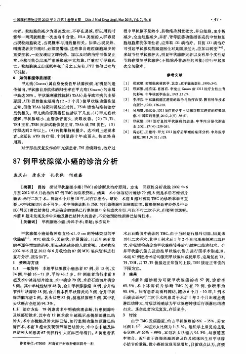


甲状腺微小癌病情说明指导书一、甲状腺微小癌概述甲状腺微小癌(thyroid microcarcinoma,TMC)又称隐匿性甲状腺癌,是指癌灶最大直径≤10mm的甲状腺恶性肿瘤,是常见的内分泌恶性肿瘤。
近年来患病率显著增加,女性明显高于男性。
甲状腺微小癌无典型临床症状和体征,发病隐匿,多合并甲状腺良性结节,术前确诊有一定难度,容易误诊及漏诊。
不伴转移者预后良好,及时治疗可以痊愈。
英文名称:thyroid microcarcinoma,TMC。
其它名称:隐匿性甲状腺癌。
相关中医疾病:暂无资料。
ICD疾病编码:暂无编码。
疾病分类:暂无资料。
是否纳入医保:部分药物、耗材、诊治项目在医保报销范围,具体报销比例请咨询当地医院医保中心。
遗传性:本病与遗传有关,发病有一定的家族史。
发病部位:甲状腺。
常见症状:一般无典型临床症状和体征,部分患者可在甲状腺部位触及肿物或肿大。
主要病因:放射线接触、碘摄入不当、淋巴性甲状腺炎、家族遗传、妊娠或外源性雌激素的使用。
检查项目:分子标志物水平检测、常规超声、超声造影、计算机断层扫描(CT)、磁共振成像(MRI)、放射性核素显像、正电子发射体层摄影术(PET/CT)、超声弹性成像、细针抽吸细胞学(FNA)检查、冰冻病理检查。
重要提醒:甲状腺微小癌无典型临床症状和体征,发病隐匿,出现异常需及时就医。
临床分类:基于临床病理分类:1、甲状腺乳头状微小癌(PTMC)甲状腺微小癌占甲状腺癌的6%~35%,PTMC 在微小癌中占65%~99%。
乳头状癌恶性程度最低,生长缓慢,可在甲状腺内局限数年。
病灶可经腺体内淋巴管扩散,血行转移较少,如有则以肺转移为主,次为骨转移。
随着年龄增大,肿瘤恶性程度增高。
2、甲状腺滤泡癌滤泡癌很少从淋巴转移,一般通过血行扩散,特别扩散至骨骼、肺、肝等脏器。
3、甲状腺髓样癌髓样癌恶性程度较滤泡癌高。
甲状腺髓样癌一般分为散发性和家族性。
癌肿易侵蚀腺体内淋巴管,扩散至腺体其他部位,也同样可以血行扩散至其他脏器。
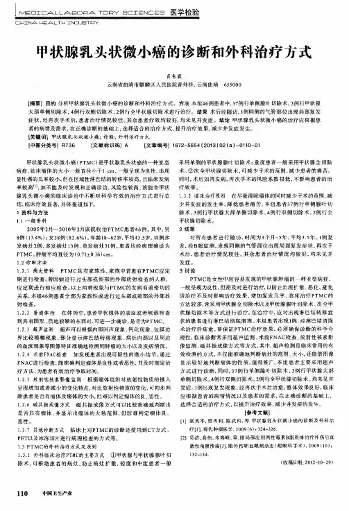
53例甲状腺微小癌的诊断和外科治疗分析作者:刘鑫来源:《中国实用医药》2013年第30期【摘要】目的探讨甲状腺微小癌的临床特点和不同术式治疗对预后的影响。
方法回顾分析本院收治的53例甲状腺微小癌病例的临床资料,对临床病理特征、手术方式和预后进行总结。
结果本组53例患者中病理组织类型为乳头状癌49例(92.45%),滤泡状癌3例(5.66%),髓样癌1例(1.88%);术后随访6个月~4年, 2例复发,行残余甲状腺全切除术,全部患者均无远处转移和死亡。
结论甲状腺微小癌手术治疗预后较好,针对不同的患者应选择合适的治疗方案,以使患者取得良好的疗效。
【关键词】甲状腺微小癌;诊断;治疗甲状腺微小癌是指肿瘤直径≤10 mm,无论有无区域淋巴结转移的甲状腺癌性病灶。
由于病灶较小,发病隐匿,且多与其他甲状腺疾病共存,因而易造成临床的漏诊和误诊[1]。
近年来,随着超声和病理诊断技术的发展和提高,甲状腺微小癌的发生率呈逐渐升高的趋势。
为探讨甲状腺微小癌的临床特点、诊治方法,以提高其诊治水平,作者回顾分析河南省郸城县人民医院普外科收治的53例甲状腺微小癌病例的临床资料,现报告如下。
1 资料与方法1. 1 一般资料 2003年5月~2012年8月本院收治并经术后病理确诊的甲状腺微小癌患者53例,男8例,女45例,男女比为1:5.63,年龄22~74岁,病程1月~5年。
术前超声检查示:肿块边界不规整,有血流较丰富的甲状腺低回声结节39例,结节伴细小、砂砾样钙化33例,伴颈部淋巴结肿大2例。
1. 2 治疗方法所有病例均给予手术治疗,术中全面探查甲状腺,切除的腺体组织送术中快速病理检查。
患侧腺叶+峡部+对侧腺体次全切除术41例,术中行颈部淋巴结清扫5例,改良颈部淋巴结清扫12例,单侧腺叶+峡部切除术5例,双侧腺叶次全切除术6例,双侧腺叶全切术1例。
患者术后均常规口服给予甲状腺素片治疗。
2 结果2. 1 术后病理和并发症 53例术后病理显示,乳头状癌49例(92.45%),滤泡状癌3例(5.66%),髓样癌1例(1.88%);瘤体直径>5 mm者15例,瘤体直径≤5 mm者38例;单发癌41例,多发癌16例;微小癌合并有结节性甲状腺肿者42例,合并有甲状腺瘤者6例,合并有桥本甲状腺肿者5例。
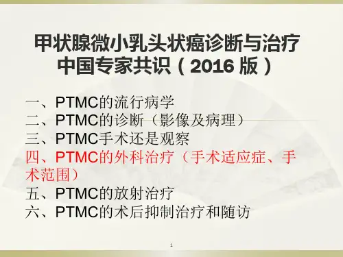
甲状腺癌微小癌的标准
一、病灶直径
甲状腺癌微小癌的病灶直径不超过1厘米。
这是判断是否为微小癌的重要指标之一。
二、病灶数量
甲状腺癌微小癌通常是单个病灶,但如果存在多个微小癌病灶,且多发的微小癌有淋巴结转移风险,也需立即治疗。
三、病灶位置
甲状腺癌微小癌的病灶生长在低风险的位置,如甲状腺腺体正中间,不能紧挨着气管等危险部位。
这意味着病灶的位置相对安全,不会对周围重要器官造成威胁。
四、淋巴结转移情况
甲状腺癌微小癌没有发生淋巴结转移。
这是判断病情的重要指标之一,因为淋巴结转移可能影响患者的预后和治疗方案。
综合以上四个方面,甲状腺癌微小癌的标准包括:病灶直径不超过1厘米,单个病灶或多个病灶但无淋巴结转移风险,病灶生长在低风险位置如甲状腺腺体正中间,以及无淋巴结转移。
这些标准有助于医生准确地诊断和制定合适的治疗方案,以保障患者的健康和预后。
学 校 代 码 10459学号或申请号密 级专业硕士学位论文甲状腺微小癌的诊断和治疗作 者 姓 名:导 师 姓 名:专业学位名称:普通外科培 养 院 系:完 成 时 间:2016年5月A thesis(dissertation) submitted toZhengzhou Universityfor the degree of Master(doctor)Diagnosis and treatment of thyroid cancerBy Zhijian MengXiuBo LUGeneral SurgeryThe First Affiliated Hospital of Zhengzhou UniversityMay,2016原创性声明本人郑重声明:所呈交的学位论文,是本人在导师的指导下,独立进行研究所取得的成果。
除文中已经注明引用的内容外,本论文不包含任何其他个人或集体已经发表或撰写过的科研成果。
对本文的研究作出重要贡献的个人和集体,均已在文中以明确方式标明。
本声明的法律责任由本人承担。
学位论文作者:日期:年月日学位论文使用授权声明本人在导师指导下完成的论文及相关的职务作品,知识产权归属郑州大学。
根据郑州大学有关保留、使用学位论文的规定,同意学校保留或向国家有关部门或机构送交论文的复印件和电子版,允许论文被查阅和借阅;本人授权郑州大学可以将本学位论文的全部或部分编入有关数据库进行检索,可以采用影印、缩印或者其他复制手段保存论文和汇编本学位论文。
本人离校后发表、使用学位论文或与该学位论文直接相关的学术论文或成果时,第一署名单位仍然为郑州大学。
保密论文在解密后应遵守此规定。
学位论文作者:日期:年月日中文摘要甲状腺微小癌的诊断和治疗中文摘要背景:甲状腺癌是较为常见的恶性肿瘤,有研究表明,在我国甲状腺癌人群发病率约为1.5/10万,但近年来发病率呈现出年轻化和升高的趋势[1、2、3]。
甲状腺微小癌属于特殊类型甲状腺癌,直径多小于1cm[4、5 ]。
随着现代医疗技术的发展,高分辨率超声的应用以及病理诊疗技术水平的进步,对于甲状腺微小癌的诊断和治疗报道逐年增加,但是许多病例确诊是在术中偶然发现或术后甲状腺病理检查中发现的,在临床诊疗的过程中漏诊率高[6]。
甲状腺微小癌起病隐匿,常与其他甲状腺疾病共存,有的病变长期处于一种亚临床的状态,其肿瘤行为被认为是一种良性病变,但是不是所有甲状腺微小癌都处于静止状态,也有可能出现肿瘤侵袭性生长和淋巴结转移现象,淋巴转移是其主要转移途径,甲状腺微小癌发生淋巴结转移后就会出现临床表现,此时必须予以治疗[7、8、9]。
既往研究表明甲状腺微小癌治疗效果较为理想,外科治疗能彻底治愈[10]。
因而,要特别重视甲状腺微小癌的诊断和治疗,降低复发和转移的发生率。
目的:回顾性分析甲状腺微小癌患者的临床特征,淋巴结转移、手术方式,以期为甲状腺微小癌的诊疗提供指导。
方法:回顾分析2012年1月-2014年1月到郑大一附院甲状腺外一科住院101 例甲状腺微小癌患者,且经术后病理证实的甲状腺微小癌患者。
在性别方面:男 26 例,女 75 例,在年龄方面,所有患者年龄均在 23-68 岁之间,其中年龄在45岁以下患者58 例,45岁以上患者43 例,平均年龄为46.39±17. 85岁,在临床分型方面,其中T1N0M0 54 例,T1N1M0 47 例。
在发病情况方面,双侧腺发病20例,单侧腺发病81例,在合并症方面,101例患者中合并其他甲状腺的良性疾病61例。
1、本次入选的101例甲状腺微小癌患者术前均行超声影像学检查及细针穿I中文摘要刺针吸细胞学检查;2例患者行放射性核素扫描;5例患者行CT 检查。
2、所入选的101 例患者均采用外科手术治疗方式。
其中行11例单侧腺叶及峡部切除术,双侧甲状腺全切术6例,患侧甲状腺切除并中央区淋巴结清扫54例,双侧甲状腺切除并患侧中央区淋巴结清扫25例,甲状腺改良根治术5例。
在淋巴结的清扫方面,84例患者行颈部淋巴结清扫术,其中79例患者行单纯VI区淋巴结清扫术。
3、所有数据通过spss19.0处理,计量资料应用t检验,计数资料应用X2检验;采用单因素方差分析对年龄、性别有无淋巴转移、肿瘤直径、病灶情况、手术方式、是否侵袭包膜等因素进行分析。
P<0.05作为有临床意义的标准。
结果:1、甲状腺微小癌的超声影像学结果:术前甲状腺结节超声根据Kwak等[1]提出的TI-RADS分级方法,在本次临床研究的101例甲状腺微小癌患者中,Ⅲ级7例、Ⅳa级 46例、Ⅳb级 32例、Ⅳc级5例、Ⅴ级5例。
其中在Ⅲ级及Ⅳ级中,有83例均行超声引导针吸细胞学检查,符合微小癌诊断的有75例。
2、在本组病例的101例均行术中冰冻病理检查,其中19例在术后病理检查中确诊,假阴性率19%;3、本组病例中出现VI区淋巴结转移的占38%(38/101),颈侧区淋巴结转移的占14%(14/101),两组具有显著差异(P<0.05),有统计学意义;4、在VI区淋巴结清扫中,术前超声检查发现转移为60.00%(30/50);未发现转移淋巴结为25.00%(8/32),具有统计学意义(P<0.05);颈侧区淋巴结清扫术中,术前超声检查发现及未发现转移淋巴结分别为63.16%和16.67%,有统计学意义(P<0.05)5、对影响预后、肿瘤复发的相关因素进行单因素方差分析及多因素Logistic回归分析,最后发现病灶为多发、有包膜以及出现淋巴转移为病人出现术后肿瘤复发的独立相关因素(P<0.05)。
6、本次临床研究中,8例患者手术后出现甲状旁腺功能低下,但在术后3II中文摘要个月内恢复,未出现喉返神经麻痹。
结论:1、目前高分辨率超声和超声引导针吸细胞学检查是术前诊断甲状腺微小癌的主要手段。
2、甲状腺微小癌的淋巴结转移率高,需要常规行患侧甲状腺全切除+VI区淋巴结清扫或者患侧腺叶加峡部切除。
结合病情开展综合个体化治疗。
3、甲状腺微小癌可能早期出现颈部淋巴结转移,所以并不都是早期癌。
关键词:甲状腺微小癌;早期诊断;超声;针吸细胞学;IIIAbstractAbstractBackground:Thyroid carcinoma is a common malignant tumor. The study shows that the incidence of thyroid carcinoma in China is about 15/100000, but in recent years, the incidence rate is younger and higher. Thyroid Mini carcinoma is a special type of thyroid carcinoma, with a diameter of more than 1cm, which is a special type of thyroid carcinoma. With the development of modern medical technology, high resolution ultrasound and pathological diagnosis and treatment level of technology progress and to report on the diagnosis and treatment of thyroid microcarcinoma increased year by year, but many cases is found accidentally discovered in the intraoperative or postoperative thyroid pathological examination, in the process of clinical diagnosis and treatment of misdiagnosis rate is high. Thyroid microcarcinoma of insidious onset, often coexist with other thyroid diseases, and some lesions in long-term a subclinical state. The tumor behavior is considered to be a benign lesion, but not all thyroid microcarcinoma in static state, may also appear tumor invasion hit growth and lymph node transfer phenomenon, lymphatic metastasis is the major route of metastasis, lymph node metastasis will appear after the clinical manifestations of thyroid microcarcinoma and must then be treated. Previous studies have indicated that the effect of thyroid Mini carcinoma treatment is more ideal, surgical treatment can be completely cured. Therefore, special attention should be paid to the diagnosis and treatment of small thyroid carcinoma, and to reduce the incidence of recurrence and metastasis.Objective: To retrospectively analyze the clinical characteristics, lymph node metastasis and surgical methods of small thyroid carcinoma patients, and to provide guidance for clinical diagnosis and treatment of thyroid carcinoma.Methods: a retrospective analysis of 101 cases of thyroid Mini carcinoma in theIVAbstractDepartment of thyroid surgery, the 1st Affiliated Hospital of Zhengzhou University, from January 2012 to January, and patients with thyroid carcinoma were confirmed by postoperative pathological examination. In terms of gender: 26 cases of male, female 75 cases, in terms of age, age of all the cases were 23-68 between the ages of. The age in 58 cases of patients under the age of 45, 43 cases of patients over 45 years old, average age for 46.39±17.85 years old, in the clinical type, including 53 cases of T1N0M0, t1n1m0 in 47 cases. In the incidence of the disease, 20 cases of bilateral gland disease, 81 cases of unilateral gland disease, in terms of the complications, 101cases of patients with other thyroid benign diseases in 61 cases.1.The selected 101 cases of patients with small thyroid carcinoma were examined by ultrasonography and fine needle aspiration cytology before operation. 2 patients underwent radionuclide scan and 5 patients underwent CT examination.2.Surgical treatment was adopted in 101 patients. Among them, 99 cases underwent subtotal thyroidectomy. 2 patients underwent unilateral lobectomy and isthmus resection. In lymph node dissection, 99 cases of patients with cervical lymph node dissection, which 51 cases of patients with pure VI lymph node dissection, 31 cases underwent VI + neck region of lateral lymph node dissection; 5 cases and bilateral lateral neck dissection.3.All the data by spss19.0 processing, measurement data using t-test, count data using X2test. The single factor variance analysis of gender, age, lymph node metastasis, tumor size, tumor, surgical approach, invasion envelope and other factors were analyzed. P<0.05 as a standard of clinical significance.Result:1.Thyroid microcarcinoma of ultrasound imaging studies showed that preoperative ultrasound of thyroid nodules by Kwak et al proposed Ti radsclassification grade 7 cases in grade III, IV A-level 46 cases, IV grade B 32 cases, IV grade C in 5 cases,VAbstractgrade V 5 cases. Among them, 83 cases of grade III and IV were all performed by ultrasound guided needle aspiration cytology in 76 cases. Cervical lymph node enlargement: VI area in 35 cases, 25 cases of cervical side area, of which 8 cases of cervical lateral lymph node needle biopsy confirmed metastatic lymph nodes for thyroid cancer. CT examination results: 4 cases all showed no significant accounting, not all of the thyroid gland density, 2 cases showed that the neck and trachea lymph nodes increased. Cytological examination of cervical lymph nodes:. Thyroid “cold nodule”.2.In this group of cases, 101 cases were diagnosed by intraoperative frozen pathological examination, of which 19 cases were confirmed by pathological examination after operation, the false negative rate was 19%;3.In this group, there were 38% (38/101) in the lymph node metastasis in the VI teral cervical lymph node metastasis was 14% (14/101), the two groups had significant difference (P<0.05), there was statistical significance;4. In VI lymph node dissection, preoperative ultrasonography found for the transfer of 60.00% (30 / 50); no metastasis was found in the lymph node was 25.00% (8 / 32), with statistical significance (P < 0.05); neck region of lateral lymph node dissection, preoperative ultrasonography was found and no metastasis was found in the lymph node was 63.16% and 16.67% respectively, was statistically significant (P < 0.05) 5.Affect the prognosis and recurrence related factors of single factor variance analysis and multiple logistic regression analysis, finally found lesions were multiple, enveloped and lymph node metastasis tumor recurrence of independent factors (P < 0.05) for the patient.6. After surgery, there were 8 cases of patients with low parathyroid function, temporary, 3 months after the recovery, no recurrent laryngeal nerve palsy. Conclusion:High resolution ultrasound and ultrasound guided needle aspiration cytological examination areVIAbstractthe main methods for the diagnosis of thyroid micro carcinoma before operation. The rate of lymph node metastasis in thyroid carcinoma is high, which requires that the +VI region lymph node dissection in the near total resection of the thyroid gland is required. Thyroid cancer can be early cervical lymph node metastasis, not all of them early cancer.Key words: thyroid cancer; early diagnosis;ultrasound;needle aspiration cytologyVII目录目录正文部分前言 (1)资料与方法 (2)结果 (3)讨论 (6)结论 (21)参考文献 (21)综述部分甲状腺乳头状微小癌的主要诊断和外科治疗 (28)参考文献 (35)附录部分个人简介 (38)攻读硕士学位期间发表论文: (38)致谢 (39)VIII目录DIRECTORYBODY PARTPREFACE (1)MATERIALS AND METHODS (2)RESULT (3)DISCUSSION (6)CONCLUSION (21)REFERENCE (21)REVIEW SECTIONPRIMARY DIAGNOSIS AND SURGICAL TREATMENT OF PAPILLARYTHYROID CARCINOMA (28)REFERENCE (35)POSTSCRIPTURAL MATTERPERSONAL PROFILE (38)STUDY FOR A MASTER'S DEGREE IN PUBLISHED PAPERS: (38)THANKS (39)IX前言前言甲状腺癌是头颈部和内分泌系统最常见的恶性肿瘤,近十年来,其发病率在世界范围内呈现明显上升的趋势,而在育龄女性中的发病率更高[1]。