条件培养液对兔骨髓基质细胞向成骨细胞分化的影响
- 格式:pdf
- 大小:142.21 KB
- 文档页数:3

短期冻存兔骨髓间充质干细胞对其生物特性的影响武成聪;李强;陈佳滨;茹嘉;蔡伟良;宁寅宽【摘要】Objective To study the effects of short-term cryopreservation on the proliferation ,bio-characteristics and osteogenic capability of rabbit BMSCs and lay the foundation for the further study .Methods The 5th generation of rabbit BMSCs were cryo-preservated by liquid nitrogen for 30 days ,and then thawing the cells to culture it to the 10th generation in vitro ,put the non-cryo-preservated and the same generation BMSCs as the control group .The MTT ,cell cycle ,cell surface marker ,the content of ALP and the immunohistochemical staining for collagen typeⅠ and alizarin red staining for calcium after differentiation induced by osteogene-sis were used to evaluate the proliferation ,bio-characteristic and osteogenic differentiation capability of rabbit BMSCs .Results The growth incubation period of BMSCs after cryopreservated was extended ,but it gradually recover through serial passage .The prolif-eration index and the proportion of BMSCs in G1 phase were 42 .9% ± 3 .4% and 57 .0% ± 3 .4% respectively .The positive rate of cell surface marker of CD44 was 93 .62% ± 1 .05% without expression of CD45 .The contents of ALP(U/gprot) were 6 .73 ± 1 .92 and 15 .99 ± 4 .36 in BMSCs after 7th day and 14th day with osteogen ic induction ,respectively .The collagen typeⅠ and alizarin red staining for calcium indicated positive at BMSCs after 14th day and 21th day with osteogenic induction ,respectively .These results showed no significant difference compared with the control group(P>0 .05) .Conclusion Short-term cryopreservation has no obvi-ous impacts on the proliferation ,bio-characteristic and osteogenic capability of BMSCs and it could be used for further study .%目的:探讨短期冻存对兔骨髓间充质干细胞(BMSCs)的增殖、生物特征及成骨能力的影响。

Bio-Gide胶原膜对兔骨髓间充质干细胞增殖和成骨分化的影响许繁;杨德圣【摘要】背景:相关实验表明Bio-Gide胶原膜与细胞有良好的生物相容性,但有关与其复合培养干细胞成骨分化能力的报道少见。
目的:观察Bio-Gide胶原膜对骨髓间充质干细胞增殖及成骨分化的影响。
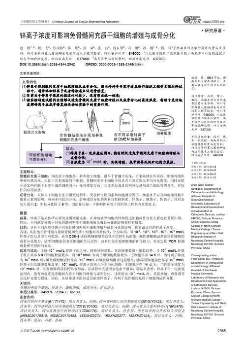
方法:全骨髓贴壁法体外分离培养兔骨髓间充质干细胞,将第3代兔骨髓间充质干细胞分别接种于覆盖Bio-Gide胶原膜的培养板(实验组)与单纯培养板(对照组)培养。
于培养1,4,7,14 d利用CCK-8试剂盒检测细胞增殖;成骨分化诱导培养1,4,7,14 d收集细胞培养液上清,检测细胞碱性磷酸酶活性。
结果与结论:两组细胞数量均随着培养时间的增加而不断增加,对照组培养7 d细胞数量明显多于实验组(P<0.05),其他时间点组间比较差异无显著性意义。
两组细胞碱性磷酸酶活性均随着培养时间的增加而不断增加,实验组成骨诱导14 d细胞碱性磷酸酶活性高于对照组(P <0.05),其他时间点组间比较差异无显著性意义。
表明Bio-Gide胶原膜可促进兔骨髓间充质干细胞的增殖及成骨分化。
%BACKGROUND:Bio-Gide col agen membranes show a good biocompatibility with stem cells. But the research on the osteogenetic differentiation of bone marrow mesenchymal stem cells cultured on theBio-Gide col agen membranes is rarely reported. OBJECTIVE:To observethe effect of Bio-Gide col agen membranes on the proliferation and the osteogenetic differentiation of bone marrow mesenchymal stem cells. METHODS:Bone marrow mesenchymal stem cells from rabbits were isolated and cultured by using the whole bone marrow adherence method in vitro. Passage 3 bone marrow mesenchymal stem cells were selectedand seeded on the Bio-Gide col agen membrane pretreated petri dish (experimental group) and simple petri dish (control group). The proliferation of bone marrow mesenchymal stem cells was detected by cellCounting Kit-8 at 1, 4, 7, 14 days. The supernatant of the cells cultured in osteogenic differentiation medium were col ected to detect the activity of alkaline phosphatase at 1, 4, 7, 14 days. RESULTS AND CONCLUSION:The number of bone marrow mesenchymal stem cells in the two groups was increased with the increasing time, and the control group had more cells than the experimental group at 7 days (P<0.05). There was no significant difference between the two groups at other time points. The alkaline phosphatase activity was increased with the increasing culture time, and the experimental group had a higher activity than the control group at 14 days (P<0.05). There was no significant difference between the two groups at other time points. Experimental findings indicate that Bio-Gide col agen membranes can promote the proliferation and the osteogenetic differentiation of bone marrow mesenchymal stem cells.【期刊名称】《中国组织工程研究》【年(卷),期】2014(000)030【总页数】7页(P4797-4803)【关键词】生物材料;骨生物材料;胶原膜;骨髓间充质干细胞;复合培养;细胞增殖;成骨分化;骨组织工程;引导骨组织再生技术【作者】许繁;杨德圣【作者单位】武警总医院口腔科,北京市 100039;武警总医院口腔科,北京市100039【正文语种】中文【中图分类】R3180 引言 Introduction引导骨组织再生技术中利用胶原膜作为屏障膜创造了一个相对封闭的组织环境,在软组织与骨缺损之间建立生物屏障,使迁移速率较软组织细胞慢、具有骨组织修复能力的骨细胞优先进入骨缺损腔[1-2],为牙种植区骨量不足、牙周病治疗及其他骨缺损的修复、骨折愈合提供新方法。

《中国组织工程研究》 Chinese Journal of Tissue Engineering Research文章编号:2095-4344(2020)19-02985-06 2985www.CRTER .org·研究原著·赵桥,男,1993年生,西南医科大学在读硕士,主要从事脊柱外科方面的研究。
通讯作者:冯刚,博士,教授,西南医科大学附属医院骨与关节科,四川省骨科置入器械研发与应用技术工程实验室,四川省泸州市 646000;川北医学院第二临床医学院·南充市中心医院组织工程与干细胞研究所,四川省南充市 637000并列通讯作者:段可,博士,副教授,西南医科大学附属医院骨与关节科,四川省骨科置入器械研发与应用技术工程实验室,四川省泸州市 646000文献标识码:A投稿日期:2019-05-08 送审日期:2019-05-22 采用日期:2019-08-07 在线日期:2019-11-25Zhao Qiao, Mastercandidate, Department of Orthopedics and Arthrology, Affiliated Hospital of Southwest MedicalUniversity, Laboratory of Research and Development and Application ofOrthopedic Devices, Luzhou 646000, Sichuan Province, China; Second Clinical College of North Sichuan Medical College • Tissue Engineering and Stem Cell Research Institute ofNanchong Central Hospital, Nanchong 637000, Sichuan Province, ChinaCorresponding author: Feng Gang, MD, Professor, Department of Orthopedics and Arthrology, Affiliated Hospital of Southwest Medical University,Laboratory of Research and Development and Application of Orthopedic Devices, Luzhou 646000, Sichuan Province, China; Second Clinical College of North Sichuan Medical College • Tissue Engineering and Stem Cell Research Institute of Nanchong Central Hospital, Nanchong 637000, Sichuan Province, China锌离子浓度可影响兔骨髓间充质干细胞的增殖与成骨分化赵 桥1,2,杨 飞3,张成栋2,陈 硕2,孙 枭2,张 波3,肖东琴2,刘 康2,冯 刚1,2,段 可1 (1西南医科大学附属医院骨与关节科,四川省骨科置入器械研发与应用技术工程实验室,四川省泸州市 646000;2川北医学院第二临床医学院·南充市中心医院组织工程与干细胞研究所,四川省南充市 637000;3南充市中心医院骨科,四川省南充市 637000) DOI:10.3969/j.issn.2095-4344.2042 ORCID: 0000-0003-1353-2148(赵桥)文章快速阅读:文题释义:骨髓间充质干细胞:间充质干细胞是一种多能干细胞,最早于骨髓中发现,后来陆续在外周血、脂肪等组织中被分离出来。

VEGF 对兔骨髓细胞分化形成破骨细胞的诱导作用孙华;宋开茂;王敏;王小琼【摘要】AIM : To observe the inducing effect of recombinant human vascular endothelial growth factor (rhVEGF) on osteoclasts differentiation from rabbit bone marrow cells.METHODS : Rabhit bone marrow cells were harvested and cultured in the presence or absence of 5ng/mL rhVECF.On day 3, 6 and 9.the cells were stained with tartrate-resistant acid phosphatase ( TRAP) and bone absorption lacunas were counted.RESULTS : On day 3 after rhVEGF induction, no difference was found in the number of TRAP-posirive cells and absorption lacunas ( P >0.05 ).However, the number of TRAP-positive cells and ahsorption lacuna were significantly increased on day 6 and dav 9 post-rhVEGF induction.CONCLUSION: VEGF can induce osteoclasts differentiation in vitro.%目的:观察血管内皮生长因子(VEGF)对兔骨髓细胞分化为破骨细胞的诱导作用.方法:应用终未浓度为5 ng/mL 的人重组rhVEGF 体外诱导培养兔骨髓细胞,并与骨片共同培养,分别于诱导培养后3、6、9 d用抗酒石酸酸性磷酸酶(tartrate-resistant acid phosphatase,TRAP)染色进行破骨细胞鉴定计数和骨片吸收陷窝数目的统计.结果:诱导培养3 d,实验组与对照组的破骨细胞数和骨吸收陷窝数无显著性差异(P>0.05),而随着诱导培养时间延长,实验组两者的数量明显增加,在诱导培养6 d和9 d时,均明显高于对照组,差异有统计学意义(P<0.05).结论:VEGF 具有体外诱导骨髓细胞形成破骨细胞的作用.【期刊名称】《牙体牙髓牙周病学杂志》【年(卷),期】2011(021)001【总页数】3页(P11-13)【关键词】人重组血管内皮生长因子;兔;破骨细胞【作者】孙华;宋开茂;王敏;王小琼【作者单位】解放军兰州军区疗养院第二疗养区口腔科,陕西,西安,710600;解放军273医院口腔科,新疆,库尔勒,841000;解放军兰州军区疗养院第二疗养区口腔科,陕西,西安,710600;西安建筑科技大学医院口腔科,陕西,西安,710055【正文语种】中文【中图分类】R780.2[牙体牙髓牙周病学杂志,2011,21(1):11][Chinese Journal of Conservativedentistry,2011,21(1):11]血管内皮生长因子(vascular endothelialgrowth factor,VEGF)是主要的血管形成因子,又是骨生长因子,与骨代谢关系密切。
兔骨髓间充质干细胞的生物学特征及其对不同生长因子的反应【摘要】为了研究兔骨髓间充质干细胞(rBM-MSC)的生物学特征及其对不同生长因子的反应,使用贴壁培养法从兔骨髓单个核细胞中获得间充质干细胞,在光学显微镜和电子显微镜下观察其基本生长行为和生物学特征;使用流式细胞仪检测rBM-MSC的免疫学表型;RT-PCR法检测其胶原表达;诱导rBM-MSC多向分化并使用特异性染色和RT-PCR予以鉴定;最后使用MTT法检测IL-1、3、8和HGF对rBM-MSC生长增殖的影响。
结果显示:贴壁生长的rBM-MSC具有典型的成纤维样细胞形态,可传15代以上,第5代rBM-MSC高表达基质受体CD44,低表达造血细胞标记CD45;RT-PCR显示高表达I型胶原,弱表达II型胶原,不表达X型胶原;在不同的诱导条件下,rBM-MSC 可被诱导分化为成骨细胞、软骨细胞、脂肪细胞和神经元样细胞。
rBM-MSC对IL-3最为敏感,10 ng/ml的低浓度IL-3可显着促进细胞增殖达32%以上,而高浓度IL-3能显着抑制其生长。
结论:分离培养了rBM-MSC,其生物学特征与人和猕猴等BM-MSC相似的生物学特性,低浓度IL-3可有效促进其增殖。
【关键词】骨髓;间充质干细胞;生长因子;兔Biological Characteristics of Rabbit Bone Marrow Mesenchymal Stem Cells and Their Response to Different Growth FactorsAbstractThis study was aimed to analyze the biological characteristics of rabbit bone marrow mesenchymal stem cells (rBM-MSCs) and their response to different growth factors. RabbitBM-MSCs were separated from bone marrow mononuclear cells by using adherent cultivation. Biologicalcharacteristics were investigated by optical and electron microscopy. Immunophenotype of rBM-MSCs was measured by flow cytometry. Theexpression of collagen was detected by RT-PCR. Differentiation potential was identified by specific staining andRT-PCR. The response of rBM-MSCs to IL-1, 3, 8 and HGF with different concentrations were tested by MTT. The results showed that the rBM-MSCs gave rise to a population of adherent cells characterized by the presence of a predominant cell type with a typical fibroblast-like morphology and could be cultured for over 15 passages. CD44 was highly expressed on F5 rBM-MSCs (32%) and CD45 was lowly expressed (%). Type I collagen was highly expressed, while type II collagen was lowly expressed and type X collagen was not detected on rBM-MSCs using RT-PCR method. In various conditions inducting differentiation, rBM-MSCs could differentiate into the osteoblast, chondrocyte, adipocyte and neuron-like cells. The rBM-MSCs weresensitive to IL-3, even low concentration (10 ng/ml) of IL-3 could promote the proliferation of rBM-MSCs effectively (32%, ), whereas high concentration IL-3 inhibited it significantly. It is concluded that rabbit BM-MSCs were successfully isolated and culture-expanded. The biological characteristics of rabbit BM-MSCs are similar to those of human and rhesus BM-MSCs. IL-3 with low concentration can promote the proliferation of rBM-MSCs effectively, but high concentration of IL-3 can inhibit their proliferation.Key wordsbone marrow; mesenchymal stem cell; growth factor; rabbit骨髓间充质干细胞具有高度自我更新和多向分化的潜能,在不同的诱导条件下,可分化为多种组织细胞类型,如骨髓基质细胞、成骨细胞、成软骨细胞和脂肪细胞[1]。
兔骨髓基质干细胞的培养、鉴定及标记的实验研究摘要目的对兔骨髓基质干细胞的培养方法、鉴定及标记技术进行初步研究。
方法采用贴壁法培养幼年兔骨髓细胞,经反复传代细胞逐渐纯化,以流式细胞术检测细胞表面抗原。
以5-溴脱氧尿嘧啶核苷(BrdU)标记骨髓基质干细胞,将骨髓基质干细胞注入兔脑内,2周后取脑作石蜡切片,进行免疫荧光染色。
结果通过贴壁法培养幼年兔骨髓细胞可获得较纯化的细胞,并能在体外长期培养。
CD31的阴性率为5.67%,CD34的阴性率为4.31%,CD45的阴性率为4.42%,CD44的阳性率为98.82%,CD71的阳性率为99.17%,CD90的阳性率为98.23%。
提示CD31、CD34、CD45 的结果为呈阴性,CD44、CD71、CD90的结果为阳性,BrdU能够掺DNA合成期的骨髓基质干细胞。
结论贴壁法可获得高纯度的骨髓基质干细胞,BrdU可作为骨髓基质干细胞体内示踪的理想标记物。
关键词骨髓基质干细胞;CD分子;5-溴脱氧尿嘧啶核苷【Abstract】Objective To briefly research culture,identification and mark of rabbit bone marrow stroma stem cells. Methods Bone marrow cells of young rabbits were cultured by adhesion method for purification by repeated passage,and cell surface antigen was detected by flow cytometry. Bone marrow stroma stem cells marked by 5-bromodeoxyuridine (BrdU)was injected into rabbit brain for immunofluorescent staining of brain paraffin section after 2 weeks. Results Bone marrow cells cultured by adhesion method provided purified cells suitable for long-term culture in vitro. CD31 had negative rate as 5.67%,CD34 had negative rate as 4.31%,CD45 had negative rate as 4.42%,CD44 had positive rate as 98.82%. CD71 had positive rate as 99.17%. CD90 had positive rate as 98.23%. CD31,CD34 and CD45 showed negative outcome,and CD44,CD71 and CD90 showed positive outcome. BrdU was suitable to be taken by bone marrow stroma stem cells in DNA synthesis. Conclusion Adhesion method provides bone marrow stroma stem cells with high purification,and BrdU can be used as ideal marker for in vivo tracking bone marrow stroma stem cells.【Key words】Bone marrow stroma stem cells;CD molecule;5-bromodeoxyuridine骨髓基质细胞是一种不具有造血功能的细胞,由于该细胞容易取材,并且可以体外扩增以及多向分化等优点,逐渐成为受到广泛研究[1-4]。
16号骨髓穿刺针接10 mL注射器,穿刺进入骨髓腔,注射器内含稀释(3 000 kU/L)的肝素钠1.0 mL ,抽取骨髓液约5.0 mL。
细胞分离培养及传代按文献[3]方法进行。
1.2.2MSC的生长曲线描绘分别取第1、3、5、8、10代MSC,用2.5 g/L胰蛋白酶消化后加入L-DMEM 完全培养液,调整细胞密度至1.0×10⁷/L,接种于5个24孔培养板中,培养板上作相应标记,每孔细胞悬液1.0 mL。
每天每板取3孔消化,形成细胞悬液,混匀,行细胞计数并计算均值,连续9 d(消化8次)。
然后以时间为横坐标,细胞数为纵坐标,绘制生长曲线。
1.3 MSC的鉴定1.3.1形态学观察在倒置显微镜下不定期观察细胞生长情况。
1.3.2超微结构观察取生长状况良好的细胞2瓶,用2.5 g/L胰酶将细胞消化下来,800 r/min离心5 min,弃上清,加入约1.5 mL的L-DMEM完全培养液重悬细胞,移至1.5 mL的Ep管中,1 500 r/min离心10 min,弃上清,可见Ep管底下方有一粒约芝麻大小灰白色团块,沿管壁小心加入25 g/L戊二醛约1.5 mL固定2 h,再用锇酸溶液固定2 h,乙醇梯度及丙酮脱水,环氧树脂包埋,做成超薄切片,醋酸钠及枸橼酸铅双重染色,透射电镜下观察其超微结构。
1.3.3细胞表面标志鉴定收集第3代生长状况良好的细胞,传代培养于6孔板中行爬片,采用免疫化学ABC法检测CD34、CD44、CD90等抗原表达。
2 结果2.1 细胞分离经1.073 kg/L的Percoll分离液分离后,可以看到管内液体分为明显的4层。
最上层约占液柱的1/2,为红色的培养液层;第3层约占液柱的1/2,为乳白色的Percoll分离液层;在两层的交界处为高约1.0 mm的灰白色云雾状层,为单个核细胞层(此层为所要的细胞层);最底层紧附试管壁,为一薄层红色的细胞层,为密度较大的红细胞等。