超声波逆转肿瘤细胞多药耐药优化参数的筛选
- 格式:pdf
- 大小:265.49 KB
- 文档页数:3
细胞筛选一嘌呤霉素(一)确定最优筛选浓度当用于筛选特定细胞的嘌呤霉素合适浓度未知时,需进行滴定,或制定针对那种细胞的嘌呤霉素杀菌曲线。
一般而言,嘌呤霉素浓度范围在2-10微克/毫升时是足以杀灭大多数未转染的哺乳动物细胞系。
1.培养待转染(而不是转染后)的细胞。
(筛选的目的是杀灭未转染的细胞)2.取对数生长期的细胞(一般在铺满培养器皿底部的70%~80%时),用新鲜无抗无血清的培养基制成1.5×10⁵个/ml 的细胞悬液。
3.向96孔培养板中加细胞悬液,每孔100微升(使每孔细胞数在1.5×10⁴个),然后向每孔加新鲜无抗无血清的培养基适量,培养箱中静置培养过夜。
(这样做是为了保证在固定细胞密度下确定最佳G418药物筛选浓度。
)4.第二天用嘌呤霉素浓度分别为0, 2, 4, 6, 8, 10微克/毫升的新鲜无抗无血清培养基溶液替换各孔中的旧的培养基。
(每个浓度可用两个复孔,相当于每个浓度测定三次)。
(注意:对于多数细胞种类而言,过量的嘌呤霉素能引起许多非必需的表型的反应。
)5.每日检查细胞活力,根据细胞活力,每三天( 即每隔两天)更换含嘌呤霉素的新鲜无抗无血清培养基溶液一次。
如细胞生长过快,可以缩短换液时间(每隔一天)。
6.在正常的实验操作规程时,一种细胞最优筛选浓度的确立时间随细胞的生长率和一般生存时间而定,大概需3到14天。
在所需时间之后,嘌呤霉素的导致所有细胞死亡的最小浓度就是应该用于该细胞和该实验的浓度。
最优浓度为在3-5天内杀死所有细胞的浓度。
(二)转染细胞1.第一天:在60mm培养皿内种植细胞,细胞密度为能使第二天细胞融合能达到70%-80%的密度,CO2孵箱过夜培养。
2.第二天:准备3ml无抗生素无血清培养基,加入Polybrene使其终浓度为8μg/ml。
将已经制备的病毒颗粒0.5 ml加至上述培养基,轻吹混匀。
去除60mm培养皿内的旧的培养基,加入含病毒培养基。
体外抗药肿瘤细胞模型的建立实验的具体步骤及方法体外抗药肿瘤细胞模型的建立实验采取长时间药物处理、诱导抗性,最终筛选出具有
一定抗性的细胞克隆。
培养条件
一、
用含10%小牛血清的DMEM培养液,5%CO2,37℃培养。
实验步骤
二、
1. 将CHO细胞培养在含10%血清的DMEM培养液中,先用DOX(阿霉素)0.01
ug/ml 诱导培养CHO细胞3个月。
2. 然后在3个月内逐步增倍DOX的浓度达0.1 ul/ml,接着进行DOX低浓度的筛选。
3. 随后用无菌钢圈套取抗性倍数较高的克隆RC1作CHO和RC1对DOX的剂量-反应
曲线,求CHO和RC1的IC50值。
4. 抗性倍数即为RC1的IC50与CHO的IC50。
三、注意事项
1. 要具备培养细胞的实验室条件,谨防污染。
2. 通过实验计算出敏感细胞对该药物的IC50值,以确定其实诱导浓度。
3. 采用长期增量的培养法可产生稳定的抗药性细胞株,对阿霉素等产生抗性的细胞株,
往往对其他天然来源的抗肿瘤药亦可产生抗药性。
mtt评价法摘要:1.MTT 评价法的定义和背景2.MTT 评价法的核心理念3.MTT 评价法的具体操作步骤4.MTT 评价法的优点和局限性5.MTT 评价法在实际应用中的案例分析正文:一、MTT 评价法的定义和背景MTT 评价法,全称“最小肿瘤杀伤浓度评价法”,是一种用于评估抗肿瘤药物活性的实验方法。
该方法起源于20 世纪60 年代,由美国国立癌症研究所(NCI)的Murray 等人首次提出。
其目的是为了在药物筛选阶段,找到对肿瘤细胞具有最佳杀伤效果的药物,同时确保药物对正常细胞的毒性较低。
二、MTT 评价法的核心理念MTT 评价法的核心理念是通过测量药物对肿瘤细胞和正常细胞的毒性来评估药物的活性。
该方法基于细胞呼吸的原理,通过检测细胞内的脱氢酶活性来判断细胞是否存活。
三、MTT 评价法的具体操作步骤1.细胞种植:首先将肿瘤细胞种植在96 孔板中,每孔细胞数量适宜。
然后将不同浓度的药物加入不同孔的培养液中。
2.培养:将96 孔板放入培养箱中,按照实验要求进行培养。
3.检测:培养一定时间后,取出96 孔板,加入MTT 溶液,继续培养1-4 小时。
4.酶标仪检测:用酶标仪检测各孔中MTT 的吸光度,以反映细胞存活率。
5.数据处理:以药物浓度为横坐标,MTT 吸光度为纵坐标,绘制剂量- 反应曲线,计算半数抑制浓度(IC50)和95% 抑制浓度(IC95)。
四、MTT 评价法的优点和局限性优点:1.操作简单,易于实现自动化;2.可以快速评估药物的活性;3.对细胞类型和药物种类适用性广泛。
局限性:1.不能完全反映药物在体内的药效和毒性;2.对细胞状态要求较高,细胞状态不佳时,结果可能不准确;3.受试剂质量、操作方法等因素影响较大。
五、MTT 评价法在实际应用中的案例分析案例:研究某种新型抗肿瘤药物X 对肺癌细胞A549 的活性。
步骤:1.将A549 细胞种植在96 孔板中,每孔细胞数量适宜。
2.将不同浓度的药物X 加入不同孔的培养液中。
G418筛选稳定表达细胞系构建技术原理原理分析转化的功能和表达需要DNA稳定转染至宿主细胞染色体。
外源基因进入细胞后,部分能够通过细胞质进入细胞核内,根据细胞类型,至多80%的进入核内的外源DNA得到瞬时表达。
极少数情况下,进入细胞的外源DNA通过系列非同源性分子间重组核连接,形成巨大的***结构最终整合进细胞染色体。
细胞基因组自由部分表达,所以整合并不一定意味着表达,只有整合到表达区的基因才会表达,而且整合到不同的染色体区段的外源基因的表达的量也是不同的。
由于摄取、整合、表达外源基因是小概率事件,通常根据新表型筛选稳定转染体。
一般情况下这种新表型由共转染的编码抗生素抗性基因提供。
细菌Tn5转座子序列(neo抗性基因)携带的氨基糖苷磷酸转移酶可以将G418转变成无毒形式。
G418是一种氨基糖类抗生素,其结构与新霉素、庆大霉素、卡那霉素相似,它通过影响80S核糖体功能而阻断蛋白质合成,对原核和真核等细胞都有毒性 ,包括细菌、酵母、植物和哺乳动物细胞,也包括原生动物和蠕虫。
是稳定转染最常用的选择试剂。
当neo基因被整合进真核细胞基因组合适的地方后,则能启动neo基因编码的序列转录为mRNA,从而获得抗性产物氨基糖苷磷酸转移酶的高效表达,使细胞获得抗性而能在含有G418的选择性培养基中生长。
G418的这一选择特性,已在基因转移、基因敲除、抗性筛选以及转基因动物等方面得以广泛应用。
在进行转染时细胞膜受到影响,抗生素可能对细胞产生较大影响,加上G418有杀菌作用,所以有人主张转让时不加其它抗生素。
其实G418本身有很好的杀菌效果,在用G418进行筛选的过程中很少会发生污染。
但有一点,其实我觉得问题也不是很大,那就是:在老外的一本实验手册中提到,在脂质体转染时所用培养基中最好不加任何抗生素。
我想他的想法可能是脂质体对细胞膜有影响,可能此时加抗生素对细胞损伤较大。
因为庆大霉素、链霉素、G418均是氨基糖甙类药物,其药理作用完全一样。

超声设备参数调整方案
超声设备是一种常用的医疗设备,用于检查人体内部的器官和组织,对于调整设备参数的方案,可以采取以下措施:
1.根据不同的检查对象和检查目的,调整超声设备的频率和探头。
对于浅层组织和婴儿检查,可以选择高频率的超声探头,而对于深层组织和肥胖患者,可以选择低频率的超声探头。
2.调整超声设备的增益和增强功能。
增益可以调整超声图像的
亮度和对比度,增强功能可以增强超声波在组织中的回声,提高图像的清晰度和分辨率。
3.调整超声设备的深度和聚焦。
深度可以控制超声波的穿透深度,聚焦可以调整超声波的聚焦点,从而获得清晰的图像和详细的结构信息。
4.根据检查部位和需求,调整超声设备的扫描模式和角度。
可
以选择B超模式、彩色多普勒模式或者三维超声模式,通过
不同的角度扫描来获取全面的图像。
5.根据患者的条件和体位,调整超声设备的参数和位置。
例如,对于肥胖患者可以适当增加超声波的功率和减小探头的触压力,对于呼吸急促的患者可以选择快速扫描模式。
6.定期校准超声设备的参数,确保其精确性和稳定性。
可以通
过与标准模型进行对比,调整设备的校准参数,保证测量结果的准确性。
总之,超声设备参数的调整方案需要根据不同的检查对象和检查目的,灵活运用超声设备的各种功能和模式,确保获取到清晰、准确的超声图像和数据,为医生提供准确的诊断依据。
Fast chromatin immunoprecipitation assayJoel D.Nelson 1,2,Oleg Denisenko 2,Pavel Sova 2and Karol Bomsztyk 1,2,*1Molecular and Cellular Biology Program and 2UW Medicine Lake Union Research,University of Washington,Seattle,WA 98109,USAReceived November 15,2005;Revised and Accepted December 12,2005ABSTRACTChromatin immunoprecipitation (ChIP)is a widely used method to explore in vivo interactions between proteins and DNA.The ChIP assay takes several days to complete,involves several tube transfers and uses either phenol–chlorophorm or spin columns to purify DNA.The traditional ChIP method becomes a chal-lenge when handling multiple samples.We have developed an efficient and rapid Chelex resin-based ChIP procedure that dramatically reduces time of the assay and uses only a single tube to isolate PCR-ready DNA.This method greatly facilitates the probing of chromatin changes over many time points with several antibodies in one experiment.INTRODUCTIONChromatin is composed of DNA,proteins and RNA (1–3).Chromatin structure is dynamic,responds to extracellular sig-nals,and controls gene expression,cell division and DNA repair (3–5).Chromatin is one of the most intensely studied structures in biology and ChIP assays have proven to be a powerful means to investigate a host of DNA-dependent pro-cesses (3,6,7).Along with other techniques (8,9),Chromatin immunoprecipitation reveals an extraordinarily rich and dynamic chromatin environment (3,9).The traditional method has several limitations,it takes several days to complete,requires DNA precipitations and involves multiple tube transfers (6).It is becoming increasingly clear that chromatin is a very dynamic structure (1,3,8,10–13).Thus,the traditional method may not be suffi-cient enough to explore the rich environment of chromatin.In the traditional ChIP protocol (6),DNA extractions and precipitations,are not only time consuming and tedious but also introduce steps for potential sample loss and contam-ination.This becomes a bigger problem in experiments invol-ving multiple chromatin samples.We set out to simplify the standard ChIP protocol by eliminating the multiple tube transfers to purify DNA.MATERIALS AND METHODS Mammalian cellsRat mesangial cells were grown in 150mm plastic cell culture dishes in RPMI 1640media supplemented with 10%FBS,2mM glutamine,penicillin (100U/ml),streptomycin (0.01%)and humidified with 7/93%of CO 2/air gas mixture (14).Yeast strainsMedia used for the growth of Saccharomyces cerevisiae were previously described (15);cells were grown at 30 C.The strain used in this study was MAT a HML a HMRa ade2-101his3-D 200leu2-D 1lys2-801trp1D -1ura3-52(16).ReagentsAntibodies and blocking peptides.The antibody to the C-terminal peptide of K protein was raised in rabbits as described before (17).Anti-Sir2p serum was a gift from Jasper Rine,University of California,Berkeley (18).The other antibodies used,anti-RNA polymerase II,anti-histone H3and rabbit IgG were from Santa Cruz (cat#sc-899),Abcam (cat#1791),and Vector Laboratories (cat#I-1000),respectively.The blocking peptide to K protein antibody was previously described (17)and the blocking peptide to the RNA polymerase II antibody was obtained from Santa Cruz (cat#sc-899p).Chelex-100was purchased from BioRad (cat#142–1253)and proteinase K from Invitrogen (cat#25530–015).In vivo cross-linking and immunoprecipitationsCell cross-linking was done by adding 0.8ml of 37%formaldehyde to 20ml of overlaying media for 15min at RT,followed by the addition of glycine to a final concentration of 125mM (6).After cross-linking,cells were harvested and then washed twice with 10ml phosphate-buffered saline (PBS).Cells from one dish were lysed with 1.0ml IP buffer [150mM NaCl,5mM EDTA,1%Triton X-100,0.5%NP-40,50mM Tris–HCl (pH 7.5)and 0.5mM DTT]containing the following inhibitors;10m g/ml leupeptin,0.5mM phenyl-methlysulfonyl fluoride (PMSF),30mM p -nitrophenyl phos-phate,10mM NaF,0.1mM Na 3VO 4,0.1mM Na 2MoO 4and*To whom correspondence should be addressed at UW Medicine Lake Union,Box 358050,University of Washington,Seattle,WA 98109,USA.Tel:+12066167949;Fax:+12066168591;Email:karolb@ÓThe Author 2006.Published by Oxford University Press.All rights reserved.The online version of this article has been published under an open access ers are entitled to use,reproduce,disseminate,or display the open access version of this article for non-commercial purposes provided that:the original authorship is properly and fully attributed;the Journal and Oxford University Press are attributed as the original place of publication with the correct citation details given;if an article is subsequently reproduced or disseminated not in its entirety but only in part or as a derivative work this must be clearly indicated.For commercial re-use,please contact journals.permissions@Nucleic Acids Research,2006,Vol.34,No.1e2doi:10.1093/nar/gnj004Published online January 5, 2006 at Library of the Third School of Clinical Medical of Peking University on September 22, 2012/Downloaded from10mM b-glycerophosphate.After one wash with1.0ml IP buffer the pellet was resuspended in1ml IP buffer(containing all inhibitors)and sheared with a sonicator microprobe for 4rounds of15,1s pulses at output level7(Misonix3000). Sheared chromatin was cleared by centrifugation(10min at 17000g,Eppendorf5403),split into two0.5ml fractions,and was used immediately or stored atÀ70 C.After adding anti-body pre-incubated(30min at room temp)with or without blocking peptide,0.5ml of the sheared chromatin fraction was incubated in an ultrasonic water bath(15min,4 C)(Bronson 3510).Tubes were centrifuged(10min at17000g,Eppendorf 5403)and the supernatant was transferred to fresh tubes con-taining20m l of washed protein A beads(Pharmacia)(19).The slurry was rotated for45min(4 C)and then the beads were washedfive times with1ml cold IP buffer containing no inhibitors.Yeast chromatin was prepared as previously described(6).Isolation of DNA using conventionalphenol–chlorophorm methodThis procedure is based on previously described methods (6,20).Briefly,DNA is eluted twice from the protein A beads with250m l of elution buffer(1%SDS and0.1M NaHCO3)for15min with periodic vortexing at room tem-perature.Cross-linking is reversed by adding20m l of5M NaCl and incubating the eluate overnight at65 C.After add-ing5m g linear acrylamide,as a carrier(21),DNA is precipi-tated with1.0ml100%EtOH.The pellet is washed with1ml 70%EtOH,and then dissolved in100m l TE buffer(pH8.0). Proteins are digested by adding11m l10·proteinase K buffer [0.1M Tris(pH7.8),50mM EDTA and5%SDS]and1m l of 20m g/m l proteinase K at50 C for30min.DNA is extracted using phenol/chlorophorm and then chloroform,precipitated with ethanol and thefinal DNA pellet is dissolved in200m l TE buffer.Isolation of PCR-ready DNA using the new methodA total of100m l of10%Chelex(10g/100ml H2O)is added directly to the washed protein A beads and vortexed.After 10min boiling,the Chelex/protein A bead suspension is allowed to cool to room temperature.Proteinase K (100m g/ml)is then added and beads are incubated for 30min at55 C while shaking,followed by another round of boiling for10min.Suspension is centrifuged and super-natant is collected.The Chelex/protein A beads fraction is vortexed with another100m l water,centrifuged again,and thefirst and the second supernatants are combined.Eluate is used directly as a template in PCR and makes up to25%of the final reaction volume.Real-time PCRThe reaction mixture contained5m l2·SYBR Green PCR Master Mix(Applied Biosystems),2.5m l DNA template and 0.3m M primers(10m lfinal volume)in384-Well Optical Reaction Plate(Applied Biosystems).Amplification(three step,40cycles),data acquisition and analysis were done using the7900HT Real-Time PCR system and SDS Enterprise Database(Applied Biosystems).CalculationsFactor density from ChIP assays are expressed as a signal ratio, R,using the following formula,R¼exp2(CT mockÀCT specific), where CT mock and CT specific are mean threshold cycles of PCR done in triplicates on DNA samples from specific and mock immunoprecipitations.RESULTS AND DISCUSSIONMethod developmentChelex-100resin has been used previously for DNA extraction from forensic specimens(22).We reasoned that addition of Chelex resin to immunoprecipitated chromatin samples could facilitate DNA extraction.To test if the Chelex-based method can efficiently extract DNA from immunoprecipitated chro-matin we used antibodies to hnRNP K(K protein).K protein is a conserved DNA/RNA-binding protein involved in gene expression including transcription(23–27).Using the tradi-tional ChIP assay,we have previously shown the binding of K protein to multiple gene loci(20).Recruitment of this factor to DNA was estimated by comparing the DNA signal obtained with the specific antibody to that obtained in mock immuno-precipitation where the antibody is blocked with a specific peptide(20).Control experiment(western blot,Figure1A) shows that K protein immunoprecipitation is blocked with the specific peptide.Sheared chromatin from3H-thymidine-labeled cells was pulled down with protein A beads and anti-K protein antibody pre-incubated with or without blocking pep-tide.Chelex-100suspension was added to the washed protein A immunoprecipitates and after boiling,the Chelex/protein A bead suspensions were treated with proteinase K.3H counts in the bead and supernatant fractions were measured by liquid scintillation.As shown in Figure1B most of the3H counts were recovered in the supernatants indicating that the DNA is efficiently extracted from the immunoprecipitated chromatin. In agreement with previous studies(20),these results also show that a fraction of chromatin binds to the antibody-loaded protein A beads non-specifically.The DNA eluted from protein A beads is typically treated with proteinase K to remove associated proteins.After proteo-lysis DNA is purified to remove the proteinase and peptide fragments(6).To avoid this DNA purification step,after treat-ment with proteinase K,we boiled the Chelex/protein A bead suspension containing genomic DNA.After centrifugation, the supernatant was used as template in real-time PCR with primers to the egr-1and b-globin genes.Figure1C illustrates that this procedure eliminated the potential inhibitory effects of proteinase K on PCR.We next estimated the concentration of proteinase K needed to isolate DNA from the chromatin precipitated with antibod-ies to hnRNP K(Figure1D).We found that DNA can be extracted even without proteinase K,however,addition of the enzyme increases DNA yield.The enhancing effect of proteinase K digestion was greater for the silent b-globin locus(Figure1D,lower panel)compared with the active egr-1 locus(Figure1D,upper panel),with a2.4-compared with 1.5-fold increase in specific DNA yield,respectively.In sub-sequent experiments,we used100m g/ml concentration of proteinase K.In addition,we found that30min incubatione2Nucleic Acids Research,2006,Vol.34,No.1P AGE2OF7at Library of the Third School of Clinical Medical of Peking University on September 22, 2012/Downloaded fromwith proteinase K is as effective as 4h (data not shown).In subsequent experiments we used 30min proteinase K treatment.We also introduced an improvement in the immunoprecipi-tation step itself.In the ChIP assay,immunoprecipitation is typically carried out over several hours (6).We found that pre-incubating sheared chromatin with the antibody in an ultra-sonic water bath (15min at 4 C)first and then binding to protein A beads (45min at 4 C)is sufficient to immunopre-cipitate chromatin.This faster method for chromatin IP works well with different antibodies (20,28).Verification of the fast ChIP assay in mammalian cells To verify the new method,we examined kinetics of the recruit-ment of RNA polymerase II and K protein to the PMA-inducible ing the traditional ChIP assay,we have previously demonstrated transient recruitment of hnRNP K to the inducibly transcribed egr-1locus (20).The recruitment of RNA polymerase II is a highly regulated key step control-ling the rate of mRNA synthesis from target gene loci (4,29).Treatment of rat mesangial cells with mitogens potently acti-vates the immediate-early egr-1gene (20).We compared kinetics of PMA-induced recruitment of RNA polymerase II and K protein to the egr-1locus to the induction of egr-1mRNA measured by real-time RT–PCR.This comparison revealed that the kinetics of RNA polymerase II recruitment to the egr-1gene parallels the PMA-induced changes in the transcript (Figure 2A).The nearly identical changes in mRNA levels (Figure 2A,top panel,blue lines)and recruitment of RNA polymerase II (Figure 2,middle panel,blue lines)vali-date our ChIP protocol.Note reproducibility of the results of two independent ChIP is similar to that of two separate RT–PCR parison of the new (Figure 2A,middleFigure 1.A Chelex-100-based method to isolate PCR-ready DNA from immunoprecipitated chromatin.(A )Anti-K protein antibody (10m g)was pre-incubated with (+)or without (À)blocking peptide (4m g)(30min,RT)(17).Lysates were incubated with the antibodies,complexes were pulled down with protein A beads (IP),and after washing,proteins were eluted by boiling in SDS–PAGE loading buffer.Proteins were resolved by SDS–PAGE and,after transfer to PVDF membranes,immunostained (IS)with anti-K protein antibody.(B )Cells were labeled with 3H-thymidine overnight (10m Ci in 10ml media).After cross-linking with formaldehyde,cells were lysed and sonicated.Half of the sonicated chromatin was incubated with antibody blocked with the peptide [(+)blocking peptide]and the other half with antibody that was not blocked [(À)blocking peptide].After five washes with 1ml of IP buffer,Chelex was added and the mixture was boiled.After cooling,proteinase K (200m g/ml)was added and the tubes were incubated at 55 C for 60min,mix was again boiled and beads were centrifuged.3H counts in the supernatant and the beads were measured using a liquid scintillation counter.(C )Purified rat genomic DNA (Total DNA)was boiled with the Chelex/protein A beads suspension.After cooling to room temperature the suspension was treated with [(+)proteinase K]or without [(À)proteinase K]proteinase K and then the mix was boiled again for 10min.The suspension was centrifuged and the supernatant was used as a template in real-time PCR using primers to the egr-1and b -globin genes.Three step real-time PCR was run for 40cycles.Results are expressed as 40-CT,(Threshold Cycle,Applied Biosystems,ABI7900manual),which directly reflects levels of amplicons.(D )Sonicated chromatin was incubated with anti-K protein antibody as before.After five washes with 1ml of IP buffer,Chelex was added and the mixture was boiled.After cooling the mix was treated without or with proteinase K (100or 200m g)for 60min (55 C),suspension was boiled again and the released DNA was used as a template in real-time PCR.Plots show values mean ±SD n ¼3.P AGE 3OF7Nucleic Acids Research,2006,Vol.34,No.1e2at Library of the Third School of Clinical Medical of Peking University on September 22, 2012/Downloaded fromFigure 2.Verification of the new ChIP protocol.(A )Serum-deprived rat mesangial cells were treated with PMA (10À7M)for indicated time points.Whole cell RNA was used in RT with random hexamer primers.Real-time PCR was carried out with primers to either egr-1(exon 1)or LAMC1(exon 28)genes.Results normalized to b -actin mRNA are shown as fold induction,mean ±SD,two experiments,PCR done in triplicates (top panel).Serum-deprived mesangial cells were treated with PMA as above (top panel).After cross-linking with formaldehyde,cells were lysed,pelleted and sonicated.Chromatin IPs were prepared with either RNA polymerase II (4m g)(Middle panel)or K protein (10m g)(Bottom panel)antibody with or without blocking peptides (4m g).Equal amounts of chromatin fraction were used in the IPs.DNA purified with either the new (solid blue line)or the conventional (dotted blue line)ChIP protocols was used as a template in real-time PCR.The results are expressed as a ratio of the level of PCR products obtained without (À)and with (+)blocking peptide.The graphs show results from two independent IPs done with the new ChIP protocol and results of one representative experiment is shown for the traditional method.PCR was done in triplicates (middle and bottom panel).(B )New ChIP protocol was used to assesses PMA-induced kinetics of RNA polymerase II and K protein recruitment to the different regions (I–VII)along the LAMC1gene in rat mesangial cells.The graph with results of RT–PCR analysis of mRNA is shown in the right panel (V).Results are are shown as mean values of two independent experiments.Diagram above the graphs represents LAMC1transcribed (rectangle)and flanking regions (lines).The arrows point at the sites of the respective pair of primers (I and II are 20and 5kb 50to the start of transcription,respectively,III is the promoter region,IV is exon 2,V is exon 28and VI and VII are 5and 20kb 30to the end of the last exon,exon 28).(C )Comparison of the density of RNA polymerase II,K protein and histone H3in the 50flanking (I)and transcribed (II)regions of egr-1,and at the silent b -globin (III)locus.Equal aliquots of sheared chromatin were used in the new ChIP assay with either anti-H3(4m g),anti-RNA polymerase II or K antibodies.For H3ChIP,purified rabbit IgG fraction (4m g)was used as a mock IP control.Diagram above the graphs represents egr-1and b -globin genes (rectangle)and flanking regions (lines).The arrows represent the sites of the respective pair of primers (I–III).e2Nucleic Acids Research,2006,Vol.34,No.1P AGE 4OF7at Library of the Third School of Clinical Medical of Peking University on September 22, 2012/Downloaded frompanel,solid blue line)and the conventional(Figure2A,dotted blue line)ChIP protocols done on the same extracts reveals similar PMA-induced kinetics of RNA polymerase II recruit-ment to the egr-1locus.Both methods also revealed similar kinetics for K protein recruitment to this locus(Figure2A, lower panel).Thus,results obtained with the new and the traditional methods were very similar.In comparison to the rapid and robust activation of the short egr-1gene(3.8kb),induction of the long laminin g1(LAMC1) (128kb)gene is slow and of small magnitude(Figure2A, upper panel,red lines);2to3-fold induction for LAMC1 mRNA levels compared with30–50-fold induction for egr-1 transcript.Results of ChIP analysis show that recruitment of RNA polymerase II and K protein to the last exon of LAMC1 (exon28,primer V,diagram in Figure2B)is much lower than to the egr-1locus and parallels low levels of mRNA induction (Figure2A,compare the red and blue lines).We next examined the inducible recruitment of hnRNP K and RNA polymerase II to the long and weakly induced LAMC1gene in greater details(Figure2B).Results of the ChIP analysis revealed that there was PMA-inducible increase in hnRNP K and RNA polymerase II recruitment to the LAMC1gene and50and30flanking regions.The highest levels were observed in the promoter region with the density not only decreasing along the transcribed region but also exhibiting different kinetics(Figure2B).Likewise,in the case of the egr-1locus(Figure2A),at most of the examined LAMC1 sites the recruitment of K protein resembled but was not ident-ical to that of RNA polymerase II(II–VI).At the intergenic sites20kb50and30from the gene there were low constitutive and PMA-inducible levels of K protein with little or no RNA polymerase II detected.Thefinding here that K protein binding differs from that of RNA polymerase II is consistent with the notion that hnRNP K is involved not only in tran-scription but also in chromatin remodeling(24).The higher density of hnRNP K at regions that encode laminin g1protein (IV and V)than at the intergenic sites(I and VII)indicates preferential K protein recruitment to domains that include LAMC1open reading frame(ORF).The analysis of the LAMC1gene illustrates that the new method has a sufficient signal to noise ratio to study genes that exhibit low levels of induction.The ability to simultaneously monitor DNA-binding of sev-eral factors enhances chromatin studies.We used equal ali-quots of chromatin from the same time course experiment to compare density of RNA polymerase II,histone H3and K protein at50-flanking and transcribed region of e gr-1gene and the silenced b-globin locus(Figure2C).These experiments revealed that at zero time point the histone H3density was comparable at transcribed(Figure2C,upper panels,II)and non-transcribed(I)regions of egr-1and at the silent b-globin locus(III).In the transcribed region of egr-1(II)there was large PMA-inducible loss of H3-DNA contact associated with RNA polymerase II recruitment to this site(Figure2C,center top and middle panel).The kinetics of K protein recruitment (Figure2C,bottom panel)is similar but not identical to that of RNA polymerase II.In the intergenic region5kb50to the egr-1gene and at the silent b-globin locus RNA polymerase II was not detected(Figure2C,I and III)and the level of H3 decreased slightly or remained the same after PMA treatment (Figure2C,top panels I and III).As in the case of the regions flanking the LAMC1gene(Figure2B,I and VII)there were low levels of K protein(Figure2C,bottom panels I and III). Finding K protein in the intergenic regions is consistent with the previous suggestion that its binding is genome-wide(20). These data confirm previous observations of the inverse rela-tionship between the density of RNA polymerase II and histones(30–36).These results also further validate the new ChIP method and show the ability to simultaneously monitor DNA-binding of several factors.The presence of RNA polymerase II in the transcribed and promoter regions but not in the intergenic(Figure2B and C) domains and the differential loss of H3-DNA contact (Figure2C,top panels,I–III)confirms the specificity of the fast ChIP assay.The high reproducibility of the results obtained with the new ChIP method is illustrated by close kinetics of changes in the density of RNA polymerase II,histone H3and hnRNP K protein at egr-1locus observed in two separate experiments (Figure2C).Also,note that in these experiments(Figure2C) the kinetics of RNA polymerase II and hnRNP K protein recruitment to the transcribed region of egr-1is similar to the set of two other experiments shown in Figure2A. These experiments illustrate that the new method yields repro-ducible results and allows probing of multiple factors in chro-matin preps from one experiment.We use one15cm($107cells)dish to prepare chromatin sufficient to IP with one antibody,with and withoutblocking Figure3.Verification of the new ChIP assay in yeast.The new and traditional ChIP methods were used to assess recruitment of the yeast Sir2p to HMR and an adjacent genomic locus.(A)Diagram of the yeast HMR locus(37).Primers for PCR analysis were designed to the indicated regions.(B)Results of ChIP analysis with either antibodies to Sir2p or no antibodies(mock IP).IPs and DNA purification using either the new(blue)or traditional(purple)methods were done in parallel with equal amounts of yeast chromatin.Purified DNA samples were analyzed as above with real-time PCR.Results are expressed as signal ratios of anti-Sir2p IP to mock IP.PCR were done in triplicates,data represented as mean±SD.P AGE5OF7Nucleic Acids Research,2006,Vol.34,No.1e2at Library of the Third School of Clinical Medical of Peking University on September 22, 2012/Downloaded frompeptide.DNA purified with this method from one chromatin IP is sufficient for80–100PCR.Verification of the fast ChIP assay in yeastS.cerevisiae has proven to be a valuable system to study chro-matin processes(6)and many heterochromatin factors were initially characterized in yeast[reviewed in(37)].Among these factors,Sir2p is a very conserved protein that binds to all major silenced domains in the yeast nucleus,including HML and HMR mating type loci(37).We estimated the den-sity of Sir2at HMR and at a control locus that does not bind this protein using the new and traditional ChIP methods.Equal amounts of extracts and antibodies were used in IPs.The results of real-time PCR analysis demonstrate that both meth-ods reveal Sir2p recruitment to the silenced HMR region but not to the control locus(Figure3).These results demonstrate that the fast ChIP method can be also used to study chromatin processes in yeast.In summary,we have developed a simple and efficient ChIP assay that is fast,and allows using of multiple antibodies in one experiment to simultaneously process many samples. The new ChIP assay will greatly facilitate experiments designed to study chromatin dynamics from yeast to mammals.ACKNOWLEDGEMENTSWe thank Dr J.Rine for antibodies,and members of KB lab for valuable discussions of the method.This work was supported by NIH DK45978,GM45134and Juvenile Diabetes Research Foundation(K.B.).Funding to pay the Open Access publica-tion charges for this article was provided by NIH DK45978. Conflict of interest statement.None declared. REFERENCES1.Bernstein,E.and Allis,C.D.(2005)RNA meets chromatin.Genes Dev.,19,1635–1655.2.Schubeler,D.and Elgin,S.C.(2005)Defining epigenetic states throughchromatin and RNA.Nature Genet.,37,917–918.3.Felsenfeld,G.and Groudine,M.(2003)Controlling the double helix.Nature,421,448–453.4.Sims,R.J.,IIIrd,Mandal,S.S.and Reinberg,D.(2004)Recent highlights ofRNA-polymerase-II-mediated transcription.Curr.Opin.Cell Biol.,16, 263–271.5.Thiriet,C.and Hayes,J.J.(2005)Chromatin in need of a fix:phosphorylation of H2AX connects chromatin to DNA repair.Mol.Cell, 18,617–622.6.Kuo,M.H.and Allis,C.D.(1999)In vivo cross-linking andimmunoprecipitation for studying dynamic Protein:DNA associations in a chromatin environment.Methods,19,425–433.7.Impey,S.,McCorkle,S.R.,Cha-Molstad,H.,Dwyer,J.M.,Yochum,G.S.,Boss,J.M.,McWeeney,S.,Dunn,J.J.,Mandel,G.and Goodman,R.H.(2004)Defining the CREB regulon:a genome-wide analysis oftranscription factor regulatory regions.Cell,119,1041–1054.8.Dundr,M.,Hoffmann-Rohrer,U.,Hu,Q.,Grummt,I.,Rothblum,L.I.,Phair,R.D.and Misteli,T.(2002)A kinetic framework for a mammalian RNA polymerase in vivo.Science,298,1623–1626.9.Cheutin,T.,McNairn,A.J.,Jenuwein,T.,Gilbert,D.M.,Singh,P.B.andMisteli,T.(2003)Maintenance of stable heterochromatin domains bydynamic HP1binding.Science,299,721–725.10.Spector,D.L.(2003)The dynamics of chromosome organization and generegulation.Annu.Rev.Biochem.,72,573–608.11.Bannister,A.J.and Kouzarides,T.(2005)Reversing histone methylation.Nature,436,1103–1106.12.Belotserkovskaya,R.,Oh,S.,Bondarenko,V.A.,Orphanides,G.,Studitsky,V.M.and Reinberg,D.(2003)FACT facilitates transcription-dependent nucleosome alteration.Science,301,1090–1093.13.Festenstein,R.,Pagakis,S.N.,Hiragami,K.,Lyon,D.,Verreault,A.,Sekkali,B.and Kioussis,D.(2003)Modulation of heterochromatinprotein1dynamics in primary Mammalian cells.Science,299,719–721.14.Suzuki,H.,O’Neill,B.C.,Suzuki,Y.,Denisenko,O.N.and Bomsztyk,K.(1996)Activation of a nuclear DNA-binding protein recognized by a transcriptional element,bcn-1,from the laminin B2chain gene promoter.J.Biol.Chem.,271,18981–18988.15.Adams,A.,Gottschling,D.E.,Kaiser,C.A.and Stearns,T.(1997)Methodsin Yeast Genetics.CSHL Press.16.Brachmann,C.B.,Davies,A.,Cost,G.J.,Caputo,E.,Li,J.,Hieter,P.andBoeke,J.D.(1998)Designer deletion strains derived fromSaccharomyces cerevisiae S288C:a useful set of strains and plasmids for PCR-mediated gene disruption and other applications.Yeast,14, 115–132.17.Van Seuningen,I.,Ostrowski,J.and Bomsztyk,K.(1995)Description of anIL-1-responsive kinase that phosphorylates the K protein.Enhancement of phosphorylation by sequence-selective DNA and RNA motifs.Biochemistry,34,5644–5650.18.Rusche,L.N.,Kirchmaier,A.L.and Rine,J.(2002)Ordered nucleation andspreading of silenced chromatin in Saccharomyces cerevisiae.Mol.Biol.Cell,13,2207–2222.19.Ostrowski,J.,Schullery,D.S.,Denisenko,O.N.,Higaki,Y.,Watts,J.,Aebersold,R.,Stempka,L.,Gschwendt,M.and Bomsztyk,K.(2000)Role of tyrosine phosphorylation in the regulation of the interaction ofheterogenous nuclear ribonucleoprotein K protein with its protein and RNA partners.J.Biol.Chem.,275,3619–3628.20.Ostrowski,J.,Kawata,Y.,Schullery,D.S.,Denisenko,O.N.andBomsztyk,K.(2003)Transient recruitment of the hnRNP K proteinto inducibly transcribed gene loci.Nucleic Acids Res.,31,3954–3962.21.Gaillard,C.and Strauss,F.(1990)Ethanol precipitation of DNA withlinear polyacrylamide as carrier.Nucleic Acids Res.,18,378.22.Walsh,P.S.,Metzger,D.A.and Higuchi,R.(1991)Chelex-100as amedium for simple extraction of DNA for PCR-based typingfrom forensic material.Biotechinques,10,506–513.23.Tomonaga,T.and Levens,D.(1995)Heterogeneous nuclearribonucleoprotein K is a DNA-binding transactivator.J.Biol.Chem.,270, 4875–4881.24.Bomsztyk,K.,Denisenko,O.and Ostrowski,J.(2004)hnRNP K:oneprotein multiple processes.Bioessays,26,629–638.25.Stains,J.P.,Lecanda,F.,Towler,D.A.and Civitelli,R.(2005)Heterogeneousnuclear ribonucleoprotein K represses transcription from a cytosine/thymidine-rich element in the osteocalcin promoter.Biochem.J., 385,613–623.26.Lynch,M.,Chen,L.,Ravitz,M.J.,Mehtani,S.,Korenblat,K.,Pazin,M.J.and Schmidt,E.V.(2005)hnRNP K binds a core polypyrimidine element in the eukaryotic translation initiation factor4E(eIF4E)promoter,and its regulation of eif4e contributes to neoplastic transformation.Mol.Cell Biol.,25,6436–6453.27.Da Silva,N.,Bharti,A.and Shelley,C.S.(2002)hnRNP-K and Pur(alpha)act together to repress the transcriptional activity of the CD43genepromoter.Blood,100,3536–3544.28.Denisenko,O.and Bomsztyk,K.(2002)Yeast hnRNP K-like genes areinvolved in regulation of the telomeric position effect and telomere length.Mol.Cell Biol.,22,286–297.29.Ptashne,M.and Gann,A.(1997)Transcriptional activation byrecruitment.Nature,386,569–577.30.Zhang,H.,Roberts,D.N.and Cairns,B.R.(2005)Genome-wide dynamicsof Htz1,a histone H2A variant that poises repressed/basal promoters for activation through histone loss.Cell,123,219–231.31.Lee,C.K.,Shibata,Y.,Rao,B.,Strahl,B.D.and Lieb,J.D.(2004)Evidencefor nucleosome depletion at active regulatory regions genome-wide.Nature Genet.,36,900–905.32.Katan-Khaykovich,Y.and Struhl,K.(2005)Heterochromatin formationinvolves changes in histone modifications over multiple cell generations.EMBO J.,24,2138–2149.33.Reinke,H.and Horz,W.(2003)Histones are first hyperacetylated and thenlose contact with the activated PHO5promoter.Mol.Cell,11,1599–1607.e2Nucleic Acids Research,2006,Vol.34,No.1P AGE6OF7at Library of the Third School of Clinical Medical of Peking University on September 22, 2012/Downloaded from。

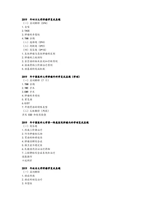
2019 年四川大学肿瘤学复试真题(一)名词解释(5*4)1.自噬2.TACE3.肿瘤的异型性4.TNM 分期(二)选择题(5*4)(三)判断题(5*2)(四)简答题(5*10)1.良性肿瘤与恶性肿瘤的区别2.肿瘤的三级预防3.食管癌的临床表现和诊断原则4.癌痛药物三阶梯治疗原则5.癌基因的发病机制2019 年中国医科大学肿瘤外科学复试真题(学硕)(一)名词解释(7 个)1.TNM 分期2.TME 手术3.EMR 手术4.肿瘤的异质性5.重复癌6.GIST7.早期胃癌的特殊类型(二)文献翻译(两段)涉及 ESD 和标本检查2019 年中国医科大学第一附属医院肿瘤内科学硕复试真题(一)简答题1.疼痛三阶梯治疗2.何为肿瘤标记物3.胃癌的转移途径4.肿瘤溶解综合症5.微卫星不稳定性6.乳腺癌内分泌治疗药物7.上腔静脉综合症表现和治疗技能操作口述胸穿2019 年延安大学肿瘤学复试真题(一)名词解释1.癌症疼痛2.癌症的姑息治疗3.异型性4.分子靶向治疗5.免疫检查点抑制剂(重点)(二)简答题1.简述肿瘤的的血管生成的基本步骤2.肿瘤标志物的化学成分和临床应用3.病理学检查的新技术以及对肿瘤治疗的意义4.化疗药的临床适应症5.免疫逃逸的机制6.肝动脉栓塞化疗的原理以及适应症(三)论述题1.肿瘤的综合治疗原则2.放射性肺炎的概念,预防以及其预后(治疗好像也有)3.哪些是肿瘤的高危人群2018 年延安大学肿瘤学复试真题(一)名词解释1.细胞凋亡2.分区放射治疗3.多药耐药性4.肿瘤的姑息治疗5.癌症性疼痛(二)简答题1.化疗的适应症2.肿瘤放疗的临床分类3.化疗的应用原则4.化疗在临床中的应用有哪些?5.癌症性疼痛的三阶梯治疗,并说出常用药物(三)论述题1.肿瘤的综合治疗原则2019 年北京协和医学院肿瘤学复试真题(一)简答题1.上消化道出血的原因2.五个高血压引起的靶器官损害3.子宫肌瘤手术指征4.小儿肾病综合征诊断指标2019 年苏州大学肿瘤内科复试真题(一)名词解释1.原位癌2.ECOG 评分3.癌基因4.第二信使5.多药耐药(二)简答题1.简述 Cancer、Carcinoma、 Adenoma、 Adenocarcinoma、 Sarcoma 各自的含义以及它们的区别2.以肺癌为例说明恶心肿瘤的诊断方法及原则3.以大肠癌为例说明肿瘤是一个多基因多步骤的发生发展过程4.基因异常的表现形式,癌基因和抑癌基因的表现异常有哪些不同5.简述肿瘤的转移机制和转移过程6.外科手术在肿瘤综合治疗中的地位和作用2018 年苏州大学肿瘤学复试真题(一)名词解释1.剂量强度2.肿瘤抗原3.多药耐药4.Oncogenes5.第二信使6.表观遗传学(二)简答题1.简述化疗药物的分类及作用机制2.以肺癌为例说明恶性诊断原则及方法3.以胃癌为例说明肿瘤发生是一个多基因多步骤的过程4.基因异常的表现形式,癌基因和抑癌基因的表现异常有哪些不同5.肿瘤外科的手术方式和应用范围6.肿瘤逃避免疫系统的机制2019 复旦大学肿瘤学一、简答题(6 选 5)ECGO 评分定义,分级及原则多学科综合性治疗的基本原则减少恶性肿瘤医源性传播和复发的措施简述放疗的实施步骤癌基因和抑癌基因的定义,各举 2-3 个例子良恶性肿瘤的区别二、文献翻译Hallmarks of Cancer 这篇文献的摘要段落翻译:体细胞突变对下游信号传导通路的影响笔试一、名词解释循环肿瘤细胞遗传易感性RECIST自噬预后因子新辅助治疗肿瘤治疗方法和适应症和自限性。
两种方法建立人宫颈癌细胞顺铂耐药细胞株及其r耐药性的评价陈晶;李祎博;邓金桂【摘要】目的:通过两种方法建立顺铂耐药的人宫颈癌细胞耐药细胞株,对其耐药性进行分析.方法:用小剂量诱导法和大剂量冲击法分别建立Hela细胞顺铂耐药细胞株.比较正常Hela细胞株和两种方法建立的顺铂耐药Hela细胞株的RI指数和相关耐药基因表达情况,评价耐药细胞株是否成功建立.绘制三种细胞的生长曲线,计算比较三种细胞的细胞倍增时间.结果:成功用两种方法建立了Hela细胞顺铂耐药细胞株,分别命名为Hela/DDP细胞株和Hela/DDPs细胞株,其耐药相关基因的表达、RI指数、细胞倍增时间表达均与亲代Hela细胞有明显差异.结论:成功用两种方法建立两株Hela细胞耐药细胞株,分别属低度或中度耐药.【期刊名称】《中国民康医学》【年(卷),期】2017(029)013【总页数】4页(P50-52,62)【关键词】Hela细胞;顺铂;耐药;小剂量法;大剂量法【作者】陈晶;李祎博;邓金桂【作者单位】沈阳医学院附属中心医院,辽宁沈阳 110024;沈阳医学院附属中心医院,辽宁沈阳 110024;沈阳医学院附属中心医院,辽宁沈阳 110024【正文语种】中文【中图分类】R737.33宫颈癌是常见妇科恶性肿瘤,我国每年新发病例为13.2万,约有3万妇女死于宫颈癌。
手术和放疗是宫颈癌的主要治疗方法,但近年来国内外通过基础和临床研究肯定了术前化疗及同步放疗、化疗在宫颈癌治疗中的作用[1,2]。
顺铂是最有效的化疗用单药,是临床上最广泛的抗癌药物之一,抗瘤谱广泛,但其固有的或获得的耐药性又降低了其疗效[3,4]。
通过体外建立顺铂耐药的人宫颈癌耐药细胞株,可为宫颈癌化疗治疗的耐药机制的研究提供基础。
本文通过两种方法建立顺铂耐药的人宫颈癌细胞耐药细胞株,对其耐药性进行分析。
1.1 材料1.1.1 细胞株 Hela细胞株由深圳市人民医院医学中心馈赠。
1.1.2 主要试剂 PRMI 1640培养基,Trizol试剂盒(Invitrogen公司),顺铂(昆明贵研药业公司),CCK8试剂盒(日本同仁公司),DNA提取试剂盒(上海生工公司),反转录试剂盒miScript II RT Kit(Qiagen公司),PCR反应试剂盒miScript PCR System(Qiagen公司),PCR引物(大连宝生物公司),其他试剂均为国内分析纯。
转染细胞的稳定筛选1、药物筛选前的准备药筛前应确定药物筛选浓度,不同细胞系具有不同的药物敏感度,因此在筛选前应该用在药物浓度范围内设定浓度梯度来确定筛选稳定克隆的药物浓度.药物浓度一般确定为能使未转染细胞在7天之内全部死亡,如G418一般需要7天,而puro一般时间很短,只需要3—4天即可。
2、药物筛选注意事项(1)药筛的时间加药时间一般为转染后48h。
但由于慢病毒表达较慢,因此利用慢病毒感染将质粒转入细胞的方法,加药时间一般为72h空白对照加药筛选时,最好设置空白对照,即未转染细胞同时加药,待空白对照中细胞全部死亡,转染组中不具有抗药性的细胞基本药杀完,但还需继续加药.(2)换液如果加药后,细胞死亡较多,需及时换液,以防死细胞释放有害物质导致具有抗药性的细胞死亡.另外,随着细胞的代谢,抗生素的活性会降低,因此,每隔3-5天应更换一次抗生素筛选培养液。
3、克隆筛选注意事项若转染的质粒带有荧光标记,不管是以下哪种筛选克隆的方法,都应该选择带有荧光较强的细胞克隆,因为加药筛选时,可能会产生耐药性的细胞,因此最好选择带有荧光较强的细胞.若是转染的质粒不带有荧光,那么只能盲挑。
不管是带有或者不带有荧光标记,都应该挑出克隆后进行验证,验证存在不成功的概率。
因此,挑克隆时,应尽量多挑几个克隆,20个左右.4、稳定克隆筛选步骤(1)有限稀释法步骤a)将药筛后的细胞(一般长满六孔板即可,若细胞生长很快药筛时可以在10cm的dish中进行)用胰酶消化下来b)对消化后的细胞悬液进行计数(如果细胞数量过大,可先稀释后计数)c)计算后,用枪头吸取约200个细胞(其中有部分为死细胞)到10ml培养液中充分混匀,剩下的大部分细胞冻存保种d)然后将以上10ml细胞悬液加到96孔板中,每孔100ul,这样有的孔就可能只有一个细胞,过程中注意不时用枪头吹打混匀细胞悬液(一个96孔板得到的克隆可能较少,可以用同样的方法做2—3个96孔板)e)待细胞贴壁后,逐孔在显微镜下观察,没有细胞或者多余一个细胞的孔划叉,只有一个细胞的孔划勾,以做好标记,如果只有一个细胞的孔数量太少,可将一个孔内有两个细胞的孔也划勾,但这样克隆就可能不纯。