
感染性心内膜炎病例共25页
- 格式:ppt
- 大小:2.79 MB
- 文档页数:25


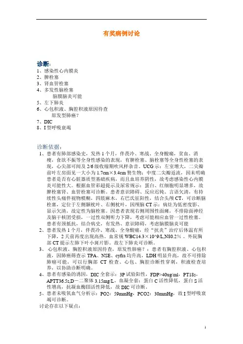
有奖病例讨论诊断:1、感染性心内膜炎2、脾栓塞3、肾血管栓塞4、多发性脑栓塞脑膜脑炎可能5、左下肺炎6、心包积液、胸腔积液原因待查原发型肺癌?7、DIC8、I型呼吸衰竭诊断依据:1、患者有肺部感染史,发热1个月,伴畏冷、寒战、全身酸痛,贫血、消瘦,食欲不振等全身性感染的表现,有脾栓塞、脑栓塞等全身性栓塞的表现,心尖部可闻及2/6级收缩期吹风样杂音。
UCG示:左室增大,二尖瓣前叶左房面见一大小为1.7cm×3.4cm赘生物;中度二尖瓣返流,因未明确患者是否有心脏器质型基础疾病,而且血培养阴性,故考虑感染性心内膜炎可能性大。
根据血管彩超提示及尿常规示:蛋白、红细胞明显增多,故脾栓塞肾、血管栓塞可诊断。
患者意识障碍、反应迟钝、言语欠清,有持续性头痛伴视物模糊,四肢麻木,右巴氏征阳性,结合头颅CT,可诊断脑栓塞,定位于左侧颞枕叶、右侧枕叶,因颅脑CT示:病灶为低密度影、显示欠清,故定性为脑栓塞。
因患者表现右侧周围性面瘫,不排除面神经及脑干核团受损,一过性双侧听力下降,考虑可能相应血管一过性栓塞。
患者有颈抵抗,结合病史,有发热、意识障碍,考虑脑膜脑炎可能2、患者发热1个月,伴畏冷、寒战、全身酸痛,经“抗炎”治疗后体温有所下降,2天前再度出现高热。
血常规WBC14.3×10^9/L,N80.2%。
外院胸部CT提示左肺下叶小斑片影。
故左下肺炎可诊断。
3、心包积液、胸腔积液原因待查,原发性肺癌?:患者有胸腔积液、心包积液,因肺癌筛查示TPA、NSE、cyfra均升高,LDH明显升高,故不可排除肺癌可能,可以行胸部CT检查、心包、胸腔诊断性穿刺,积液检查培养,以协助诊断明确。
4、患者有感染的诱因,DIC全套示:3P试验阳性,FDP>40ug/ml,PT18s,APTT36.5s,D-二聚体3.15mg/L。
血凝全套:蛋白C活性降低,蛋白S活性增高,抗凝血酶Ⅲ活性降低,故DIC可诊断。
5、患者未吸氧血气分析示:PO2:59mmHg,PCO2:30mmHg,故I型呼吸衰竭可诊断。

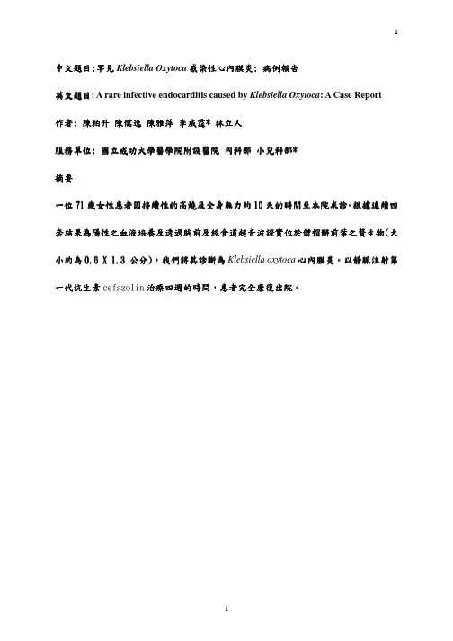
中文題目:罕見Klebsiella Oxytoca 感染性感染性心內膜炎心內膜炎: 病例報告英文題目: A rare infective endocarditis caused by Klebsiella Oxytoca : A Case Report 作者: 陳柏升 陳儒逸 陳雅萍 李威霆* 林立人服務單位: 國立成功大學醫學院附設醫院 內科部 小兒科部*摘要一位71歲女性歲女性患者患者患者因持續性的高燒及全身無力約因持續性的高燒及全身無力約10天的時間至本院求診。
根據根據連續四連續四套結果為陽性之血液培養套結果為陽性之血液培養及透過胸前及經食道超音波證實位於僧帽瓣前葉之贅生物及透過胸前及經食道超音波證實位於僧帽瓣前葉之贅生物(大小約為0.6 X 1.3 公分),我們將其診斷為Klebsiella oxytoca 心內心內膜炎膜炎。
以靜脈注射第一代抗生素cefazolin 治療治療四週的時間四週的時間,患者患者完全康復出院完全康復出院。
A rare infective endocarditis caused by Klebsiella Oxytoca: A Case ReportPo-Sheng Chen, Ju-Yi Chen, Ya-Ping Chen, Wei-Ting Lee, Li-Jen LinDivision of Cardiology, Department of Medicine, National Cheng Kung University Medical Center, Tainan, Taiwan.Conflicts of interest: noneRunning title: Community-acquired Klebsiella oxytoca endocarditisKey words: Infective endocarditis; Klebsiella oxytocaReprint requests and correspondence to:Li-Jen Lin, MDDivision of Cardiology, Department of MedicineNational Cheng Kung University Medical Center138 Sheng-Li Road, Tainan 70428, TaiwanE-mail: linl@.tw; Tel: 886-6-235-3535 ext, 2382; Fax: 886-6-275-3834AbstractWe report the case of a 71-year-old woman who presented with persistent high fever and progressive weakness for 10 days. Klebsiella oxytoca infective endocarditis was diagnosed based on four sets of positive blood culture of Klebsiella oxytoca together with fluttering vegetation (0.6 cm in diameter × 1.3 cm long) on the base of the anterior mitral leaflet. The diagnosis was documented using transthoracic and transesophageal echocardiograms. After 4 weeks of intravenous cefazolin therapy, the patient completely recovered.INTRODUCTIONKlebsiella species are rare causal microorganisms that may account for 1.2% of native valve endocarditis,[1-7] among which, Klebsiella oxytoca endocarditis is extremely rare. There has been only one such case reported: in 1985, Klebsiella oxytoca endocarditis occurred after transurethral resection of a patient’s prostate gland.[1] Here we report the first case of community-acquired Klebsiella oxytoca endocarditis.CASE REPORTA 71-year-old woman presented with persistent fever and generalized weakness for 10 days. She also had deteriorating shortness of breath. She had not traveled abroad. Her past medical history included diabetes mellitus, hypertension, and mild chronic renal insufficiency, all which had been regularly treated. When the patient arrived at the emergency room, her body temperature was 39.4°C and blood pressure was 178/78 mmHg. Her respiratory rate was 18 per minute, her conjunctiva appeared slightly pale, and her jugular veins were not engorged. She had a grade 2/6 diastolic murmur at the left upper sternal border and a grade2/6 pansystolic murmur at the apex. Chest auscultation showed some crackles in both lower lung fields. Her liver and spleen were not palpable. She had no conjunctival hemorrhage, Janeway’s lesion, Osler node, or splinter hemorrhage. Her eye-ground checkup showed no Roth’s spots.Hemograms showed hemoglobin at 9.42 gm/dl, white blood cells at 14,000/cmm with 95% segmented forms, monocytes at 3%, and lymphocytes at 2%; platelets at 309,000/cmm.A blood biochemistry showed serum creatinine at 2.7 mg/dl, blood urine nitrogen at 41 mg/dl, serum sodium concentration at 147 mmol/l, serum potassium level at 3.5 mmol/l, serumC-reactive protein at 64 mg/l, AST at 25 mg/dl, and ALT at 26 mg/dl. Her urinalysis was normal. A chest X-ray showed mild pulmonary congestion and cardiomegaly. Twelve-lead electrocardiograms revealed a normal sinus rhythm and left ventricular hypertrophy with secondary ST-T changes. Arterial blood gas showed a pH of 7.395, paO2 at 95 mm Hg, paCO2 at 36 mm Hg, and HCO3− at 22 mEq/l. The patient also had mild mitral regurgitation, aortic regurgitation, and concentric left ventricular hypertrophy. The cardiac chambers were not dilated, and the left ventricular systolic function was normal. Transesophageal echocardiograms clearly revealed fluttering vegetation (0.6 cm in diameter × 1.3 cm long) with a fine connection to the mitral valve on the ventricular side of the anterior mitral leaflet base (Fig. 1). Ultrasonograms of the liver and kidneys were unrevealing. Four sets of blood cultures showed Klebsiella oxytoca, which is susceptible to cefazolin, amikacin, ceftriaxone, cefuroxime, levofloxacin, and imipenem, but resistant to gentamicin, sulfamethoxazole, and trimethoprim according to a standard disk susceptibility test. The patient’s urine culture was negative.The patient was given 1 gram of intravenous cefazolin every 8 hours for 4 weeks. Herfever subsided 4 days after antibiotic treatment began. Blood cultures repeated after 4 weeks of antibiotic treatment were negative. The patient was in good condition at her 4-month follow-up visit, when echocardiograms showed negligible residual vegetation.DISCUSSIONThis is the first case report of community-acquired Klebsiella oxytoca endocarditis. The diagnosis was based on one major (oscillating intracardiac mass on the mitral valve) and three minor (predisposing cardiac mitral and aortic valvular regurgitation, fever ≥ 38°C, and positive blood cultures) modified Duke criteria for infective endocarditis.[8] This is also the first reported case of Klebsiella oxytoca endocarditis that completely meets the modified Duke criteria.Klebsiella species, the same as other Gram-negative bacteria, are an uncommon but ominous cause of bacterial endocarditis.[9] Klebsiella endocarditis accounts for 1.2% of native valve endocarditis and 4.1% of prosthetic valve endocarditis.[9] Klebsiella oxytoca endocarditis, however, is less often encountered. The mortality of Klebsiella endocarditis is higher than for other Gram-negative endocarditis.[9,10,11] Similar to other Gram-negative bacterial endocarditis, aggressive antibiotic therapy is necessary to treat Klebsiella endocarditis.Third-generation cephalosporin and aminoglycosides are often used in combination,[9]and a 6-week treatment course seems prudent for Klebsiella endocarditis. Inthe present case of Klebsiella oxytoca endocarditis, however, 4 weeks of cefazolin treatment eliminated the pathogen. This is quite different from reported experience with other types of Klebsiella endocarditis.The portals of entry for Klebsiella oxytoca bacteremia include, in decreasing order of frequency, the hepatobiliary tract (58-65%), an intravascular or urinary catheter (7-12%), the urinary tract (5%), the skin and soft tissues (5%), and the peritoneal cavity (2%).[5,7] We were not able to specify the portal of entry in the present case.SUMMARYThis was a rare case of Klebsiella oxytoca endocarditis diagnosed using modified Duke criteria. The patient was given 4 weeks of cefazolin treatment and recovered completely.References1.Watanakunakorn C. 1985. Klebsiella oxytoca endocarditis after transurethral resection ofthe prostate gland. Southern Medical Journal.78:356-7.2.Dhawan VK. 2002. Infective endocarditis in elderly patients. Clinical Infectious Diseases.34:806-12.3.Werner GS, Schulz R, Fuchs JB, et al. 1996. Infective endocarditis in the elderly in theera of transesophageal echocardiography: clinical features and prognosis compared with younger patients. Am J Med.100:90-7.4.Leport C, Bure A, Leport J, Vilde JL. 1987. Incidence of colonic lesions in Streptococcusbovis and enterococcal endocarditis. Lancet.1(8535):748.5.Kim BN, Kim JR, Woo JH. 2002. Retrospective analysis of clinical and microbiologicalaspects of Klebsiella oxytoca bacteremia over a 10-year period. Eur J Clin Microbiol Infect Dis.21:419-26.6.Podschun R, Ullmann U. 1998. Klebsiella spp. as nosocomial pathogens: epidemiology,taxonomy, typing methods, and pathogenicity factors. Clin Microbiol Review. 11:589-603.7.Lin RD, Hsueh PR, Chang SC, Chen YC, Hsieh WC, Luh KT. 1997. Bacteremia due toKlebsiella oxytoca: clinical features of patients and antimicrobial susceptibilities of the isolates. Clin Infect Dis.24:1217-22.8.Li JS, Sexton DJ, Mick N, et al. 2000. Proposed modifications to the Duke criteria for thediagnosis of infective endocarditis. Clin Infect Dis. 30:633-8.9.Michael J. Anderson, Edward N. Janoff. 1998. Klebsiella endocarditis: report of twocases and reviews. Clin Infect Dis. 26: 468-74.10.Geraci JE, Wilson WR. 1982. Endocarditis due to gram-negative bacteria. Mayo ClinProc. 57: 145-8.11.Polk RE. 1997. Infectious endocarditis. In: Pharmacotherapy: a pathophysiologicapproach. 3rd ed. Stamford, Connecticut: Appleton & Lange.Figure LegendFig. 1. Transesophageal echocardiography revealed fluttering vegetation originating from the base of the anterior mitral leaflet (white arrow).。