椎间盘源性下腰痛诊断-英文-翻译1
- 格式:doc
- 大小:55.00 KB
- 文档页数:4
椎间盘源性腰痛护理常规
一、椎间盘源性腰痛的概述
椎间盘源性腰痛(lumbar intervertebral discogenic backache)
是指椎间盘病变所致的腰痛,其病因及病理病程主要包括椎间盘破裂、椎
间盘积液、椎间盘退变等。
病症表现为腰背部疼痛、痛引伸至腰部、大腿
后侧、膝关节及脚跟,伴有侧弯、屈腰等反射性活动障碍。
1、预防与教育
(1)宣传正确的生活习惯,如保证充足的睡眠、定期活动锻炼、戒
烟限酒等,以减轻椎间盘源性腰痛发病的危险因素。
(2)学习正确的体位姿势和活动方法,及时调整不良姿势,加强身
体的活动训练,增强腰腹部的力量,注意避免单一体位作业或坐姿作业,
尽量减少腰部的负荷。
(3)加强饮食管理。
多加食用富含维生素A、C、E、B6、钙、铁等
营养的食物,减少脂肪、膳食纤维及水分的摄入,注意控制体重,加强免
疫力,积极预防腰痛发作。
2、药物治疗
(1)选用复方丹参颗粒、脑淫羊藿、乌鸡白凤丸等中药具有临床疗效,可减轻卒中性腰痛、肌肉拘束及炎性症状,增强机体对疾病的抵抗力。
(2)应用止痛药、抗炎药、肌肉松弛剂等西药。

提要目的:探讨HIZ(high intensity zone)与椎间盘源性下腰痛的相关性。
方法:随机选取30例椎间盘源性下腰痛患者做为实验组,随机选取30例无椎间盘源性下腰痛患者(其中仅有根性症状的腰椎间盘突出症11例,查体10例,腰椎外伤9例)做为对照组,两组患者下腰痛VAS评分均在5~8分之间;所有患者行MRI检查,观察分析两组HIZ的出现率并进行统计学分析。
结果:实验组HIZ的出现率为63.33%,对照组为16.67%,经统计学分析两组HIZ的出现率有非常显著性差异(P<0.01)。
结论:HIZ与椎间盘源性下腰痛存在明显的正相关,可以做为椎间盘源性下腰痛诊断的重要依据。
关键词椎间盘源性下腰痛;高信号区;磁共振The research of the relationship between high intensity zone anddiscogenic low back painSpecialty: Orthopaedics of TCMAuthor: Zhao XinyouTutor: Dong JianwenAbstractObjective: To research the relationship between high intensity zone and discogenic low back pain. Methods: 30 cases of discogenic low back pain patients were randomly selected as the experimental group, and 30 cases of non-discogenic low back pain patients were randomly selected as the control group (11 cases of only radicular symptoms of lumbar disc herniation, 10 cases of investigation , 9 cases of spinal trauma) .The Visual Analogue Score of two groups was 5~8 points; The lumbar spinal magnetic resonance imaging examinations were taken in the two groups of patients. And the statistical analysis was taken on the HIZ occurrence rate of the two groups. Results: The occurrence rate of HIZ in the experimental group was 63.33%.And it in the control group was 16.67%. By statistical analysis on HIZ occurrence rate of the two groups, it had significant difference in the statistical significance (P<0.01). Conclusions: The HIZ and discogenic low back pain showed direct relationship, and it can be used as a marker for diagnosis of discogenic low back pain.Key words Discogenic low back pain; High intensity zone; Magnetic resonance imaging目录引言 (1)临床研究 (3)一、材料与方法 (3)(一)诊断标准 (3)(二)临床资料 (5)(三)检查方法 (6)(四)观察指标 (6)(五)统计学方法 (6)二、研究结果及分析 (7)(一)两组HIZ的出现数比较(见表4) (7)(二)实验组中男、女性患者HIZ的出现数比较(见表5) (7)(三)HIZ出现位置的比较(见表6) (7)(四)HIZ的节段分布(见表7) (8)(五)HIZ的形态特点(见表8) (8)讨论 (9)一、HIZ的临床发现 (9)二、HIZ的临床意义 (9)三、HIZ的解剖部位和信号特征 (11)四、HIZ的病理特点 (12)结语 (14)参考文献 (15)综述 (17)附录 (34)致谢 (38)引言下腰痛正日益成为严重影响人体健康的一种疾病,60%~80%的成年人均经历过不同程度的下腰痛。

椎间盘源性下腰痛物理治疗研究进展李猛;蒋戈利;石晓明;周金生【期刊名称】《解放军医药杂志》【年(卷),期】2016(000)002【总页数】4页(P28-31)【关键词】椎间盘源性下腰痛;手法治疗;物理治疗【作者】李猛;蒋戈利;石晓明;周金生【作者单位】天津 300319,解放军天津疗养院全军中医针灸康复中心国家中医药管理局颈腰椎脊柱疾病中心;天津 300319,解放军天津疗养院全军中医针灸康复中心国家中医药管理局颈腰椎脊柱疾病中心;天津 300319,解放军天津疗养院全军中医针灸康复中心国家中医药管理局颈腰椎脊柱疾病中心;天津 300319,解放军天津疗养院全军中医针灸康复中心国家中医药管理局颈腰椎脊柱疾病中心【正文语种】中文【中图分类】R681.533.1;R242[作者单位]天津 300319,解放军天津疗养院全军中医针灸康复中心国家中医药管理局颈腰椎脊柱疾病中心[DOI]10.3969/j.issn.2095-140X.2016.02.008椎间盘源性下腰痛(discgenic low back pain, DLBP)是一种常见的慢性腰痛疾病,由于椎间盘内部的物理结构和代谢功能异常所致,不伴神经根性症状或节段间过度活动的影像学表现[1]。
据流行病学调查,DLBP在世界范围内普遍存在[2],发病率仅次于普通感冒,每个人一生中的某个时期出现腰痛的比率70%~85%,其中约18%的人一直都在遭受腰痛困扰[3]。
同时,90%的急性腰痛发作者采取自然恢复的方式并在1个月内回到工作岗位[4],7%的急性腰痛逐渐发展成为慢性下腰痛[5],并引发持续失能3~12个月[6],每年超过25%的患者都要去医院接受治疗[7],至今已形成严重的医学和社会经济问题。
因此,其治疗和预防显得尤为重要。
本文将从最常见且患者最易接受的无创物理治疗等方法进行文献综述。
1.1 现代康复手法治疗现代康复手法治疗DLBP,主要以McKenzie治疗理念为主,强调自我运动训练在腰部疾患治疗中的作用及重要性,通过姿势矫正、医疗体操及健康教育为脊柱疾患的康复提供一种全新的诊疗思维和途径。
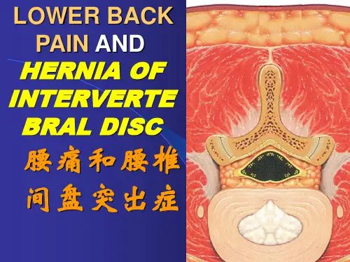
Many patients with back pain, leg pain, or weakness of the lower extremity muscles arediagnosed with a herniated disc.许多患腰腿疼痛,下肢肌端乏力的病患均为椎间盘突出症。
When a disc herniation occurs, the cushion that sits between the spinal vertebra is pushedoutside its normal position.椎间盘突出发生时,脊柱间的缓冲带将发生侧突。
A hrniated disc would not be a problem if it werent for the spinal nerves that are very close tothe edge of these spinal discs.如果脊神经不是离椎间盘特别近的话,椎间盘突出就不是什么大问题了。
HOW ARE THE SPINE AND ITS DISCS *****D脊柱与椎间盘The vertebras are the bony building blocks of the spine.脊椎是建造脊柱的构件。
Between each of the largest parts (bodies) of the vertebrae are the discs.各椎骨之间为椎间盘。
Ligaments are situated around the spine and discs.脊椎和椎间盘周围散布着韧带。
The spine has seven vertebrae in the neck (cervical vertebrae), 12 vertebrae in the mid-back(thoracic vertebrae) , and five vertebrae in the low back (lumbar vertebrae).颈部有7条椎骨,胸部为12条,腰部有5条。
脊柱外科常用英语词汇LUMBARSPINELumbardischerniationsyndrome腰椎间盘突出症FenestrationandDiscectomyLovemethod(开窗髓核摘除术Love法)minimallyinvasivediscectomy微创椎间盘切除术MED(micro-endoscopicdiscectomy)内镜椎间盘切除术Burst(Compression)fractureofT1-12,L1-5胸腰椎爆裂(压缩)骨折Openreductionandpediclescrewinternalfixation切开复位椎弓根螺钉内固定Decompressionandpediclescrewsysteminternalfixationwithbonegraft减压植骨椎弓根钉内固定Lumbarspinecanalstenosis腰椎管狭窄症spinalstenosisFenestrationdecompression(开窗减压)laminectomy椎板切除术Degenerativespondylolisthesis假性(退变性)脊柱滑脱Spondyloliticspondylolisthesis真性(峡部裂性)滑脱Reductionandinterbodyfusion(复位椎间植骨融合)anteriorapproach前入路posteriorapproach后入路Removalofinternalfixation内固定取出术ALIF:anteriorlumberinterbodyfusion前路椎间融合PLIF:posteriorlumbarinterbodyfusion后路椎间融合TLIF:transforaminallumbarinterbodyfusion经椎间孔融合Endoscopicoperation内镜手术spinalfusion脊柱融合pseudarthrosis假关节chemonucleolysis髓核融解术discogenicbackpain椎间盘源性腰痛sacroiliacjoint骶髂关节spondylolisthesis脊柱滑脱Removal取出(术)backpaininchildrenandadolescents儿童和青少年的下腰痛discitis间盘炎CERVICALSPINEAnold_ChariDeformity颅底畸形C1laminectomyandOccipital_C3Fusion(颈1后弓切除加枕颈融合术)JeffersonFracture环椎爆裂骨折Halo-vestexternalfixation(头环背心外固定)Atlantoaxialsubluxation环枢椎脱位MargelScrew(C1-2关节突螺钉)Trans-oralapproach(经口咽入路手术)DensFracture齿状突骨折DensScrewinternalfixation(齿状突螺钉内固定)HangmanFractureC2椎弓骨折AnteriorC2-3Fusion(前路C2-3融合)PosteriorC2-3pediclescrewinternalfixation(后路C2-3椎弓根内固定)Cervicalspinedischerniation颈椎间盘突出症Anteriordiscectomyandfusion(前路间盘摘除植骨融合)Cervicalspinecanalstenosis颈椎管狭窄症CervicalspineOPLL(OsificationofPosteriorLongitudenalLigarment)颈椎后纵韧带骨化症Cervicalspinedislocationandfracture颈椎骨折脱位Posterioropenreductionandanteriorfusion(后路切开复位-前路植骨融合术)Over-extensioninjuryofCervicalspine颈椎过伸性损伤C3-6laminoplasty(C3-6椎板成形椎管扩大术)KurokawamethodFrenchdoor-open(双开门)HirabayashimethodEnglishdoor-open(单开门)cervicalspondyloticradiculopathy神经根型颈椎病cervicalspondyloticmyelopathy脊髓型颈椎病cervicallaminoplasty颈椎椎板成形术uppercervicalfracture(Occiput,AtlasandAxis.C0~C1andC2)lowercervicalfracture(C3~C7andT1)Thoracicspinecanalstenosis胸椎管狭窄症thoracoscopyofthespine胸腔镜下脊柱手术larproscopicspinalfusion内镜下脊柱融合ThoracicspineOLF(OsificationofLigarmentumFlavum)胸椎黄韧带骨化症DEFORMITYTetherdcordsyndrome脊髓拴系综合征Spineshorteningosteotomy(脊柱短缩截骨术)DegenerativeScoliosis退变性侧弯CongenitalScoliosis先天性侧弯IdiopathicScoliosis特发性侧弯Deformitycorrection(畸形矫正术Hemivertabra半椎体)congenitalanomaliesofthespinalcord脊髓先天畸形myelomeningocele脊髓脊膜膨出occultspinaldysraphismandtetheredcord隐性脊柱裂和脊髓栓系spinaldisordersassociatedwithskeletaldysplasiasandmetabolicdiseases脊柱疾病合并骨骼发育不良及代谢性疾病juvenileandadolescentscoliosis青少年脊柱侧弯congenitalscoliosis先天性脊柱侧弯idiopathicscoliosis特发性脊柱侧弯neuromuscularscoliosis神经肌肉性脊柱侧弯juvenilekyphosis青年型脊柱后凸TUMORSpinalcordtumor:Neurinoma神经鞘瘤Neurifibroma神经纤维瘤Meningioma脊膜瘤Giantcelltumor巨细胞瘤Chordoma脊索瘤INFECTIONTuberculosis结核Pott’sdisease脊(柱结核)Debridment清创(术)OTHERNominatureofthedisease:spineinjury脊柱损伤spinalcordinjury脊髓损伤spinalorthoses矫形器spinaltumor脊柱肿瘤spinalinfection脊柱感染ankylosingspondylitis强直性脊柱炎rheumatoidarthritis类风湿性关节炎intraduraltumors硬膜内肿瘤intraspinalinfections椎管内感染,epidural硬膜外,intradural硬膜内arteriovenousmalformationsofthespinalcord脊髓动静脉畸形syringomyelia脊髓空洞Chronic,acute,haematogenousosteomyelitis急,慢性,血源性骨髓炎dislocation,subluxation脱位,半脱位cervicalspinalstenosis颈椎管狭窄scoliosis侧弯,kyphosis后凸,lordosis前凸bonecement骨水泥OPLL:ossificationofposteriorlongitudinalligment后纵韧带骨化(钙化calcium)OLF::ossificationofligmentflavum黄韧带骨化Operativemethods:Expansiveopen-doorlaminoplasty单开门椎管扩大成形术hirabayashi1977Double-doorlaminoplasty双开门椎管成形术Kurokawa1982Plaster石膏correctiveoperationofscoliosisandkyphosis脊柱侧突及后凸畸形矫形术THA(bipolar):totalhiparthroplasty全髋关节置换(双极头)TKA(arthroplasty,replacement)全膝关节置换术ORIF(openreductionandinternalfixation)切开复位内固定Clavicalfracture锁骨骨折Acromioclavical肩锁关节Humeralfracture(distal,proximal,middle,shaft)Radius,ulna,femur,tibia,fibulaepiphyseal,diaphyseal(shaft),metaphyseal干骺端的bonegraftCDH(congenitaldislocationofthehip)DDH(developmentaldysplasiaofthehip)DDH(developmentaldislocationofthehip)ACL:anteriorcruciateligmentPCL:posteriorcruciateligmentMCL:mdialcollateralligmentLCL:lateralcollateralligmentMeniscus:半月板,repairmentAVN:avascularnecrosis缺血性坏死Coredecompressionandmesenchymalstemcelltransplantation髓心减压间充质干细胞移植。
椎间盘源性下腰痛的诊断椎间盘源性下腰痛是一种腰椎间盘退变性疾病引起的慢性顽固性下腰痛,临床表现常不典型,诊断较困难,容易被误诊,导致患者不能得到及时、有效的治疗,增加了经济负担及痛苦。
因此作一综述,希望能引起临床医师的注意。
一概念Crock[1] 1970年提出“椎间盘源性下腰痛”的概念,又称“椎间盘内紊乱”,指由于一个或者多个椎间盘内部结构和代谢功能出现异常,如退变、终板损伤或无菌性炎症释放出某些因子,刺激椎间盘内疼痛感受器所引起的腰痛,它一般不伴神经根性症状或节段间过度活动的放射学证据。
二发病机制确切发病机制尚未研究清楚。
目前认为椎间盘源性腰痛是由纤维环破裂(Internal disc disruption,IDD)引起,不伴有椎间盘突出、脱出、节段不稳或其他影像学上的异常[1,2]。
Bogduk[3]认为椎间盘外层纤维环存在P 物质、降钙素相关基因肽以及血管肠肽,这些物质都属于神经传递素,与痛觉有关。
Shinohara认为:在变性的椎间盘,神经纤维可能伴随肉芽组织沿破裂的纤维环深入到椎间盘深层,无髓纤维裸露在间质液中可以感受到间质内成分的变化而产生疼痛。
目前最新的研究结果倾向于:椎间盘源性腰痛以及椎间盘造影诱发痛不仅与椎间盘承受的压力刺激椎间盘内部的压力传导器引起痛觉,同时与椎间盘内部的物质变化特别是炎症因子的增加引起痛觉的敏感性增加有关。
三临床表现椎间盘源性腰痛的临床表现为:多数病人有久坐、久弯腰、提重物史或腰椎直接暴力外伤史。
疼痛部位:L4/5、L5/S1的棘间、髂后、臀部、腹股沟、股前、股后、大转子等处的自发性胀痛,多痛不过膝;以腰痛为主,不耐受久坐、久弯腰或久站,腰椎前屈、旋转、侧屈加重或诱发疼痛,卧床休息缓解。
典型的病人是保守治疗无效或反复发作,逐渐加重。
查体可见腰椎因疼痛活动受限或正常,腰痛部位深在,压痛轻微,叩击痛明显。
腿痛往往发生于腰痛之后,常常没有精确的定位,通常难以言表,多主诉为臀部和下肢的酸胀、沉重感或抽筋,而且疼痛区域缺乏神经分布的特点,神经系统检查正常,没有皮肤感觉过敏和缺失。
临床下腰痛患者较多见,常需要MRI检查来诊断有无椎间盘盘膨出、突出,有时临床症状较明显,临床手术发现椎间盘纤维环断裂,即使未发现明确椎间盘突出,MRI显示腰椎间盘局限性高信号区(high⁃intensity zone,HIZ),即在矢状位MRI T2WI中见椎间盘后缘出现小而圆的局限性高信号区,提示纤维环破裂,是诊断椎间盘源性腰痛的重要MRI征象[1,2]。
近些年来国内外文献也逐渐报道了该征象,笔者回顾性35例下腰痛患者伴有腰椎间盘MRI局限性高信号区的影像学表现特征进行分析、诊断,以提高该征象对临床诊断的意见。
1资料与方法1.1一般资料35例下腰痛患者伴有腰椎间盘MRI局限性高信号区中,男18例,女17例,年龄38~68岁,平均48.5岁。
在下腰痛患者中,右下肢麻木并疼痛5例,左下肢麻木并疼痛7例,下腰痛无下肢症状23例。
腰椎间盘MRI局限性高信号对下腰痛诊断的临床意义何永忠,刘爽,张文俊[摘要]目的分析腰椎间盘MRI局限性高信号区的影像学表现与临床对照,以提高对该征象的临床意义。
方法回顾分析35例下腰痛患者伴有腰椎间盘MRI局限性高信号区的影像学表现特征进行分析、诊断,对照临床资料及手术病理对照。
结果腰椎间盘MRI局限性高信号区MR矢状位T2WI图像呈圆形(71.4%),弧形(20.0%)或放射状(8.6%),病灶表现以圆形常见。
轴位像为条状或梭形,与椎间盘纤维环破裂一致;高信号出现的位置与临床症状相符合。
结论腰椎间盘MRI局限性高信号区对诊断腰椎间盘性下腰痛有一定的特异性,出现该征象可提示椎间盘纤维环撕裂,为下腰痛患者的诊治提供参考。
[关键词]椎间盘;磁共振成像;高信号区doi:10.3969/j.issn.1009⁃976X.2020.06.018中图分类号:R681.53;R445文献标识码:ACorrelation between lumbar disc high⁃intensity zone on MRI and low back pain and its correlat⁃ed clinical significanceHE Yong⁃zhong,LIU Shuang,ZHANG Wen⁃junDepartment of Radiology,Conghua District Hospital of Traditional Chinese Medicine,Guangzhou510900,China[Abstract]Objective To analyze the imaging manifestations and clinical comparison of the localized high⁃signal area of lumbar disc MRI,so as to improve the clinical significance of this sign.Methods Retrospective analysis of35patients with low back pain associated with lumbar disc MRI localization high signal area imaging characteristics were analyzed,diagnosis,clinical data and surgical pathology control.Results MR sagittal T2WI images in the localized high signal area of lumbar disc MRI were round(71.4%),arc⁃shaped(20.0%)or radial(8.6%).The axial image was striate or fusiform,consis⁃tent with the annulus rupture of intervertebral disc.The location of the high signal is consistent with clin⁃ical symptoms.Conclusion The localized high signal area of LUMBAR disc MRI has a certain specificity for the diagnosis of lumbar intervertebral disc low back pain.The presence of this sign may indicate disc annulus tear and provide reference for the diagnosis and treatment of patients with low back pain.[Key words]intervetebral disc;magnetic resonance imaging;high⁃intensity zone作者单位:广州市从化区中医医院放射科,广州510900通讯作者:何永忠,EmaiI:*****************1.2检查方法采用Siemen Essenza1.5T MR成像设备。
扶他林乳胶剂超声透入治疗椎间盘源性下腰痛的疗效观察随着人们对椎间盘退变疾病的深入研究,腰椎间盘突出症引起的腰痛越来越被医疗工作者所了解,但是椎间盘内部出现病变而髓核没有突出引起下腰痛的病例被人们发现,称为椎间盘源性下腰痛(discogenic low back pain, DLBP)。
Crock在1986年将DLBP定义为一种椎间盘外周部基本保持完整,而内部各种病理(退变、终板损伤、炎症等)刺激椎间盘内疼痛感受器引起的功能丧失性下腰痛,并且不伴有根性症状,无神经或节段过度活动的放射学证据[1]。
DLBP是慢性下腰痛中最常见的疾病,占到其发病率的39%,而椎间盘突出症不到30%,其他疾病如小关节源性疼痛的发病率就更低[2]。
笔者所在科室从2007年11月至2009年2月诊治的DLBP患者共335例,除201例经过微创和开放性手术治疗,其他因不能手术的134例分别采用扶他林乳胶剂超声透入或单独的超声波治疗。
临床效果满意,总结报告如下。
1 资料和方法1.1 一般资料本组病例134例:男85例,女49例;年龄最小24岁,最大57岁;病程最短6个月,最长9年; L3~4间盘病变16例,L4~5者69例,L5~S1者32例,两个以上17例;所有病例均经X线、CT、MRI检查,132例有黑间盘,78例有HIZ,94例有Modic 改变;所有病例均进行椎间盘造影术检查为阳性。
1.2 诊断依据①反复发作下腰痛, 持续时间>6个月, 不能久坐、久站,不能弯腰,部分患者体检有疼痛的中心化现象和棘突骨震动试验阳性,不伴下肢放射痛和间歇性跛行, 无神经根受压体征;②X线检查排除腰椎峡部裂、滑脱和不稳,CT检查无椎间盘突出及椎管狭窄,MRI检查在T2加权像上病变椎间盘呈低信号改变(黑间盘),或者有椎间盘后方有高信号区,或者终板有Modic改变;③椎间盘造影可诱发出一致性下腰痛,V AS≥5,间盘形态异常,注射剂量≥3 ml,有相邻间盘阴性对照。
盘源性下腰痛的鉴别诊断
With so many possible causes of lower back pain, physicians must pinpoint the source(s) of pain to recommend appropriate action or no action. The IDET procedure is not suitable for every form of lower back pain. Chronic discogenic lower back pain unresponsive to aggressive non-operative therapy is the principal indication for the IDET procedure.
IDET不是适合于任何下腰痛患者。
盘源性下腰痛才是IDET的手术指征。
∙Differential diagnosis starts with a detailed history, physical examination and an MRI.
鉴别诊断要从相信的病史、体检及MRI检查开始。
∙Non-musculoskeletal causes of back pain such as tumor, infection, aneurysm and renal and vascular pathology should be considered and ruled out.
排除非肌肉骨骼原因引起的下腰痛,如:肿瘤、感染、动脉瘤、肾脏疾病等,都需要被考虑到并排除。
∙Fractures and ankylosing spondylitis should also be ruled out.
排除骨折及强直性脊柱炎。
∙Discogenic pain should be differentiated from radicular pain, which may also be present.
还需与神经根痛相鉴别。
盘源痛与神经根痛的比较
∙
∙
∙
∙
∙
∙
∙
∙
∙
∙
∙
∙
∙
∙
∙Pain which is unilateral and predominantly below lumbar vertebra 5 is probably not discogenic. 单侧疼痛而且疼痛点低于腰5的情况下大多不是盘源性的。
∙Diagnostic joint blocks can be used to trace the source of pain, which can be attributed to the sacroiliac joint in about 20% of patients and lumbar zygapophysial joint pain in up to 40% of patients.
压痛点有助于医师发现疼痛的根源,大约20%的病人压痛点在骶髂关节,而40%以上的患者压痛点在腰椎小关节处。
∙Patients with discogenic pain may have no abnormalities visible on the MRI or may have a bright spot in the posterior anulus referred to as the high intensity zone or HIZ. This is best seen on sagittal views and indicates a tear in the disc.
大多数盘源性疼痛患者MRI图像没有明显的异常。
或在纤维环后方有高强度区域。
这种情况在矢状面时比较容易看到。
常表明椎间盘有微小的裂纹。
椎间盘源性痛可以通过椎间盘造影刺激并重建症状。
Discography is a minimally invasive diagnostic procedure performed while the subject is awake. Sedation and analgesia can be provided to minimize discomfort. Contrast medium is injected into the three or four discs under investigation as the source of the low back pain.
椎间盘造影是一种微创的复制患者症状的检查方法。
操作中可适当给以镇静止痛药物以减轻患者不安,将对比造影剂注射入3-4个椎间盘内以确定疼痛源。
∙The injection of fluid into each disc increases the fluid volume and pressure in the disc and may reproduce the subject’s usual pain pattern.
将造影剂注射入每个椎间盘内可以增加椎间盘内液体体积及压力可以重建患者的疼痛症状。
∙ A positive response in one disc confirmed by the absence of a pain response in one or more other adjacent discs confirms the origin of the pain.
椎间盘内注射出现的阳性反应与患者疼痛区域相同可以证实某个椎间盘为症状根源。
∙In addition, the contrast medium allows the morphology of the disc to be visualized fluoroscopically.
另外,造影剂可以通过透视显示椎间盘的形状。
Since provocation discography is minimally invasive and produces some discomfort, it should only be performed when a definitive diagnosis will lead to treatment that would otherwise not be performed.
尽管椎间盘造影是微创过程仅有较少不适,但这一检查方式仅应在为确定诊断或确定手术部位时才能使用。
确定疼痛源
The disc which is the source of the low back pain can be confirmed when:
如下步骤以证实引起下腰痛的椎间盘:
∙The pain elicited is similar to patient’s usual pain pattern.
复制出的症状与患者平时的症状相同或相似。
∙At least one adjacent disc (preferably two) does not reproduce pain. 至少一个相近的椎间盘不能复制出症状。