云杉针叶的扫描电镜观察 Scanning Electron Microscopic Observation of the Needle Leavesin Pic
- 格式:pdf
- 大小:252.79 KB
- 文档页数:3

扫描电镜图像分析仪在矿物鉴定中的应用郭 嘉(山东省第一地质矿产勘查院,山东 济南 250013)摘 要:在传统的矿物鉴定中,难以立体地描述矿石样本中的矿物类型及所在区域,因此将扫描电镜图像分析仪应用于矿物鉴定中。
论述扫描电镜图像分析技术原理,归纳总结电子束击打在矿石样本表面后形成的分散电子类型,并分别描述其性质,分析该技术的优势,包括分辨率高、具备三维立体结构等。
论述扫描电镜图像分析仪在矿物鉴定中的应用方法,通过矿石自身的导电性能,区分所需扫描电镜种类及参数,分析不同矿石中的元素组成含量,推断矿石具体成分,寻找页岩结构中的微小孔隙。
关键词:扫描电镜图像分析仪;矿物鉴定;岩石矿物鉴定;扫描电镜;电镜图像分析中图分类号:P575.4 文献标识码:A 文章编号:1002-5065(2021)17-0209-2Application of Scanning Electron Microscope Image Analyzer in Mineral IdentificationGUO Jia(Shandong First Geological and Mineral Exploration Institute, Ji’nan 250013,China)Abstract: In traditional mineral identification, it is difficult to three-dimensionally describe the types and areas of minerals in ore samples. Therefore, scanning electron microscope image analyzers are used in mineral identification. Discuss the principle of scanning electron microscope image analysis technology, summarize and summarize the types of scattered electrons formed after the electron beam hits the surface of the ore sample, and describe their properties respectively, and analyze the advantages of this technology, including high resolution and three-dimensional structure. Discuss the application method of scanning electron microscope image analyzer in mineral identification. Through the conductivity of the ore itself, distinguish the required scanning electron microscope types and parameters, analyze the element composition content of different ore, infer the specific composition of the ore, and look for the shale structure. Tiny pores.Keywords: scanning electron microscope image analyzer; mineral identification; rock mineral identification; scanning electron microscope; electron microscope image analysis我国的工业发展对矿石有极大的需求,因此合理并及时地大范围开采矿物资源是满足人们生产和生活的前提。
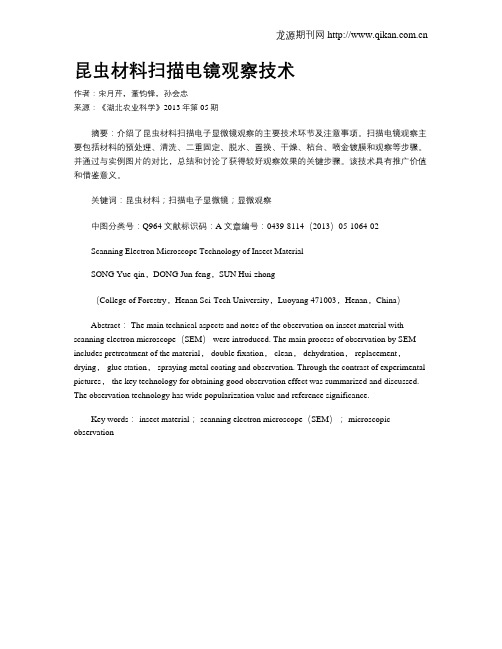
龙源期刊网 昆虫材料扫描电镜观察技术作者:宋月芹,董钧锋,孙会忠来源:《湖北农业科学》2013年第05期摘要:介绍了昆虫材料扫描电子显微镜观察的主要技术环节及注意事项。
扫描电镜观察主要包括材料的预处理、清洗、二重固定、脱水、置换、干燥、粘台、喷金镀膜和观察等步骤。
并通过与实例图片的对比,总结和讨论了获得较好观察效果的关键步骤。
该技术具有推广价值和借鉴意义。
关键词:昆虫材料;扫描电子显微镜;显微观察中图分类号:Q964 文献标识码:A 文章编号:0439-8114(2013)05-1064-02Scanning Electron Microscope Technology of Insect MaterialSONG Yue-qin,DONG Jun-feng,SUN Hui-zhong(College of Forestry,Henan Sci-Tech University,Luoyang 471003,Henan,China)Abstract: The main technical aspects and notes of the observation on insect material with scanning electron microscope(SEM) were introduced. The main process of observation by SEM includes pretreatment of the material, double fixation, clean, dehydration, replacement,drying, glue station, spraying metal coating and observation. Through the contrast of experimental pictures, the key technology for obtaining good observation effect was summarized and discussed. The observation technology has wide popularization value and reference significance.Key words: insect material; scanning electron microscope(SEM); microscopic observation。

扫描电镜实验报告扫描电镜(Scanning Electron Microscope,SEM)是一种应用广泛的高分辨率显微镜,能够对样品进行表面形貌和微观结构的观测和分析。
本实验旨在通过扫描电镜对不同样品的表面形貌和微观结构进行观察和分析,从而加深对扫描电镜原理和应用的理解。
首先,我们准备了几种不同的样品,包括金属材料、植物组织和昆虫外骨骼等。
在实验过程中,我们首先对样品进行了表面处理,包括金属样品的金属镀膜处理、植物组织的冷冻干燥处理以及昆虫外骨骼的金属喷镀处理,以保证样品在扫描电镜下的观察效果。
接下来,我们将样品放置在扫描电镜的样品台上,并调整好合适的观察条件。
在观察过程中,我们发现扫描电镜能够清晰地显示样品的表面形貌和微观结构,包括金属样品的晶粒结构、植物组织的细胞结构以及昆虫外骨骼的纹理结构等。
通过对这些结构的观察和分析,我们不仅可以直观地了解样品的表面特征,还可以深入地研究样品的微观结构和性质。
在实验中,我们还发现扫描电镜具有较高的分辨率和深度信息,能够对样品进行三维观察和分析。
通过调整扫描电镜的工作参数,我们成功地获得了不同角度和深度的样品图像,进一步揭示了样品的微观结构和表面形貌。
这为我们深入理解样品的微观特征提供了重要的信息和依据。
总的来说,通过本次实验,我们深入了解了扫描电镜的原理和应用,掌握了样品的表面形貌和微观结构的观察方法,提高了对样品性质和特征的认识。
扫描电镜作为一种重要的分析工具,将在材料科学、生物学、医学等领域发挥重要作用,为科学研究和工程应用提供有力支持。
通过本次实验,我们不仅提高了对扫描电镜的认识,还对不同样品的表面形貌和微观结构有了更深入的理解。
扫描电镜的高分辨率和深度信息为我们提供了更多的观察和分析角度,有助于我们更全面地认识样品的特性和性能。
希望通过今后的实践和研究,能够更好地利用扫描电镜这一强大的工具,为科学研究和工程应用做出更多的贡献。
第23卷 第1期2005年3月 贵 州 科 学GU IZHOU SCIENCE Vol.23,No.1Mar.2005 收稿日期:2004-06-18基金项目:国家攻关子项目:编号2001BA 606A -09-05、贵州省“十五攻关”项目:编号黔科合农社字(2001)114号)、国家基金等的部分内容。
作者简介:黄丽华(1980-),女,在读硕士研究生。
研究方向:资源植物学。
陈训,男,教授,博士生导师。
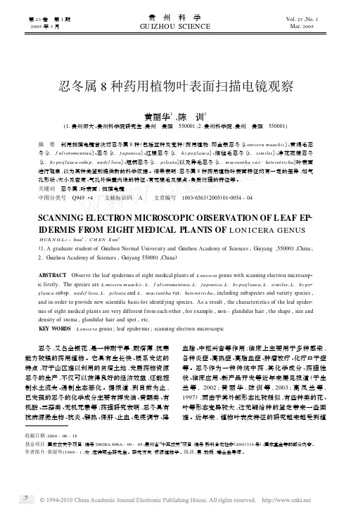
忍冬属8种药用植物叶表面扫描电镜观察黄丽华1,陈 训2(1.贵州师大、贵州科学院研究生,贵州 贵阳 550001;2.贵州科学院,贵州 贵阳 550001)摘 要 利用扫描电镜首次对忍冬属8种(包括亚种及变种)药用植物,即金银忍冬〔L onicera maackii 〕、黄褐毛忍冬〔L.f ulvotomentosa 〕、忍冬〔L.j a ponica 〕、红腺忍冬〔L.hy poglauca 〕、细毡毛忍冬〔L.similes 〕、净花菰腺忍冬〔L.hy poglauca subs p.nudi f lora 〕、短柄忍冬〔L.pileata 〕以及异毛忍冬〔L.macrantha var.heterot richa 〕叶表面进行观察,以为其种类鉴别提供新的科学依据。
结果表明:忍冬属8种药用植物叶表面特征均有一定的差异,如气孔形状、大小及密度、气孔外拱盖内缘的特征、有无腺毛及腺点、角质纹理的特征等。
关键词 忍冬属;叶表面;扫描电镜中图分类号 Q949・4 文献标识码 A 文章编号 100326563(2005)0120054-04SCANNING E L ECTRON MICROSCOPIC OBSERVATION OF L EAF EP 2IDERMIS FROM EIGHT MEDICAL PLANTS OF LON I CERA GENUSHUA N G L i -hua 1,C H EN X un 2(1.A graduate student of Guizhou Normal University and Guizhou Academy of Sciences ,Guiyang ,550001,China ;2.Guizhou Academy of Sciences ,Guiyang 550001,China )ABSTRACT Observe the leaf epidermis of eight medical plants of L onicera genus with scanning electron microscop 2ic firstly.The species are L onicera maackii ,L.f ulvotomentosa ,L.j aponica ,L.hy poglauca ,L.similes ,L.hy po 2glauca subsp.nudi f lora ,L.pileata and L.macrantha var.heterot richa ,including subspecies and variety species ,and in order to provide new scientific basis for identifying species.As a result ,the characteristics of the leaf epider 2mis of eight medical plants are very different f rom each other ,for example ,non -glandular hair ,the shape ,size and density of stoma ,glandular hair and spot ,etc.KE Y WOR DS L onicera genus ;leaf epidermis ;scanning electron microscopic 忍冬,又名金银花,是一种耐干旱,耐瘠薄,抗寒能力较强的药用植物。


功能化介孔二氧化硅微球的药物输送行为分析唐亮琛;单晓茜;杨盛兵;魏杰;孟正宇;曹勇斌;刘思达【摘要】介孔二氧化硅( MSNs)作为一类新型的药物载体在药物缓控释领域的应用方兴未艾。
采用模板诱导和自组装方法合成MCM-41型介孔二氧化硅纳米颗粒,并通过聚乙二醇(PEG)的表面修饰,制备出一类具有高亲水性、高孔隙率、高比表面积的MSNs修饰微球。
采用氮吸附( BET)、扫描电镜( SEM)、透射电镜( TEM)、粒径分析等手段表征并对比了聚乙二醇( PEG)修饰前后的MSNs基础理化性能。
在此基础上,选用阿霉素作为模型药物,包埋于介孔材料中,对比了PEG修饰前后MSNs中阿霉素的装载量、包封率及释放行为。
选用乳腺癌细胞MCF-7为模型癌细胞,选用人血清蛋白( HAS)作为模型蛋白,考察了两种材料对细胞的生物安全性及对蛋白的吸附情况,并将两种材料所对应的载药微球与MCF-7进行共培养,探究两种载药系统对癌细胞的灭杀情况。
%As a new type of drug carriers,mesoporous silica nanoparticles (MSNs) is in the ascendant in the field of drug control and release.Both the template induced and self-assembling method was employed to synthesize the MCM-41 type of mesoporous silica nanoparticles.With the co-polymerization with polyethylene glycol ( PEG) , MSNs with wormhole pore structure were obtained with high hydrophilic, high porosity and specific surface area.The modification of PEG to the MSNs was characterized by nitrogen adsorption ( BET) , scanning electron microsco-py ( SEM) , transmission electron microscopy ( TEM ) and particle size analysis.Besides, the influence of PEG modification to the drug loading content, the encapsulation efficiency and the release behavior of the MSNs were al-soanalyzed with the doxorubicin as a model drug.To compare the biosecurity and protein adsorption of the two kinds of materials, the human breast cancer cell MCF-7 and the human serum albumin ( HAS) were selected as the model cancer cells and the model protein, respectively.On the other hand, to investigate the anti-cancer effect of these two drug delivery systems, the materials were co-cultured with MCF-7 cells and determined the quantity of cells by means of Microplate Reader.【期刊名称】《科学技术与工程》【年(卷),期】2015(000)032【总页数】6页(P111-116)【关键词】介孔二氧化硅;PEG表面修饰;药物缓控释系统;生物安全性;抗癌【作者】唐亮琛;单晓茜;杨盛兵;魏杰;孟正宇;曹勇斌;刘思达【作者单位】上海应用技术学院材料科学与工程学院,上海201418;上海应用技术学院材料科学与工程学院,上海201418;上海交通大学医学院附属第九人民医院骨科实验室,上海 200011;华东理工大学材料科学与工程学院,上海200237;上海应用技术学院材料科学与工程学院,上海201418;上海应用技术学院材料科学与工程学院,上海201418;上海应用技术学院材料科学与工程学院,上海201418【正文语种】中文【中图分类】O631表面修饰的纳米介孔二氧化硅(mesoporous silica nanoparticles,MSNs)可以提高对药物的控释,促进药效的发挥。
扫描电子显微镜(ScanningElectronMicroscope) 基础知识一、扫描电子显微镜的工作原理扫描电镜是用聚焦电子束在试样表面逐点扫描成像。
试样为块状或粉末颗粒,成像信号可以是二次电子、背散射电子或吸收电子。
其中二次电子是最主要的成像信号。
由电子枪发射的能量为5〜35keV的电子,以其交叉斑作为电子源,经二级聚光镜及物镜的缩小形成具有一定能量、一定束流强度和束斑直径的微细电子束,在扫描线圈驱动下,于试样表面按一定时间、空间顺序作栅网式扫描。
聚焦电子束与试样相互作用,产生二次电子发射(以及其它物理信号),二次电子发射量随试样表面形貌而变化。
二次电子信号被探测器收集转换成电讯号,经视频放大后输入到显像管栅极,调制与入射电子束同步扫描的显像管亮度,得到反映试样表面形貌的二次电子像。
二、扫描电镜具有以下的特点(1)可以观察直径为0 〜30mm 的大块试样(在半导体工业可以观察更大直径) ,制样方法简单。
(2)场深大、三百倍于光学显微镜,适用于粗糙表面和断口的分析观察;图像富有立体感、真实感、易于识别和解释。
(3)放大倍数变化范围大,一般为15 〜200000 倍,对于多相、多组成的非均匀材料便于低倍下的普查和高倍下的观察分析。
(4)具有相当高的分辨率,一般为 3.5 〜6nm。
(5)可以通过电子学方法有效地控制和改善图像的质量,如通过调制可改善图像反差的宽容度,使图像各部分亮暗适中。
采用双放大倍数装置或图像选择器,可在荧光屏上同时观察不同放大倍数的图像或不同形式的图像。
(6)可进行多种功能的分析。
与X 射线谱仪配接,可在观察形貌的同时进行微区成分分析;配有光学显微镜和单色仪等附件时,可观察阴极荧光图像和进行阴极荧光光谱分析等。
(7)可使用加热、冷却和拉伸等样品台进行动态试验,观察在不同环境条件下的相变及形态变化等。
三、扫描电镜的主要结构1.电子光学系统:电子枪;聚光镜(第一、第二聚光镜和物镜) ;物镜光阑。
扫描电镜在金属材料检测中的应用冯 柳,彭庆梁(山东理工大学分析测试中心,山东 淄博 255000)摘 要:扫描电镜是材料研究与分析中的通用设备,在材料、化工、机械等领域的微观形貌分析中起着重要的作用。
本文主要从微观组织观察、材料断口分析、结合能谱仪的微区成分分析、显微织构分析四个方面简述扫描电镜的发展历程和特点以及扫描电镜及其附件在金属材料检测中的应用,为金属材料行业用扫描电镜分析提供了一定的参考依据。
关键词:扫描电镜;金属材料;微观分析中图分类号:TG115 文献标识码:A 文章编号:1002-5065(2020)16-0208-2Application of scanning electron microscope in metal material detectionFENG Liu, PENG Qing-liang(Analysis and testing center of Shandong University of Technology,Zibo 255000,China)Abstract: SEM is a general equipment in material research and analysis, which plays an important role in the micro morphology analysis of materials, chemical industry, machinery and other fields. In this paper, the development process and characteristics of SEM and the application of SEM and its accessories in metal material detection are briefly introduced from four aspects: microstructure observation, material fracture analysis, micro zone composition analysis combined with energy dispersive spectrometer, and micro texture analysis. The application of SEM and its accessories in metal material detection provides a certain reference for the use of SEM in metal material industry.Keywords: scanning electron microscope; metal materials; microscopic analysis1965年在英国诞生了世界上第一台扫描电镜,截止至今,扫描电镜的技术指标和功能性也发生的质的飞跃[1]。
扫描电镜在观察溃疡性结肠炎果蝇肠道超微结构中的应用研究王博】%车畅2关肠1王林燕1"1.浙江中医药大学中医药科学院,杭州310053$.中国计量大学生命科学学院,杭州310018)摘要:为阐明溃疡性结肠炎对肠道超微结构的影响,本研究以黑腹果蝇为研究对象,通过扫描电镜分别对正常组和肠炎组肠道进行观察与比较。
结果表明:肠炎组肠道微绒毛出现损伤,肠上皮细胞内细胞器膜性结构遭到破坏,肠道干细胞数量增多,未见肠分泌细胞,肠道微生物数量激增。
结论:溃疡性结肠炎会使得肠道超微结构发生改变,部分改变可能是由于肠道启动保护机制所致,用于应对肠炎引起的不利影响。
关键词:溃疡性结肠炎扫描电镜黑腹果蝇超微结构DOI:10.3969/j.issn.1001—232x.2021.02.028Observation of the intestinal ultrastructure of drosophila melanogaster with ulcerative colitis by scanning electron microscope・Wang Bo1%#Che Chang2#Quan Yang#Wang Linyan1(1.Academy of Chinese Medical Sciences#Zhejiang Chinese Medical University#HangZhou310053,China; 2.College of Life Sciences#China JUaing University,HangZhou310018,China)Abstract:The intestines of Drosophila,melanogaster with UC were observed by scanning electron mi-croscop1andcompar1dwiththatofnormalDrosophilam1lanogast1r.Th1r1sultsshow1dthatth1int1sti-nalmicrovi l iofDrosophila.m1lanogast1rwithUCw1r1damag1d,th1organ1l1m1mbran1structur1ofin-t1stinal1pith1lialc1l swasd1stroy1d,th1numb1rofint1stinalst1mc1l sincr1as1d,noint1stinal1nt1ro1n-docrin1c1l sw1r1found,andth1numb1rofint1stinalmicroorganismsincr1as1dsharply.Thusitcanb1 s11nthatulc1rativ1colitiscanchang1th1int1stinalultrastructur1,andsom1ofth1chang1smayb1du1to th1int1stinalprot1ctiv1m1chanismtod1alwithth1adv1rs11f1ctscaus1dbyulc1rativ1colitis.Key words:Ulcerative colitis(UC);Scanning electron microscope;Drosophila melanogaster;Ultra-structur1溃疡性结肠炎作为临床常见的慢性特异性肠道炎症,近年来发病率急剧上升,其病因至今仍不明确,可能是由环境及遗传等因素相互作用形成’其病理特征主要包括肠道黏膜及下层出现炎症反应等⑴。
扫描电镜下神奇的花粉微观世界
佚名
【期刊名称】《自然杂志》
【年(卷),期】1998(20)6
【总页数】1页(Pf004-f004)
【关键词】扫描电镜;现代花粉学;微观结构;边缘学科;化石花粉学;花粉多糖
【正文语种】中文
【中图分类】Q944.42
【相关文献】
1.高真空扫描电镜低电压条件下花粉的显微组织研究 [J], 覃丽禄;刘莹莹;张露
2.不同处理条件下几种松树花粉的扫描电镜观察 [J], 张国云;张雯婷;姚娟妮;裴国亮
3.不同制样方法下四种豆科植物花粉的扫描电镜观察 [J], 赵颖
4.两种处理条件下拟南芥花粉的扫描电镜比较观察 [J], 何乐平;滕慧娟;张蕾;杨玲钰
5.扫描电镜下的花粉粒 [J],
因版权原因,仅展示原文概要,查看原文内容请购买。