胆囊小细胞神经内分泌癌一例
- 格式:pdf
- 大小:584.99 KB
- 文档页数:2
197投稿邮箱:zuixinyixue@世界最新医学信息文摘 2019年 第19卷 第51期·病例报告·1例原发性肝神经内分泌肿瘤病例报告王怡君,邱文生(青岛大学,山东 青岛 266021)1 临床资料患者女,60岁,因“查体发现肝占位性病变3月余”入院。
既往体健,查体:腹部平坦,对称,腹软,无压痛及反跳痛,未触及异常包块,Murphy 氏征阴性,肝、肾区无叩击痛。
辅助检查:AFP :2.72 ng/mL ,CEA :<0.2 ng/ml 。
增强CT 示:肝脏大小、形态可,肝左内叶实质内见囊性低密度影,囊壁轻度强化,范围约44.3 mm ×46.1 mm 。
MR 示:肝脏大小形态可,肝左内叶见团块样稍长T1稍长T2信号,内见斑片样长T1长T2信号,大小约52 mm ×49 mm 。
肝内外胆管未见明显扩张。
行腹腔镜下肝脏IVb 段部分切除,术后病理示:1.(肝)神经内分泌肿瘤(G1级,范围5×4 cm ),核分裂像<2个/10HPF ,未累及肝断端。
免疫组化:CKpan (+),Syn (+),CgA (+),CD56(+),CD34血管(+),Hepatocyte (-),Ki-67(+)约1%,CD99(+),CK7(-),CD31血管(+)。
术后3个月随访未见肿瘤复发。
2 讨论2.1 概论。
许多器官中存在具有内分泌功能的细胞,神经内分泌肿瘤(neuroendocrine tumor ,NET )就起源于这些细胞[1],可分为类癌和神经内分泌癌。
原发于肝脏的神经内分泌癌极为罕见,我国1994至2010年仅报道47 例,国内外发病情况、男女发病率均未见明显差异(男:女为0.96),青年至老年均有发病,平均年龄51 岁[2]。
PNEC 发病率低,且临床上无明显特异性,只有 6.8% 的 PHNET 患者出现经典类癌综合征[3],诊治过程中易发生误诊、漏诊情况,诊出时一般已出现腹痛、乏力和消瘦等临床症状[4],因此早期诊断、治疗极为重要。

1例胆囊小细胞癌报告并文献复习张群;王丽;吴波;陈一天;陈龙邦【摘要】Objective Small cell cancer of the gallbladder is a very rare tumor with insidious onset and poor prognosis. This study aims to gain a deeper insight into small cell cancer of the gallbladder. Methods We reported a case of small cell cancer of the gallbladder confirmed by histopathology after surgery, which was followed by chemotherapy. We analyzed the clinical manifestations, histological features and treatment principles of the disease with a review of the literature. Results There were no typical clinical symptoms and imaging manifestations. Histopathology and immunohistochemistry revealed positive expressions of neuron-specific eno-lase ( NSE ), synaptophysin ( Syn ), and chromogranin A ( CgA ) in the cytoplasm. Conclusion Small cell cancer of the gallbladder, as a rare malignancy, is easily misdiagnosed. Histopathology and immunohistochemistry are essential for its diagnosis, and the combination of surgery and chemotherapy can improve the survival of the patient.%目的胆囊小细胞癌罕见,起病隐匿,预后差.文中结合临床病例并复习文献,以提高对胆囊小细胞癌的认识.方法报道1例胆囊小细胞癌,经手术切除及组织病理学证实,术后行全身化疗.结合文献对胆囊小细胞癌的发生、临床表现、组织学特点、治疗原则等进行讨论.结果胆囊小细胞癌临床症状以及影像学检查无特征性表现,需经病理组织以及免疫组化方法明确,细胞质内常表达神经元特异性烯醇化酶、突触素或嗜铬蛋白.结论该病是一种罕见的恶性肿瘤,容易误诊,组织病理和免疫组化对该病诊断具有重要价值,联合手术和化疗可以提高生存期.【期刊名称】《医学研究生学报》【年(卷),期】2012(025)009【总页数】4页(P938-941)【关键词】胆囊小细胞癌;诊断;治疗【作者】张群;王丽;吴波;陈一天;陈龙邦【作者单位】210002 南京,南京军区南京总医院肿瘤内科;210002 南京,南京军区南京总医院肿瘤内科;210002 南京,南京军区南京总医院病理科;210002 南京,南京军区南京总医院肿瘤内科;210002 南京,南京军区南京总医院肿瘤内科【正文语种】中文【中图分类】R735.80 引言胆囊小细胞癌非常罕见,其发病隐匿,进展迅速,早期即可发生淋巴结、肝、肺等处的转移,预后差。

医疗纠纷:未及时告知患⽅病理为恶性肿瘤,构成⼀级甲等医疗事故【摘要】杨某某因胆囊息⾁,⾏腹腔镜下胆囊切除⼿术,⼿术后未收到相关病理切⽚报告,后杨某某被诊断为胆囊神经内分泌癌,死亡。
⼆〇⼀九年三⽉⼋⽇法院经审理认为,医⽅对于⼤于1厘⽶的胆囊息⾁有恶变的可能认识不⾜,医⽅在患者出院后,未及时通知患⽅家属病理结果,未尽到相应的告知义务三⽅⾯过失。
依据鉴定意见,被告x市医院对三原告的合理损失应承担30%的责任,赔偿209905.47元。
本⽂取材于司法裁判案例。
【关键词】病理诊断,恶性肿瘤,医疗纠纷,误诊,医疗事故⼀.基本案情杨某某⽣前于2016年5⽉13⽇在x市医院门诊超声检查,诊断为胆囊息⾁。
杨某某于2016年5⽉18⽇⼊住x市医院,x 市医院于2016年5⽉19⽇⾏腹腔镜下胆囊切除⼿术,住院治疗5⽇。
⼿术后病⼈及其家属并未收到相关病理切⽚报告。
杨某某于2017年3⽉2⽇在临河区体检中⼼体检肝、胆、脾、胰、双肾检查时,彩超超声诊断:脂肪肝(中度);胆囊切除术后,胆总管未见扩张;腹主动脉右前⽅实性结节,考虑源于后腹膜,建议CT增强。
DR胸正位影像学诊断:两肺未见明显活动性病变。
颈部⾎管彩超超声诊断:颈部⾎管未见明显异常。
2017年7⽉12⽇杨某某在临河区体检中⼼再次体检肝、胆、脾、胰、双肾检查时,彩超超声诊断:脂肪肝(中度)胆囊切除术后胆总管未见扩张脾囊肿腹主动脉右前⽅实性肿物考虑源于胰头。
2017年3⽉9⽇⾄2017年3⽉23⽇在x医科⼤学总医院检查为腹主动脉上⽅实性结节, 2017年3⽉17⽇在三级医院门诊治疗。
2017年3⽉27⽇,杨某某在x市肿瘤医院被诊断为胆囊神经内分泌癌。
杨某某因病于2017年11⽉11⽇死亡。
⼆.患⽅观点杨某某⽣前因胆囊息⾁在x市医院住院治疗,并做了胆囊切除⼿术,⼿术后并未接到相关病理切⽚报告。
杨某某⼜于2017年3⽉2⽇在临河区体检中⼼检查出腹主动脉附近淋巴结肿⼤,后经患者家属到x市医院取得当初⼿术时切除的病理切⽚,去x肿瘤医院做切⽚化验,结果显⽰为胆囊神经内分泌癌。

第47卷第3期2021年5月吉林大学学报(医学版)Journal of Jilin University(Medicine Edition)Vol.47No.3May2021747[文章编号]1671-587X(2021)03-0747-06DOI:10.13481/j.1671-587X.20210327胆囊小细胞癌伴肝脏和腹膜后淋巴结转移1例报告及文献复习周东奎1,鲁明骞1,林雅欣1,冯雪松1,高嫣1,宋浩2(1.三峡大学第一临床医学院湖北省宜昌市中心人民医院肿瘤科,湖北宜昌443000; 2.三峡大学第一临床医学院湖北省宜昌市中心人民医院放射影像科,湖北宜昌443000)[摘要]目的:分析胆囊小细胞癌(SCC)患者的临床表现、影像学和病理学特征和治疗方法,提高临床医师对该疾病的认识。
方法:收集1例胆囊SCC患者的临床资料,并结合相关文献复习,探讨胆囊SCC的临床特点、诊断和治疗方法。
结果:患者,男性,51岁,因“上腹部胀痛伴腰背部放射痛半月余”入院。
腹部CT提示胆囊占位。
肝脏动脉和静脉血管成像CT检查,胆囊新生物侵犯胆总管,胆总管及肝内胆管扩张,肝脏多发转移,肝门区腹膜后淋巴结转移。
肿瘤相关抗原CA125水平为65.2U・mL-1,神经元特异性烯醇化酶(NSE)水平为23.5L-】,两者均升高。
行超声引导下肝脏占位性病变穿刺活检,病理诊断为转移性SCC。
结合病史和临床资料诊断为胆囊SCC伴肝脏及腹膜后淋巴结转移。
患者确诊后未行手术治疗,接受2个周期依托泊苷口服化疗,4个周期奈达铂+依托泊苷(EP)静脉化疗,复查腹部CT发现病灶明显缩小。
目前患者病情稳定,密切随访中。
结论:胆囊SCC临床表现特异性较差,且容易发生远处转移,病理诊断和免疫组织化学法是诊断本病的金标准,本例胆囊SCC患者EP方案治疗有效,肿瘤体积明显缩小。
胆囊SCC患者治疗过程中应定期随访。
[关键词]胆囊小细胞癌;肝脏;腹膜;淋巴结转移;预后[中图分类号]R735.8[文献标志码]BSmall cell carcinoma of gallbladder with liver and retroperitoneal lymph node metastasis:A case report andliterature reviewZHOU Dongkui1,LU Mingqian1,LIN Yaxin1,FENG Xuesong1,GAO Yan1,SONG Hao2(1.Department of Oncology,First College of Clinical Medical Science,China Three Gorges University,Yichang Central People's Hospital,Hubei Province,Yichang443000,China;2.Department of Radiology,First College of Clinical Medical Science,China Three Gorges University,Yichang CentralPeople's Hospital,Hubei Province,Yichang443000,China)ABSTRACT Objective:To analyze the clinical manifestations,imagical and pathological features and treatment methods of one patient with small cell carcinoma(SCC)of gallbladder,and to improve the clinicians'understanding of SCC.Methods:The clinical materials of one patient with gallbladder SCC were collected,and the clinical features,diagnosis and treatment methods of gallbladder SCC were discussed combined with the related literature review.Results:The male patient aged51years old was admitted to hospital because of“upper abdominal pain with radiating pain in the lower back for more than[收稿日期]2020-10-07[基金项目]湖北省教育厅科学研究计划指导性项目(B2019018)[作者简介]周东奎(1995—),男,湖北省宜昌市人,在读医学硕士,主要从事肿瘤基础和临床方面的研究[通信作者]鲁明骞,教授,主任医师,硕士研究生导师(E-mail:*********************)748吉林大学学报(医学版)第47卷第3期2021年5月half a month”.The abdominal CT results indicated the gallbladder mass.The liver artery and vein angiography CT examination results showed that the gallbladder neoplasm invaded the common bile duct with common bile duct and intrahepatic bile duct dilatation,multiple liver metastasis and retroperitoneal lymph node metastasis in hilar region.The tumor-associated antigen CA125level was65.2U*m L_1and neuron-specific enolase(NSE)level was23.5^g*L_1,and both the levels were increased.The patient underwent ultrasound-guided puncture biopsy of liver space-occupying lesions and was pathologically diagnosed as metastatic bined with the medical history and clinical data,gallbladder SCC with liver and retroperitoneal lymph node metastasis was diagnosed.After diagnosis,the patient received 2cycles of etoposide oral chemotherapy,4cycles of nedaplatin+etoposide(EP)intravenous chemotherapy rather than the surgical treatment.The re-examination of abdominal CT results revealed that the lesion was significantly reduced.The patient was currently in stable condition and was under close follow-up.Conclusion The clinical manifestations of gallbladder SCC are poorly specific and prone to distant metastasis.Pathological diagnosis and immunohistochemistry are the golden standards for the diagnosis of gallbladder SCC.In this case,the EP regimen of gallbladder SCC is effective and the tumor volume is significantly reduced.The patients with gallbladder SCC should be followed up regularly during the treatment process.KEYWORDS small cell carcinoma of gallbladder;小细胞癌(small cell carcinoma,SCC)属于神经内分泌癌,主要见于肺、胰腺和胃肠道等组织中[1]o临床上发生于胆囊的SCC极为罕见,发生率约为5.25/10万[2],胆囊SCC最早于1981年由ALBORES-SAAVEDRA等⑷首次报道并描述,到目前为止国内外文献报道的病例不足100例。
海南医学2023年12月第34卷第23期Hainan Med J,Dec.2023,Vol.34,No.23[9]B öckenhoff P,Kupczyk P,Lindner K,et al.Uterine artery pseudoan-eurysm after an uncomplicated vaginal delivery:a case report [J].Clin Pract,2022,12(5):826-831.[10]Jullie AC,De Oliveira Larissa C,Liziane L,et al.Uterine artery pseu-doaneurysm after cesarean section treated with superselective emboli-zation:a case report [J].Advanced Ultrasound Diagnosis Therapy,2022,6(4):210.[11]Wayson J,Allen JT,Laks S,et al.Case of postpartum uterine arterypseudoaneurysm associated with von Willebrand disease [J].BMJ Case Rep,2022,15(12):e253804.[12]Dohan A,Soyer P ,Subhani A,et al.Postpartum hemorrhage resultingfrom pelvic pseudoaneurysm:a retrospective analysis of 588consecu-tive cases treated by arterial embolization [J].Cardiovasc Intervent Radiol,2013,36(5):1247-1255.[13]Song JQ,Xiao B,Hu ZC,et al.Value of color Doppler ultrasound indiagnosis of uterine artery pseudoaneurysm [J].Journal of Ultra-sound in Clinical Medicine,2015,17(3):205-206.宋建琼,肖兵,胡张春,等.彩色多普勒超声对子宫假性动脉瘤的诊断价值[J].临床超声医学杂志,2015,17(3):205-206.[14]Partridge E,Zwirewich CV ,Salvian AJ.Facial artery pseudoaneu-rysm:diagnosis by colour doppler ultrasonography [J].Can Assoc Radiol J,1995,46(6):458-460.[15]Ogoyama M,Nakamura H,Ugajin A,et efulness of dynamiccomputed tomography for diagnosing and evaluating uterine artery pseudoaneurysms in women with late post-partum hemorrhage notcomplicated by retained products of conception [J].J Obstet Gynae-col Res,2020,46(2):249-255.[16]Moon G,Jeon S,Nam KH,et al.Pseudoaneurysm of uterine arterycausing intra-abdominal and vaginal bleeding after cervical coniza-tion [J].Obstet Gynecol Sci,2015,58(3):256-259.[17]Kumar M,Quiresi S,Singh U.Management of a ruptured pseudoan-eurysm of the uterine artery using a modified percutaneous emboliza-tion technique [J].Int J Gynecol Obstet,2015,129(2):170-171.[18]Wen H,Chen L,He J,et al.A case report of late postpartum hemor-rhage caused by uterine pseudoaneurysm after cesarean section and literature review [J].Chin J Obstet Gynecol,2014,49(9):694-696.温弘,陈璐,贺晶,等.剖宫产术后子宫假性动脉瘤致晚期产后出血一例报告并文献复习[J].中华妇产科杂志,2014,49(9):694-696.[19]Matsubara S,Takahashi Y ,Usui R,et al.Uterine artery pseudoaneu-rysm manifesting as postpartum hemorrhage after uneventful sec-ond-trimester pregnancy termination [J].J Obstet Gynaecol Res,2010,36(4):856-860.[20]Pellerin O,Bats AS,Di Primio M,et al.Postpartum hemorrhage treat-ed with gelfoam slurry embolization using the superselective tech-nique:Immediate results and 1-month MRI follow-up [J].Cardio-vasc Intervent Radiol,2013,36(1):98-104.[21]Y un SY ,Lee DH,Cho KH,et al.Delayed postpartum hemorrhage re-sulting from uterine artery pseudoaneurysm rupture [J].J Emerg Med,2012,42(1):e11-e14.(收稿日期:2023-07-05)INSM1阳性宫颈小细胞神经内分泌癌一例李娟,姚宁铜仁市人民医院病理科,贵州铜仁554300【摘要】本文报道一例34岁女性宫颈小细胞神经内分泌癌病例,镜下见弥漫分布的小圆形细胞,胞质少,核深染,核仁不明显;免疫组织化学显示胰岛素瘤相关蛋白1(insulinoma-associated protein 1,INSM1)、CD56等特异性的标志物阳性,病理诊断为宫颈小细胞神经内分泌癌,此报道强调了INSM1在诊断小细胞神经内分泌癌中的潜在价值。
胆管小细胞神经内分泌癌1例报道及文献复习牛猛;丁军;李云鹏飞【期刊名称】《中国实验诊断学》【年(卷),期】2017(021)002【总页数】2页(P324-325)【作者】牛猛;丁军;李云鹏飞【作者单位】吉林大学中日联谊医院,吉林长春130033;吉林大学中日联谊医院,吉林长春 130033;吉林大学中日联谊医院,吉林长春 130033【正文语种】中文神经内分泌肿瘤起源于肽能神经元和神经内分泌细胞的异质性肿瘤,常常发生于胃肠道,发生于胆管的神经内分泌肿瘤十分罕见[1,2],因其临床及影像表现缺乏特异性,常被误诊为胆管癌,现将笔者收集的1例报道如下。
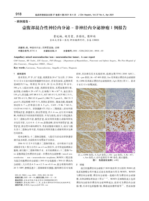
患者,女,66岁,皮肤及巩膜黄染1月,无寒战及发热,小便豆油样,大便如常,患有胆囊炎并发胆囊结石,行胆囊切除,于当地医院行ERCP检查(图1),术中取出胆管结石,并行鼻管引流,症状未见明显缓解。
门诊以“胆管占位”收入院。
CT平扫及增强(图2-6):胆总管近胆囊管起始部,胆管壁局限性增厚,可见结节样腔内突起,呈宽基底与胆管壁相连,最厚处达12.8 mm,累及长度约为16.0 mm,CT值约为39.5 HU,其以上左右肝管及胆囊管不同程度扩张。
动脉期病灶呈显著不均匀强化,CT值约115 HU,门脉期强化持续,CT值约为123.0 HU,静脉期强化减退,CT值约85.0 HU。
腹主动脉周围及肝门区可见淋巴结影,并可见环形强化,较大者短径约为9.2 mm。
CT拟诊为胆管肿瘤伴腹腔淋巴结转移。
手术所见:肝门部大量网膜组织粘连。
胆囊缺如。
肝十二指肠韧带内有肿大淋巴结。
于胆囊管起始部,紧邻胆总管处可见肿块大小约为20 mm*20 mm,质硬。
病理诊断为胆管小细胞神经内分泌癌。
图1 ERCP示胆总管上段管腔局限性充盈不良图2 CT胆总管近胆囊管起始部管壁局限性增厚,可见结节样腔内突起,呈宽基底与胆管壁相连,其以上左右胆管及胆囊管不同程度扩张神经内分泌肿瘤主要起源于Kulchitsky细胞,而Kulchitsky细胞多存在与胃肠道及呼吸系统内,因此较为常见,而发生在肝外胆管内分泌肿瘤十分少见[1],其发生率约占神经内分泌肿瘤的0.2%到2%[3,4],占全部消化系统神经内分泌肿瘤的0.67%[5]。
23原发性肝胆系统神经内分泌肿瘤病例回顾及文献复习葛大壮,毕新宇,赵宏,李智宇,张业繁,罗治文,魏哲文,陈启晨,李星辰,蔡建强*(国家癌症中心/国家肿瘤临床研究中心/中国医学科学院北京协和医学院肿瘤医院肝胆外科,北京 100021)葛大壮 博士研究生中国医学科学院北京协和医学院肿瘤医院麻醉科目的:分析原发性肝胆神经内分泌肿瘤的临床特点,并进一步明确其诊断方法和有效的治疗策略。
方法:对16例原发性肝胆神经内分泌肿瘤患者的临床病例资料进行回顾性的分析;结合文献综述,探讨原发性肝胆神经内分泌肿瘤的疾病临床特点及治疗选择。
结果:所有患者甲胎蛋白(alpha fetoprotein, AFP)、癌胚抗原(carcinoembryonic antigen,CEA)均无明显升高。
计算机断层扫描(computed tomography,CT)上动脉期明显增强,门脉期和延迟期减弱。
在磁共振成像(magnetic resonance imaging,MRI)上,肿瘤在T1WI 上表现为低信号,T2WI 上表现为高信号,DWI 上表现为局限性扩散,动脉期表现为明显的环状或不均匀增强,门脉期和延迟期表现为低信号。
所有患者均经病理证实为原发性肝胆神经内分泌肿瘤。
免疫组化结果显示所有肿瘤嗜铬粒蛋白A(chromograninA,CgA)均为阳性、11例突触素(synaptophysin, Syn)为阳性。
随访3~96个月,14例无复发。
无病生存期最长为96个月。
患者在随访过程中,全部患者均未发现其他部位神经内分泌瘤(neuroendocrine tumor, NET)。
结论:目前对于肝胆系统神经内分泌肿瘤患者,应根据患者的具体情况制定治疗方案,并充分完善相关检查,减少误诊的发生。
对于可切除的病变,手术切除是首选。
关键词:原发性肝胆系统神经内分泌肿瘤;影像学;治疗;预后摘要Primary hepatobiliary neuroendocrine tumors:a case review and literature reviewAbstractObjective: To investigate the clinical features, diagnosis and effective treatment strategies of primary hepatobiliary neuroendocrine tumor.Methods: The clinical data of 16 patients with primary hepatobiliary neuroendocrine tumor were analyzed retrospectively, and the clinical characteristics and treatment options of primary hepatobiliary neuroendocrine tumor were discussed combined with literature review.Results: There was no obvious increase in AFP and CEA in all patients. On CT, the arterial phase was markedly enhanced, while the portal phase and the delayed phase were weakened. On MRI, the tumors showed low signal on T1-weighted MRI, high signal on T2-weighted MRI, limited diffusion on DWI, and marked annular or heterogeneous enhancement in arterial phase, and decreased enhancement in portal phase and delayed phase. All patients were diagnosed as primary hepatobiliary neuroendocrine tumors by pathological results. The results of immunohistochemistry showed that chromogranin A (CgA) wasGe Dazhuang, Bi Xinyu, Zhao Hong, Li Zhiyu, Zhang Yefan, Luo Zhiwen, Wei Zhewen, Chen Qichen,Li Xingchen, Cai Jianqiang *(Department of Hepatobiliary Surgery, National Cancer Center/National Clinical Research Center for Cancer/Cancer Hospital, Chinese Academy of Medical Sciences and Peking Union Medical College, Beijing 100021, China )*通信作者:蔡建强 Email :神经内分泌肿瘤(neuroendocrine neoplasm, NEN)是来源于弥散神经内分泌细胞的一类肿瘤。