肝脏常见占位性病变及其MR鉴别
- 格式:ppt
- 大小:9.36 MB
- 文档页数:37


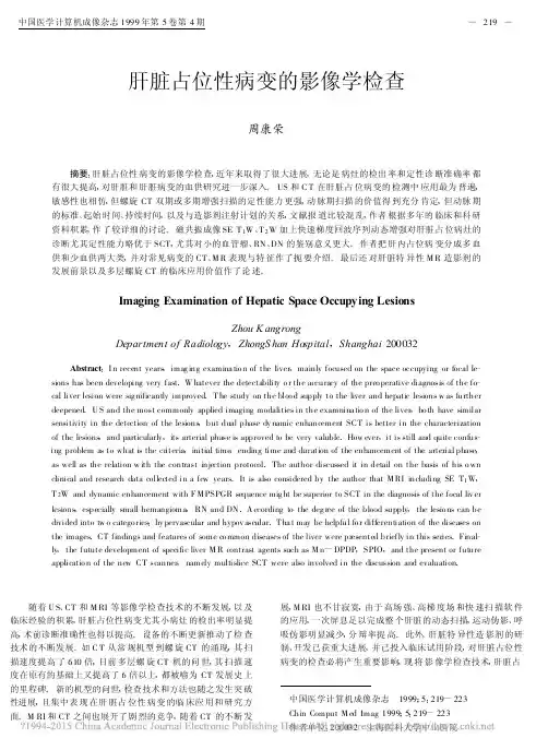
肝脏占位性病变的影像学检查周康荣 摘要:肝脏占位性病变的影像学检查,近年来取得了很大进展,无论是病灶的检出率和定性诊断准确率都有很大提高,对肝脏和肝脏病变的血供研究进一步深入。
US和C T在肝脏占位病变的检测中应用最为普遍,敏感性也相仿,但螺旋CT双期或多期增强扫描的定性能力更强,动脉期扫描的价值得到充分肯定,但动脉期的标准、起始时间、持续时间,以及与造影剂注射计划的关系,文献报道比较混乱,作者根据多年的临床和科研资料积累,作了较详细的讨论。
磁共振成像SE T1W、T2W加上快速梯度回波序列动态增强对肝脏占位病灶的诊断尤其定性能力略优于SCT,尤其对小的血管瘤、RN、DN的鉴别意义更大。
作者把肝内占位病变分成多血供和少血供两大类,并对常见病变的CT、M R表现与特征作了扼要介绍。
最后还对肝脏特异性M R造影剂的发展前景以及多层螺旋CT的临床应用价值作了论述。
Imaging Examination of Hepatic Space Occupying LesionsZhou K angrongDepartment of Radiology,ZhongS han Hospital,Shanghai200032Abstract:I n recent years,imag ing examina tio n of the liver,mainly focused on the space occupying or focal le-sions has been developing very fast.W hatever the detectability o r the accuracy of the preo perative diagnosis of the fo-cal liver lesion were sig nificantly improved.T he study on the bloo d supply to the liver and hepatic lesions w as further deepened.U S and the most commonly applied imaging modalities in the examnina tio n of the liver,bo th have similar sensitivity in the detection of the lesions,but dual phase dy namic enhancement SCT is better in the characterization of the lesions,and particularly,its arterial phase is approved to be very valuble.How ever,it is still and quite confus-ing problem as to what is the criteria,initial time,ending time and duration of the enhancement of the arterial phase, as well as the relation w ith the contrast injection protocol.The author discussed it in detail on the basis of his o wn clinical and research data collected in a few years.It is also considered by the author that M RI including SE T1W, T2W and dynamic enhancement with F M PSPGR sequence mig ht be superior to SCT in the diagnosis of the focal liv er lesions,especially small hemangioma,RN and DN.A ccording to the deg ree of the blood supply,the lesio ns can be divided into tw o catego ries:hy pervascular and hypov ascular.T ha t may be helpful fo r differentiation of the diseases on the images.CT findings and features of some co mmon diseases of the liver were presented briefly in this series.Final-ly,the futute development of specific liver M R contrast agents such as M n-DPDP,SPIO,and the present or future applicatio n of the new CT scanner,namely multislice SCT were also involved in the discussion and evaluation. 随着U S、CT和M RI等影像学检查技术的不断发展,以及临床经验的积累,肝脏占位性病变尤其小病灶的检出率明显提高,术前诊断准确性也得以提高。

肝脏MRI报告内容1.肝血管瘤MRI平扫肝表面光滑,各叶比例协调,T1WI像于肝叶可见一类圆形低信号病灶,大小×cm,边缘较清晰。
T2WI像上病灶为高信号,呈灯泡征。
T1WI增强扫描早期可见病灶边缘呈结节状强化,其后强化向中央扩展,延迟期病灶大部分强化呈等信号。
胆囊不大,壁不厚,胰腺、脾脏形态正常。
意见:肝叶占位性病变,肝血管瘤可能性大。
2.肝细胞癌MRI平扫示肝表面不光整,左叶、尾叶增大,肝裂增宽,T1WI像于肝叶可见一类圆形稍低信号病灶,边界欠清,大小× cm。
T2WI像上病灶信号稍高于正常肝实质组织。
增强扫描动脉期病灶明显不均匀强化,门脉期和肝实质期病灶信号迅速下降,逐渐低于正常肝实质信号。
延迟期包膜强化,门脉左支内可见低信号影,注入造影剂后也见强化。
胆囊不大,壁有增厚,胰腺正常,脾脏增大,达个肋单元。
食管下段与胃底静脉增粗,迂曲。
腹腔内未见肿大淋巴结。
意见:肝叶占位性病变,肝cancer可能性大3.肝转移瘤MRI平扫示肝表面光滑,各叶比例协调,肝内多发大小不等类圆形结节影,T1WI像上病灶呈边缘较清楚的均匀低信号灶,T2WI像上病灶呈高信号。
最大者位于肝叶,大小× cm。
增强扫描病灶边缘环形强化,中心强化不明显,呈牛眼征。
胆囊不大,壁不厚,胰腺、脾脏形态正常。
腹腔内未见肿大淋巴结。
意见:肝内多发占位性病变,肝转移cancer可能性大4.肝细胞腺瘤MR平扫肝表面光滑,各叶比例协调,T1WI像上于肝叶可见一类圆形略低信号灶,大小×cm,边缘较清,T2WI像上病灶呈高信号。
增强扫描病灶明显强化,延迟扫描信号接近肝脏。
胆囊不大,壁不厚,胰腺、脾脏形态正常。
腹腔内未见肿大淋巴结。
意见:肝叶占位性病变,肝细胞腺瘤可能性大5.局灶性结节增生MR平扫肝脏表面光滑,各叶比例协调,于肝叶可见一类圆形T1WI像上呈低信号,T2WI 像上呈高信号病灶,大小× cm,边缘较清晰,其内可见星芒状更长T1长T2信号影。