microRNA在动脉粥样硬化中的角色
- 格式:pdf
- 大小:268.77 KB
- 文档页数:10


MicroRNAs与动脉粥样硬化的关系微小分子核糖核酸(MicroRNAs ,miRNAs)是一类长约21-25nt 的非编码的内源性单链RNA 分子,序列在物种间具有高度保守性,其主要通过与靶mRNA 的3’非翻译区的碱基形成完全或不完全配对,从而达到直接降解或抑制靶mRNA,进而发挥沉默基因或调控的作用,而这一作用使其广泛参与了细胞的增殖、分化、凋亡及肿瘤的发生等过程。
近来越来越多的研究表明,microRNAs 在动脉粥样硬化(atherosclerosis,AS)的发生发展过程中也发挥了极其重要的作用。
另外一些微小核糖核酸可能是动脉粥样硬化的诊断标志物或者治疗目靶。
现就最近的研究阐述miRNAs與动脉粥样硬化发生过程的关系。
标签:微小分子核糖核酸;动脉粥样硬化从上世纪中叶开始,心脑血管疾病已成为导致世界人口死亡的主要疾病之一,其发病率及死亡率逐年升高,而据WHO 估计,至2020 年全球因心血管疾病死亡的人数将占到总死亡人数的36%[1]:在我国心血管疾病的发展趋势同样不容乐观,而在众多致残致死的心血管疾病当中,动脉粥样硬化更是占到了75%以上[2];目前对于该病的发病机制大部分研究者都较为认同自身炎症反应学说与免疫机制,但是在发病的整个过程中,血管与炎症跟免疫等等各个环节都需要进行相互协调与作用,关于这方面的关系尚未得到相应说明。
近些年关于内源性非编码的微小RNA的研究为这方面的深入研究提出了新的方向与思路。
本文就miRNAs 分子的概述以及近年有关miRNAs 与动脉粥样硬化关系研究的部分进展作一综述。
1 miRNAs的形成和作用机制MiRNAs是一类大小为19~25个碱基的内源性非编码小RNA 分子,通常与靶基因mRNA的3’-非编码区(3’-untranslated region,3’- UTR)相结合调控翻译,在生物进化过程中是一类高度保守的非编码小RNAs,通过碱基互补或部分碱基互补与目标mRNA 结合,降解目标mRNAs和抑制目标mRNAs 的翻译,在转录后水平调节基因表达。

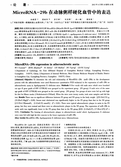
MicroRNAs与动脉粥样硬化的关系杜映荣;李红娟【期刊名称】《医学信息》【年(卷),期】2014(000)009【摘要】MicroRNAs is one kind of endogenous noncoding,single-stranded, evolution arily conserved RNAs of 21-25 nucleotides. MiRNAs that bind to 3'UTR of mRNA with imperfect complementarity block protein translation. In contrast, miRNAs that bind mRNA with perfect complementarity induce targeted mRNA cleavage. Therefore just forthis ef ect, microRNAs can act as an important regulator for cellproliferation, dif erentiation, apoptosis and the development of cancer. In addition, an increasing number of recent studies have shown that microRNAs may play important roles in atherosclerosis. This article summarizes the current studies related to the disease correlations and functional roles of miRNAs participating in atherosclerosis development process.%微小分子核糖核酸(MicroRNAs , miRNAs)是一类长约21-25nt 的非编码的内源性单链 RNA 分子,序列在物种间具有高度保守性,其主要通过与靶mRNA 的3'非翻译区的碱基形成完全或不完全配对,从而达到直接降解或抑制靶 mRNA,进而发挥沉默基因或调控的作用,而这一作用使其广泛参与了细胞的增殖、分化、凋亡及肿瘤的发生等过程。

MicmRNA作为动脉粥样硬化的诊断生物标志物的研究进展耿春晖关秀茹(哈尔滨医科大学附属第一医院检验科,黑龙江哈尔滨150001)【摘要】动脉粥样硬化是由脂质代谢紊乱和慢性炎症导致的疾病。
随着个体化和精准医疗的发展,早期精确诊断动脉粥样硬化对临床工作开展意义重大。
由于小的非编码RNAs能调节炎症蛋白含量干扰脂质的形成,因此研究小的非编码RNAs作为生物学标志物给动脉粥样硬化的快速诊断带来了新的希望%整合近年文献,通过探讨小的非编码RNAs对动脉粥样硬化的病理机制的影响,阐明其作为动脉粥样硬化生物标志物的意义%【关键词】动脉粥样硬化#小的非编码RNAs;生物标志物【DOI】10.16806/j.JnkU ion.1004-3934.2019.07.008MicroRNA as a Diagnostic Biomarker for AtherosclerosisGENGChunhuo,GUANXoueu(Aerartment o'Clnical Laboratory,Thr First Afiliated Hospital o'Harbm Merical Uniersity,Harbm150001, Heilongjiang,China%【Abstract】Athemsc/msis(AS)is a disease caused by disorders of iipid metabolism and chronic inOammation.With the development of individualization and precision meddine,accurate early diagnosis of AS is of great significrce.Small non-aoding RNAs(micmRNAs) regu/te iid/mmatoy protein levels and interfere with lipid formation,thus micmRNAs as biomarkers has brought new insights foo tie early diagnosis for AS.This papeo integrates recent literature and explores tie significance of microRNAs as a biomarkeo for AS.【Key words t Athemsc/msis;MOroRNAs;Biomarkeo动脉粥样硬化(atherosc/rosis,AS%是由脂质代谢紊乱和慢性炎症导致的疾病[1],其病理机制复杂涉及一系列病理改变[2]*AS导致的心血管疾病是全世界发病率和死亡率增高的主要原因[3]*近几年越来越多的证据表明,非编码RNA在组织稳态和病理生理状态的调节中起重要作用*其中小的非编码RNAs (micmRNAs)会干扰不同阶段的基因表达和信号通路。

外泌体microRNA在动脉粥样硬化性脑梗死中的研究进展引言动脉粥样硬化性脑梗死(ASD)是一种常见的疾病,其发展与多种因素密切相关。
研究表明,外泌体microRNA在ASD的发生和发展中发挥着重要作用。
本文将综述外泌体microRNA在ASD中的研究进展,以期为ASD的预防和治疗提供新的思路和方法。
外泌体及其microRNA简介外泌体是一类直径约30-150纳米的囊泡,由细胞内分泌物包裹而成,广泛存在于体液中,包括血浆、尿液、唾液等。
外泌体内含有蛋白质、核酸等多种生物活性成分,其中包括microRNA。
microRNA是一类长度约22核苷酸的非编码RNA,可以调控基因表达,参与细胞生长、凋亡、代谢等多个生物过程。
外泌体中的microRNA被认为是一种重要的细胞间信号传递介质。
外泌体microRNA与ASD的关联ASD的发生和发展是一个复杂的过程,其中包括血管内皮细胞的异常增殖、炎症反应的持续激活、斑块的形成等多个阶段。
研究表明,外泌体microRNA参与了ASD相关病理过程的调控,影响了血管内皮细胞的功能、炎症反应的平衡、斑块的形成等多个环节。
外泌体microRNA对血管内皮细胞的影响血管内皮细胞是血管壁的重要组成部分,其功能异常与ASD的发生密切相关。
研究表明,外泌体中的一些microRNA可以通过转移至血管内皮细胞,调控细胞增殖、凋亡、迁移等多个生物学过程,进而影响血管内皮细胞的功能和结构。
miR-126在外泌体中的含量下调与ASD患者血管内皮细胞异常增殖相关,miR-92a的上调与血管内皮细胞凋亡增加相关,这些外泌体microRNA的异常表达都影响了血管内皮细胞的健康状况,从而参与了ASD的发生和发展。
外泌体microRNA在ASD炎症反应中的作用炎症反应在ASD的发生和发展中起着重要作用。
外泌体中的microRNA可以通过转移至炎症相关细胞,如单核细胞、巨噬细胞等,调控这些细胞的炎症反应。

microRNA在动脉粥样硬化发病机制中的作用于瑞杰【摘要】微小核糖核酸(microRNA,miRNA) 是一类非编码的小分子RNA,主要在转录后水平调控基因表达.近来,miRNA与动脉粥样硬化(atherosclerosis,As)发病机制的关系成为研究热点.目前已证实miRNA在调节As病变进程相关的血管壁炎性反应、免疫细胞分化和胆固醇代谢等方面均发挥重要作用.miR-155和miR-146a参与调控血管壁的炎性反应以及辅助性T细胞(helper T cell,Th)的分化;miR-29参与调控血管平滑肌细胞(vascular smooth muscle cell,VSMC)迁移以及相关炎性反应;miR-33参与对胆固醇代谢的调控.此外,miR-365、miR-222等分别通过调控内皮细胞的凋亡和新生血管的形成参与As的发生、发展.文中就上述miRNA在As发生、发展过程中的作用机制进行综述,旨在为进一步揭示As的发病机制提供思路.【期刊名称】《医学研究生学报》【年(卷),期】2013(026)009【总页数】4页(P970-973)【关键词】microRNA;动脉粥样硬化;作用机制【作者】于瑞杰【作者单位】210002,南京,南京大学医学院临床学院(南京军区南京总医院)解放军临床检验医学研究所【正文语种】中文【中图分类】R543.120 引言miRNA是长约22个核苷酸的单链、内源性的非编码小RNA,通过与靶mRNA 结合而对基因表达进行转录后水平的调控。
目前,在已发现的miRNA中,大约有721种人源性miRNA和579种鼠源性miRNA[1]。
人类约30%的编码蛋白基因受其调控,1个miRNA可调控多个靶基因,而同一基因可由多个miRNA共同调节[2]。
越来越多的证据表明,miRNA在多种疾病的病理生理过程中起重要作用,尤其与心血管疾病的发生、发展密切相关[3-5]。
As作为心血管疾病的主要病理生理基础,是一种动脉血管壁的慢性炎症性病变[6-7]。
MicroRNA在动脉粥样硬化进展中的作用发表时间:2015-12-08T15:19:46.993Z 来源:《航空军医》2015年5期供稿作者:武胜勇[导读] 第二军医大学 MicroRNA是一类进化上高度保守、小分子非编码RNA基因产物,来源于细胞内源性转录体,与靶mRNA完全配对,介导靶mRNA降解、抑制其翻译,发挥沉默特定靶基因作用。
武胜勇第二军医大学 200433【摘要】MicroRNA是一类小分子非编码RNA,在生物发育时序调控、疾病发生中发挥重要作用,研究证实其与血管发生、免疫炎症密切相关,在动脉粥样硬化基因治疗、相关疾病诊断、冠脉事件风险预测中具有广阔的应用前景。
【关键词】动脉粥样硬化;MicroRNA;表达调控动脉粥样硬化病理生理机制尚不清楚,是冠心病重要病理基础,是脑梗死、心肌梗死等高致死性疾病重要危险因素,动脉粥样硬化形成的斑块成为重要的栓子来源,还影响血流供应,据统计约有20%~25%脑梗死与动脉粥样硬化斑块活动有关。
目前认为,动脉粥样硬化是多基因、多因素、多阶段疾病,与免疫炎症反应、氧化应激、细胞凋亡等因素有关。
MicroRNA是一类小分子非编码RNA,参与调控各类细胞生长、分化、凋亡等,成为近年来生物学研究热点,在生物发育时序调控、疾病发生中发挥重要作用。
本次研究试概述MicroRNA在动脉粥样硬化进展中的作用研究进展。
1 MicroRNA基本特点MicroRNA是一类进化上高度保守、小分子非编码RNA基因产物,来源于细胞内源性转录体,与靶mRNA完全配对,介导靶mRNA降解、抑制其翻译,发挥沉默特定靶基因作用。
某些miRNA还可调控mRNA半衰期。
一种miRNA可调节多种基因、蛋白表达,故在生物体基因表达中发挥重要作用,调控范围十分广泛,有学者认为microRNA可调控人类1/3以上基因,现被证实的miRNA序列有400多个,其中多数序列在细胞生长、分化、凋亡过程中发挥作用,可调控人体正常发育、生理活动。
MicroRNAs对动脉粥样硬化关键信号分子的影响袁绪胜;戴敏【期刊名称】《中国药理学通报》【年(卷),期】2012(028)001【摘要】microRNAs是近年来发现的内源性非编码单链RNA小分子,在心血管系统中具有特异性的表达谱,通过降解mRNA或抑制蛋白质翻译调控转录后基因的表达.microRNAs在动脉粥样硬化形成的病理过程中,参与血管内皮细胞的损伤与修复;细胞炎性因子、生长因子等信号分子的表达与释放和血管平滑肌细胞的增殖与迁移等环节,从而调控动脉粥样硬化的发生发展.%Recently, microRNAs have been discovered as a small endogenous non-coding single-stranded RNA molecule, which has a specific expression profile in the cardiovascular system, and regulates gene expression at the transcriptional level through degradating mRNA or inhibiting protein translation. In the atherosclerosis, microRNAs attends not only vascular endothelial cell injury and repair, but also expression and release of cytokines,growth factors and other active substances, meanwhile, microRNAs attends vascular smooth muscle cell proliferation and migration and other aspects, thereby regulates atherosclerotic formation and development.【总页数】4页(P20-23)【作者】袁绪胜;戴敏【作者单位】安徽中医学院药学院,安徽,合肥,230031;安徽中医学院药学院,安徽,合肥,230031;安徽省中药研究与开发重点实验室,安徽,合肥,230031;省部共建新安医学教育部重点实验室,安徽,合肥,230031【正文语种】中文【中图分类】R329.24;R342.2;R394.2;R543.5【相关文献】1.microRNA-185下调AKT1信号通路对淋巴母细胞淋巴瘤细胞增殖和凋亡的影响及相关的分子机制的研究 [J], 王金燕;刘春霞2.MicroRNA-153对靶基因下游信号分子GSK-3β表达水平及细胞抗损伤能力的影响 [J], 梁春联;朱华;黄澜;许艳峰;邓巍;马春梅;刘颖;秦川3.泛素基因沉默对TLR4/NF-κB信号通路中关键分子表达的影响 [J], 刘超英;董骎;高洪;严玉霖;富国文;单春兰4.散结通脉方对ApoE-/-小鼠动脉粥样硬化主动脉钙库操纵性钙通道信号分子及血清炎症因子的影响 [J], 丁云录;李驰坤;欧喜燕;李晓兵;成光宇;刘爱东5.电针心包经穴对急性脑缺血大鼠脑组织神经生长抑制信号通路关键分子基因表达的影响 [J], 肖豆;谢峥嵘;唐雅妮;潘江;曹越;娄必丹;章薇;陈成因版权原因,仅展示原文概要,查看原文内容请购买。
微小RNA在心血管疾病中的作用及其临床应用研究心血管疾病是当前影响人类健康的主要疾病之一,致死率和疾病负担率极高。
近年来,微小RNA(microRNA,miRNA)作为一种新型的调控生物分子,受到了广泛的研究和关注。
miRNA是一类长度约为20~22个核苷酸的非编码RNA,能够通过结合到目标mRNA靶标上,调节mRNA的翻译和降解,从而影响基因的表达。
miRNA在心血管疾病的发生和发展中扮演着重要的作用。
在细胞凋亡、细胞增殖、细胞迁移、血管生成等过程中,miRNA都发挥着不同的作用。
miRNA-1、miRNA-133、miRNA-208、miRNA-499等与心肌细胞的功能、心肌损伤修复、心肌肥厚等紧密相关。
miRNA-21、miRNA-29、miRNA-155等与心血管平滑肌细胞的增殖、内皮细胞功能、血管通透性等相关。
同时,miRNA-17~92和miRNA-106b~25家族在心血管疾病的发生和发展中也发挥着非常重要的作用。
目前,miRNA已成为心血管疾病研究的热点之一。
miRNA与心血管疾病之间的相互关系已经被广泛研究。
miRNA在心血管疾病中的作用机制是复杂的,这一点在其与心血管疾病之间的关系研究中也有所体现。
miRNA与心血管疾病之间的关系研究主要分为两个方面:一是 miRNA在心血管疾病中的作用与应用;另一方面是在 miRNA在诊断和治疗心血管疾病方面的研究。
一、miRNA在心血管疾病中的作用和应用1. miRNA在心血管疾病的预测和诊断中的应用临床上,miRNA已被应用于心血管疾病的预测和诊断。
例如miRNA-1、miRNA-133、miRNA-208、miRNA-499等miRNA的表达水平与心肌损伤相关。
研究表明,在急性心肌梗死的发病初期,miRNA-208和miRNA-499的表达水平会明显升高。
同时,miRNA-133的表达水平随着心肌梗死的恢复而逐渐升高,可以在一定程度上预测心肌损伤的恢复程度。
外泌体microRNA在动脉粥样硬化性脑梗死中的研究进展动脉粥样硬化性脑梗死是一种常见的脑血管疾病,其发病原因复杂,治疗难度较大。
近年来,外泌体microRNA在动脉粥样硬化性脑梗死中的研究引起了广泛关注。
外泌体是一种直径约30-100 nm的一种小囊泡,其富含脂质和蛋白质,可释放到细胞外,通过膜融合或内吞受体介导内吞进入受体细胞,发挥调控功能。
而microRNA是一类长度约22nt的小RNA,通过与靶基因mRNA结合,调控基因的表达水平,参与多种生物学过程。
外泌体中的microRNA被发现在调控细胞增殖、分化、凋亡等生物学过程中起着重要作用。
动脉粥样硬化性脑梗死是由于动脉粥样硬化斑块破裂、溶解而释放大量斑块物质,导致颅内血栓形成或既有脑血栓栓塞所致。
研究表明外泌体中的microRNA能够通过调控斑块物质的释放、促进或抑制血栓形成等作用参与了动脉粥样硬化性脑梗死的发生发展。
近年来,研究者发现在动脉粥样硬化性脑梗死患者的血浆和脑组织中存在一些具有调控作用的microRNA,例如miR-155、miR-146a、miR-21等。
这些microRNA可以通过靶向调控炎症因子、脂质代谢相关基因等参与了动脉粥样硬化性脑梗死的发病过程。
miR-155在炎症反应中发挥重要作用,miR-146a则参与了调控免疫应答和抗炎反应,而miR-21则能够调控血管平滑肌细胞增殖和迁移,在斑块形成和破裂中发挥关键作用。
一些研究还发现外泌体中的microRNA可以通过调控细胞凋亡、增殖等生物学过程参与了动脉粥样硬化性脑梗死的发展。
miR-126在血管内皮细胞中高表达,可以调控血管生成和维持血管完整性,而miR-92a则能够促进血管平滑肌细胞增殖和迁移,对血管重建过程有重要影响。
除了研究外泌体中的microRNA在动脉粥样硬化性脑梗死的作用外,研究者还开始探索通过调控外泌体中microRNA的方法来治疗动脉粥样硬化性脑梗死。
近期的研究发现,通过将特定的microRNA载体包裹到外泌体中,可以有效地将其释放到病变部位,并发挥治疗作用。
REVIEW ARTICLEMicroRNAs in atherosclerosisKu-Chung Chen a ,b ,Suh-Hang Hank Juo a ,b ,*a Department of Medical Research,Kaohsiung Medical University Hospital,Kaohsiung,Taiwan bDepartment of Medical Genetics,Medicine,Kaohsiung Medical University,Kaohsiung,TaiwanReceived 3January 2012;accepted 20February 2012Available online 5August 2012KEYWORDSAtherosclerosis;Cardiovascular disease;MicroRNAAbstract MicroRNAs are endogenously expressed small noncoding RNAs that regulate gene expression at the post-transcriptional level.MicroRNAs have emerged as key regulators of several physiological and pathophysiological processes in the cardiovascular system.Aberrant expression of microRNAs has been implicated in the pathophysiological processes underlying the development of atherosclerosis and cardiovascular disease,including change in endothe-lial function,vascular smooth muscle cell proliferation and migration,macrophage function,and foam cell formation.In this review,we summarize the recent data showing the roles of microRNAs in cell studies,studies on atherosclerotic mice,and human studies.Copyright ª2012,Elsevier Taiwan LLC.All rights reserved.IntroductionAtherosclerosis is a major risk factor for cardiovascular disease [1].Endothelial cells dysfunction often initiates a series of changes that leads to the development of atherosclerosis.Atherosclerotic plaques are composed of accumulated lipid particles in foam cells,proliferated vascular smooth muscle cells (VSMCs),and extracellular matrix.These plaques are landmarks of advanced athero-sclerotic lesions in the artery.Therefore,endothelial cells,VSMCs,and macrophage-derived foam cells are critical players in the development of plaques [2e 5].Several genes and cytokines have been identified as risk factors foratherosclerosis [6,7],and recent studies have suggested that microRNAs (miRNAs)may play a role in regulating the atherosclerotic process [8,9].miRNAs are a novel class of endogenous noncoding,single-stranded RNAs with a length of approximately 22nucleotides.A miRNA controls gene expression by binding to its target genes’messenger RNAs (mRNAs)for degrada-tion and/or translational repression [10].In animals (including humans),this gene-silencing effect is mediated by a complementary base pairing between the 30untrans-lated region of the mRNA and a particular region of a miRNA.This particular region of miRNA is called the “seed region”and is located at positions 2e 8of a miRNA.miRNAs can be expressed in a tissue-and cell-type specific manner and affect multiple cell functions such as proliferation,apoptosis,migration,differentiation,and also develop-ment [11].Accumulating evidence has demonstrated the role and function of miRNAs in maintaining normal physio-logical conditions and disease development.*Corresponding author.Kaohsiung Medical University,100TzYou 1st Road,Kaohsiung 807,Taiwan.E-mail address:hjuo@.tw (S.-H.Hank Juo).1607-551X/$36Copyright ª2012,Elsevier Taiwan LLC.All rights reserved.doi:10.1016/j.kjms.2012.04.001Available online atjournal homepage:Kaohsiung Journal of Medical Sciences (2012)28,631e 640Although most miRNA research has focused on cancer, the role of miRNAs in the cardiovascular system has been gradually revealed.Hence,this is a pertinent time to review and summarize the recentfindings on miRNAs in the context of atherosclerosis.This review article describes the role of miRNAs in three areas d endothelial cells,VSMCs, and macrophages(Table1).miRNAs in endothelial cellsThe endothelium has a pivotal role in maintaining vascular homeostasis and in providing a physical barrier between the blood components and the subendothelial space [12e14].Endothelial dysfunction has recently been demonstrated to be an early step in the development of632K.-C.Chen,S.-H.Hank Juoatherosclerosis,and it has also been shown to contribute to the formation,progression,and complications of atherosclerotic plaques[14].Importantly,endothelial dysfunction represents a common link among all known cardiovascular risk factors,including dyslipidemia, smoking,diabetes,hypertension,obesity,and mental stress[15].Several features can characterize endothelial dysfunction,including a reduced bioavailability of nitric oxide,increased expression of adhesion molecules, increased levels of proinflammatory and prothrombotic factors,oxidative stress,and abnormal modulation of vascular tone.Several miRNAs have been reported to be related to endothelial dysfunction,and these are summarized below.miR-21miR-21is one of the most dynamically regulated miRNAs in various pathological processes,ranging from cancer to cardiovascular disease[16].Aberrant expression of miR-21 is found in vascular neointimal lesion formation and acute myocardial infarction[17,18].miR-21is significantly upre-gulated in atherosclerotic plaques compared with non-atherosclerotic arteries[19].Using the shear stress treatment on endothelial cells,the expression levels of miR-21are greatly increased[20].In addition,the same study[20]reported that miR-21causes an antiapoptotic effect,increased endothelial nitric oxide synthase phos-phorylation,and nitric oxide production in endothelial cells by directly targeting to PTEN,a known tumor suppressor gene.However,another study using shear stress in endo-thelial cells reported that overexpressed miR-21may inhibit the expression of peroxisome proliferator-activated receptor-alpha(PPAR a),leading to enhanced expressions of vascular cell adhesion molecule-1(VCAM-1)and mono-cyte chemotactic protein-1[21].The studies mentioned indicate a critical role of miR-21in the development of atherosclerosis,but whether an increase in miR-21in endothelial cells is beneficial or deleterious remains to be clarified.miR-126miR-126is expressed only in endothelial cells,and thus it is recognized as an endothelium-specific microRNA for regu-lating angiogenesis and vascular inflammation.A low level of circulating miR-126is associated with peripheral vascular complications in patients with type2diabetes and myocardial infarction[22,23].miR-126possesses an ability to regulate monocyte adhesion by directly targeting VCAM-1,and thus it controls vascular inflammation[24].A potential E26transformation-specific sequence(Ets) binding site is located between e71and e100bp upstream of the miR-126gene[25].Either transcription factor Ets-1 or Ets-2may activate the expression of miR-126,resulting in depression of VCAM-1.Additionally,administration of miR-126in mouse models of atherosclerosis may reduce macrophage and apoptotic cell content,thereby limiting lesions size and conferring a milder inflammatory reaction [23].Accordingly,miR-126may exert an antiatherosclerotic effect.miR-217Aging is a major risk factor for the development of atherosclerosis and coronary artery disease.Since endo-thelial cell senescence contributes to age-related impair-ment of angiogenesis,miRNAs involved in senescence processes may be also related to atherosclerosis formation. Menghini et al demonstrated that miR-217is largely expressed in aged but not young endothelial cells[26]. They also identified that silent information regulator-1 (SirT1),a major regulator in metabolic disorders,is a direct target gene of miR-217.Overexpression of miR-217 can induce endothelial cell senescence formation. Conversely,inhibition of miR-217in elderly endothelial cells ultimately reduces senescence and increases angio-genic activity.Although miR-217functions as a potential tumor suppressor by targeting KRAS[27],miR-217exerts negative effects on the vascular endothelium by enhancing endothelial senescence,which is related to pathogenesis of cardiovascular diseases.miR-221/222miR-221and miR-222(miR-221/222)belong to the same family,and their genes are located in the same cluster gene on Xp11.3chromosome.miR-221/222were identified as antiangiogenic miRNAs by inhibiting c-Kit-mediated migra-tion in endothelial cells[28].Furthermore,levels of miR-221/222are significantly higher in endothelial progenitor cells from individuals with coronary artery disease than in those without[29,30].Notably,circulating endothelial progenitor cells play an important role in the maintenance of endothelial integrity and vascular repair[31].Several proteins have been identified as target genes of miR-221/ 222,including signal transducer and activator of tran-scription5A(STAT5A),a transcription factor involved in the regulation of cell proliferation and migration.STAT5A has been shown to increase along with reduced miR-222in endothelial cells from advanced neovascularized athero-sclerotic lesions[32].Overexpression of miR-221/222 effectively decreases the adhesion of Jurkat T cells to Angiotensin II-stimulated endothelial cells by inhibiting expression of Ets-1,a key endothelial transcription factor for inflammation and tube formation[33].Thus,miR-221 and miR-222have effects on regulation of angiogenesis during the development of atherosclerosis.miR-10aDisturbedflow in arteries may lead to atherosclerosis formation.Conversely,the regions of undisturbed laminar flow are more resistant to atherosclerosis[34].The endo-thelium in athero-susceptible regions displays different characteristics,including those related to sensitization of inflammatory regulators,redox/lipid balance,and endo-plasmic reticulum stress,from epithelium located at athero-resistant sites[35e37].The expression of endothe-lial miR-10a is lower in the athero-susceptible regions of the inner aortic arch and aortorenal branches than else-where[38].Knockdown of miR-10a in cultured human aortic endothelial cells can induce inflammatory processesMicroRNAs in atherosclerosis633mediated by inhibitor of kappa B/nuclear factor kappa B (I k B/NF-kB).The genes encoding mitogen-activated protein kinase kinase kinase7(MAP3K7;TAK1)and beta-transducin repeat-containing(b TRC),which are two key regulators of I k B a degradation,have been identified as direct target genes of miR-10a.Both MAP3K7and b TRC are upregulated when miR-10a is knocked down,which leads to enhanced NF k B activation.miR-365Endothelial cells apoptosis can contribute to the develop-ment of atherosclerosis.The presence of oxidized low-density lipoprotein(oxLDL)is an important risk factor for atherosclerosis and contributes greatly to the apoptosis of endothelial cells.miR-365has been identified to be upre-gulated in endothelial cells upon oxLDL treatment[39]. miR-365directly targets the antiapoptotic protein B-cell CLL/lymphoma2(Bcl-2)and promotes cell death.Knock-down of miR-365represses oxLDL-mediated apoptosis of endothelial cells,suggesting that miR-365may be a poten-tial novel therapeutic target for atherosclerosis.miR-663Recently,upregulation of miR-663by oscillatory shear stress has been identified as playing a role in inflammatory response of endothelial cells[40].miR-663mediates oscil-latory shear stress-induced monocyte adhesion to the endothelial cells via tumor necrosis factor alpha(TNF-a) activation,but it does not affect the oscillatory shear stress-mediated apoptosis of endothelial cells.Several miR-663target genes such as IL8,ATF3,and KLF4regulate important physiological processes including cell prolifera-tion and inflammatory response that are involved in atherosclerosis.Accordingly,miR-663is likely to play a role in the proatherosclerotic process.miR-125a-5p/125b-5pEndothelin-1(ET-1)is a21-amino-acid peptide that acts on seven transmembrane G protein-coupled receptors to elicit a plethora of responses[41].Elevated levels of circulating ET-1have been shown to be associated with endothelial dysfunction and dysregulation of vascular function[42].ET-1is a direct target gene of miR-125a-5p and miR-125b-5p, and both miRNAs are highly expressed in vascular endo-thelial cells[43].miR-125a/125b-5p can suppress oxLDL-induced ET-1expression,and thus both miRNAs play a protective role against development of atherosclerosis. miR-34amiR-34a is highly expressed in endothelial cells[44],and elevated miR-34a expression can be observed in senescent endothelial cells,heart,and spleen of aged mice.The overexpression of miR-34a induces endothelial cell senes-cence and also suppresses cell proliferation by inhibiting the cell cycle via directly inhibiting SirT1protein expression [45].miR-34a expression is significantly upregulated in atherosclerotic arteries versus nonatherosclerotic left internal thoracic arteries[46].The current evidence suggests that miR-34a can promote the formation of atherosclerosis.miR-146amiR-146a also has a modulatory effect in endothelial cell senescence[47].miR-146a is downregulated in aged endothelial cells.It can directly target NADPH oxidase-4 (NOX4),which implicates it in cell senescence and aging. The expression of miR-146a is decreased under oxLDL stimulation[48],while miR-146a significantly reduces intracellular LDL cholesterol content and secretion of interleukin-6(IL-6)and IL-8,chemokine(C-C motif)ligand-2,and matrix metallopeptidase-9(MMP-9)in macrophages [48].In addition,miR-146a can inhibit the activation of Toll-like receptor4(TLR4)-dependent intracellular signaling pathways,which are involved in cytoskeleton rearrangement,lipid uptake,and inflammatory cytokine secretion[48].miR-146a also regulates the maturation process and proinflammatory cytokine secretion by target-ing CD40L in ox-LDL-stimulated dendritic cells[49].Furthermore,overexpression of miR-146a in peripheral blood mononuclear cells significantly upregulates the function of T helper type1cells[50].The upregulation of these cells has been suggested to have an essential function in the development of atherosclerosis.Overexpression of miR-146a may therefore prevent atherosclerosis.miR-200Recently,the miR-200family has been shown to inhibit cell migration and modulate epithelial e mesenchymal transition in cancer cells[51].Members of the family also play a causal role in a variety of cardiovascular diseases.miR-200c is upregulated in oxidative stress-mediated endothe-lial cells[52].Overexpression of miR-200c induces growth arrest,apoptosis,and senescence via directly targeting zincfinger E-box binding homeobox-1(ZEB1),a prosurvival protein.Hypoxia induces downregulation of miR-200b and promotes angiogenesis[53].Delivery of miR-200b mimic to endothelial cells can suppress the angiogenic response through downregulation of v-ets erythroblastosis virus E26 oncogene homolog1(Ets-1),a crucial angiogenesis-related transcription factor.miR-200b can also reduce protein levels of vascular endothelial growth factor(VEGF),fms-related tyrosine kinase-1(Flt-1),and kinase insert domain receptor(KDR),resulting in an inhibition of endothelial angiogenesis[54].These results suggest that the miR-200 family may be a therapeutic target for the prevention of atherosclerosis.miR-210Hypoxia can induce the development of atherosclerosis. The hypoxia-inducible factor pathway is essential for cell survival under conditions of low oxygen and enhances endothelial cell angiogenesis[55].miR-210has been shown to be a prominent miRNA that is upregulated by hypoxia. Overexpression of miR-210in normoxic endothelial cells stimulates the formation of capillary-like structures and634K.-C.Chen,S.-H.Hank JuoVEGF-mediated cell migration via ephrin-A3down-regulation[56].Anti-miR-210inhibits cell growth and induces apoptosis in both normoxic and hypoxic conditions. In our unpublished data,we also found that dysregulation of miR-210is involved in the formation of atherosclerosis. Thus,miR-210may be another therapeutic target in atherosclerosis therapy.In addition to the miRNAs mentioned above,several miRNAs are also reported to be involved in endothelial dysfunctions related to angiogenesis,including miR-424, miR-373,miR-16,and miR-296[55,57,58].miRNAs regulate vascular smooth muscle cells VSMC plasticity plays an important role in vascular pathol-ogies during the development of atherosclerosis.VSMCs maintain an organized,differentiated,and contractile phenotype under physiological conditions.Upon inappro-priate stimulation,these contractile cells may be switched to a proliferative and migratory(synthetic migratory) phenotype[59].In atherosclerosis,the switch of VSMC phenotype contributes to neointima and plaque formation.A wide variety of growth factors,such as platelet-derived growth factor(PDGF)and insulin-like growth factor-I,have been identified to participate in the regula-tion of VSMC proliferation and migration[60].In particular, recentfindings have also demonstrated that miRNAs play a critical role in the switch of VSMC phenotype.Several endothelial miRNAs also show their effects on VSMC.Increased miR-21regulates VSMC function via targeting tropomyosin-1,programmed cell death protein-4(PDCD4), sprouty-2(SPRY2),and PPAR a in arteriosclerosis[61e63]. miR-146a and Kruppel-life factor-4(KLF4)form a feedback loop to regulate each other’s expression and VSMC prolif-eration[64].miR-10a is identified as a novel regulator in VSMC differentiation from embryonic stem cells via tar-geting histone deacetylase-4(HDAC4)[65].The expression of miR-221and miR-222is upregulated and localized in the VSMCs of injured vascular walls,resulting in VSMC prolif-eration and neointimal hyperplasia[66,67].Below,we introduce more miRNAs whose functions are specifically identified in the regulation of VSMC phenotype.miR-145/143miR-143and miR-145are contained in a bicistronic primary transcript from chromosome5.The genes for miR-143/145 are also identified as a cluster gene.Zhang and coworkers demonstrated that miR-145was abundant in normal arteries and VSMCs[68,69].The expression of miR-145is significantly downregulated in dedifferentiated VSMCs and in balloon-injured arteries.These authors also identified that miR-145was a novel VSMC phenotypic marker and modulator that is able to control vascular neointimal lesion formation.Several VSMC differentiation marker genes,such as smooth muscle alpha-actin,calponin,and smooth muscle-myosin heavy chain(SM-MHC),are upregulated by miR-145via targeting KLF5.Cordes et al also reported that miR-145and miR-143are direct transcriptional targets of serum response factor, myocardin,and NK2transcription factor related,locus5(Nkx2-5),and that they are downregulated in injured or atherosclerotic vessels containing proliferating,less differentiated VSMCs[70].Furthermore,miR-145and miR-143cooperatively target a network of transcription factors,including KLF4,myocardin,and ELK1,member of ETS oncogene family(Elk-1),to promote differentiation and repress proliferation of VSMCs.Transforming growth factor beta(TGF-b)and bone morphogenetic protein-4 (BMP4)downregulate KLF4expression through upregula-tion of miR-143and miR-145,which results in the inhibition of VSMC growth and migration[71,72].TGF-b activates transcription of the miR-143/145gene cluster through the CArG box via myocardin expression,while BMP4exerts a similar effect via nuclear translocation of myocardin-related transcription factor-A.Moreover,loss of miR-143/145results in the formation of podosomes,which are actin-rich membrane protrusions that are involved in VSMC migration[73].PDGF can mediate podosome formation in VSMCs through the regulation of miR-143/145expression via a pathway involving Src and p53.All the evidence indicates that miR-143/145play important roles in controlling VSMC cell fate and plasticity during atherosclerosis.miR-26aRecently,miR-26a has been identified as a novel regulator of VSMC function[74].Inhibition of miR-26a promotes VSMC differentiation,enhances cell apoptosis,and increases expression of the SMAD1and SMAD4genes.miR-26a is significantly downregulated in murine abdominal aortic aneurysm in which VSMCs show dedifferentiation and apoptosis.However,reports of miR-26a in VSMC are very limited,and more studies are needed to elucidate its role in VSMC and atherosclerosis.miR-29The miR-29family contains miR-29a,miR-29b,and miR-29c. miR-29reverts aberrant methylation in cancer formation by targeting DNA methyltransferases(DNMT)3A and3B,two key enzymes that are involved in DNA methylation[75]. miR-29also inhibits the expression of several extracellular matrix-related proteins,including multiple collagens,fibrillins,and elastin[76].In senescent and aged cells,the expressions of the miR-29family are upregulated[77,78]. Previously,we showed that oxLDL could enhance miR-29b expression through the lectin-like oxidized low-density lipoprotein receptor1(LOX-1)-mediated reactive oxygen species(ROS)generation pathway[79].We further showed that miR-29b indirectly promotes MMP2/MMP9gene hypo-methylation via targeting DNMT3b,which results in gene upregulation.The current data suggest that an increase in miR-29may lead to cardiovascular diseases.miR-1/133miR-1and miR-133are cluster genes,and they play a crucial role in the biology and pathophysiology of skel-etal/cardiac muscle[80].They also regulate myocardial differentiation of mouse embryonic stem cells via targetingMicroRNAs in atherosclerosis635to cyclin-dependent kinase-9[81].Although the expressions of miR-1/133are decreased during skeletal muscle hyper-trophy[82],overexpression of these two miRNAs inhibits VSMC proliferation via the target genes KLF4and Sp1, respectively[83e85].miR-24Both BMP and TGF-b signaling pathways promote a contractile phenotype in the VSMC,while the platelet-derived growth factor beta polypeptide(PDGF-BB)signaling pathway promotes a switch to the synthetic phenotype. Chan et al demonstrated that PDGF-BB induces miR-24 upregulation via its target gene of Tribbles-like protein-3 (Trb3)[86].Repression of Trb3causes VSMC to become a synthetic phenotype,and it is the synthetic phenotype that underlies the generation of atherosclerotic lesions.miR-208Aberrant expression of miR-208affects VSMC proliferation [87].Zhang et al demonstrated that insulin upregulated miR-208expression and increased VSMC proliferation.They also showed that miR-208downregulated its target gene, p21,to promote insulin-induced VSMC proliferation.Let-7miRNA let-7has nine family members in humans.The let-7 family play a pivotal role in cancer development,being tumor-suppressive miRNAs[88].However,most of their functions in cardiovascular diseases have only recently been identified.Kuehbacher et al have reported that let-7f can promote angiogenesis by targeting the antiangiogenic gene for thrombospondin-1[89],and Yu et al have reported that let-7d can directly target KRAS to inhibit VSMC prolif-eration[90].Our laboratory recently reported that oxLDL inhibited let-7g expression via a LOX-1/ROS/ERK/AP-1pathway[91] and,inversely,let-7g could directly target LOX-1to comprise a negative feedback regulation that was involved in VSMC proliferation and migration.Our unpublished data also indicate that downregulation of let-7g may be involved in the specific inflammatory pathway,leading to chronic inflammation in the cardiovascular system.miRNAs in macrophagesMacrophages are cells involved in the innate immune system,aiding the clearance of apoptotic cells and the removal of cellular debris generated by tissue remodeling and cellular necrosis[92].Macrophages can release inflammatory substances that influence atherosclerosis. Macrophages are derived from monocytes under the control of specific cytokine signals.The lipid-laden macrophages are named foam cells,and they contribute significantly to the formation of atherosclerotic lesions.The process of foam cell formation involves retention of LDL in the vascular wall and its modification via oxidation,followed by excessive uptake by macrophages that eventually become foam cells.The mechanism(s)underlying macrophage differentiation into foam cells are complex and not yet fully characterized.Recently,several studies have reported that miRNAs play important roles in foam cell formation and inflammation in atherosclerosis.miR-125a-5pmiR-125a-5p plays a role in endothelial dysfunction as well as in foam cell formation.By using miRNA microarray analysis,Chen et al found thatfive miRNAs d miR-125a-5p, miR-9,miR-146a,miR-146b-5p,and miR-155d were upre-gulated in oxLDL-stimulated monocytes[93],whereas miR-128and miR-15a were downregulated.Furthermore,miR-125a-5p was found to mediate lipid uptake and to decrease the secretion of some inflammatory cytokines(IL-2,IL-6,TNF-a,TGF-b)from monocyte-derived macrophages via targeting oxysterol binding protein-like9(ORP9).miR-155miR-155is an important regulator in the immune system and has been shown to be involved in the acute inflam-matory response[94].The dose-dependent upregulation of miR-155was found in oxLDL-treated macrophages[95]. Silencing of endogenous miR-155in macrophages signifi-cantly enhances lipid uptake,upregulates the expression of scavenger receptors,and promotes the release of several cytokines including IL-6,IL-8,and TNF-a[95].miR-155 can promote NF k B nuclear translocation and activate the NF k B pathway.Thesefindings demonstrate that miR-155 serves as a negative feedback regulator for inflammatory responses and lipid uptake in macrophages.miR-150miR-150can be selectively packaged into microvesicles and actively secreted from cells of the human THP-1monocyte cell line[96].These THP-1-derived microvesicles can enter and deliver miR-150into the endothelial cells.The elevated exogenous miR-150effectively reduces v-myb myelo-blastosis viral oncogene homolog(c-Myb)expression and enhances cell migration in endothelial cells.Furthermore, microvesicles isolated from the plasma of patients with atherosclerosis contained higher levels of miR-150than did those of controls.These results not only demonstrate the role of miR-150in atherosclerosis development,but also suggest that secreted miRNAs can be delivered into recip-ient cells to influence recipient cell function.miR-147Recently,the stimulation of TLR signaling by bacterial lipopolysaccharides has been shown to promote the accu-mulation of lipid in macrophages and consequently foam cell formation[97].Understanding the mechanisms of TLR signaling pathways may provide more insight into foam cell formation.Several miRNAs have been reported to regulate TLR signaling pathways.miR-146a inhibits accumulation of oxLDL-induced lipid and the inflammatory response via targeting TLR4[48],and miR-147can be induced upon stimulation of multiple TLRs to function as a negative636K.-C.Chen,S.-H.Hank Juoregulator of TLR-associated signaling events in macrophages [98].The TLR pathways and miRNAs regulation may consti-tute novel therapeutic targets for atherosclerosis. MicroRNA in proatherosclerotic miceHigh concentrations of dietary cholesterol and cholic acid are required to develop atherosclerotic plaques in rats[99]. However,mice respond poorly to cholesterol-rich diets and only develop small fatty streaks in the aortic root[100].By using the gene knockout techniques,apolipoprotein E(ApoE) knockout and LDL receptor(LDLR)knockout mice have been developed as a mouse model to study atherosclerosis [101,102].The high-fat diet is used to induce atherosclerosis in ApoE knockout mice due to the absence of the ApoE ligand on chylomicron remnants and very low-density lipoproteins. In the absence of hepatic LDL receptors,LDLR knockout mice become hypercholesterolemic after being fed a potent cholesterol-rich diet.It is useful to identify the functions of the gene using these animal models.Several studies have also used these knockout animal models to further explore the roles of miRNAs in atheroscle-rosis.The levels of miR-143and miR-145markedly decreased in the aorta of ApoE knockout mice when they were put on a high-fat diet[103].Because miR-143and miR-145originate from the same transcriptional unit,the miR-143/145knockout mice demonstrated that this unit plays a critical role in the reversion of the VSMC differentiation phenotype[103].Other miRNAs that have been tested in animal studies are briefly described below:levels of miR-125b decreased in the aortas of ApoE knockout mice fed a high-fat diet[104];anti-miR33showed an increase in circulating high-density lipoprotein levels and enhanced reverse cholesterol transport to the plasma,liver, and feces[105]in LDLR knockout mice;and administration of miR-126into mouse models limited atherosclerosis formation and also stabilized the atherosclerotic plaque[23]. MicroRNA in human atherosclerosis studies Although several miRNAs have been reported from cellular and animal studies on atherosclerosis,human data provide more direct and useful information.Cipollone et al re-ported thatfive miRNAs d miR-100,miR-127,miR-145,miR-133a,and miR-133b d were overexpressed in symptomatic plaques[106].Among thesefive miRNAs,miR-145and miR-133a were further shown to modulate stroke-related proteins in vitro.Li et al demonstrated that the levels of miR-21,miR-130a,miR-27b,let-7f,and miR-210increased significantly in the sclerotic intimal samples compared with normal intimal samples from the same patients with atherosclerotic oblit-erans[107],whereas miR-221and miR-222decreased significantly.Furthermore,significant increases in miR-130a,miR-27b,and miR-210expression were observed in the serum.Another study reported that the expressions of miR-21,miR-34a,miR-146a,miR-146b-5p,and miR-210 were significantly upregulated in human atherosclerotic arteries versus nonatherosclerotic arteries[46].Studies have reported that stable miRNAs can be detected in serum and other bodyfluids[108].Thus, investigating the profiles of circulating miRNAs that are related to disease formation may lead to their use as novel noninvasive biomarkers[109].Fichtlscherer et al demon-strated that the circulating levels of miR-126,miR-17,miR-92a,miR-145,and miR-155were significantly reduced in patients with coronary artery disease compared with healthy controls[110].In contrast,miR-133a and miR-208a levels tend to be higher in patients with coronary artery disease.Yao et al.also reported that the expression of miR-155was decreased by approximately60%in the plasma and peripheral blood mononuclear cells of patients with acute coronary syndrome[111].Conversely,the levels of miR-21 and miR-146a were significantly increased.Similar to other human association studies,some miRNAs have been reported in one study but could not be repli-cated in others.It is clear that more studies are warranted to clarify the role of these potential miRNAs as either biomarkers of or functional molecules in human athero-sclerotic diseases.AcknowledgmentsThis work was supported by the following grants:NSC99-2628-B-037-037-MY3from the National Science Council (Taiwan,R.O.C.),NHRI-Ex101-10107PI from National Health Research Institutes(Taiwan,R.O.C.),Kaohsiung Medical University Hospital intramural grants(KMUH98-8I11). References[1]Libby P,Ridker PM,Hansson GK.Progress and challenges intranslating the biology of atherosclerosis.Nature2011;473: 317e25.[2]Albiero M,Menegazzo L,Fadini GP.Circulating smoothmuscle progenitors and atherosclerosis.Trends Cardiovasc Med2010;20:133e40.[3]Onat D,Brillon D,Colombo PC,Schmidt AM.Human vascularendothelial cells:a model system for studying vascular inflammation in diabetes and atherosclerosis.Curr Diab Rep 2011;11:193e202.[4]Moore KJ,Tabas I.Macrophages in the pathogenesis ofatherosclerosis.Cell2011;145:341e55.[5]Cecchettini A,Rocchiccioli S,Boccardi C,Citti L.Vascularsmooth-muscle-cell activation:proteomics point of view.Int Rev Cell Mol Biol2011;288:43e99.[6]Ait-Oufella H,Taleb S,Mallat Z,Tedgui A.Recent advanceson the role of cytokines in atherosclerosis.Arterioscler Thromb Vasc Biol2011;31:969e79.[7]Ramsey SA,Gold ES,Aderem A.A systems biology approachto understanding atherosclerosis.EMBO Mol Med2010;2: 79e89.[8]Zhang C.MicroRNAs in vascular biology and vascular disease.J Cardiovasc Transl Res2010;3:235e40.[9]Zhang C.MicroRNAs:role in cardiovascular biology anddisease.Clin Sci(Lond)2008;114:699e706.[10]Carthew RW,Sontheimer EJ.Origins and mechanisms ofmiRNAs and siRNAs.Cell2009;136:642e55.[11]Chen D,Farwell MA,Zhang B.MicroRNA as a new player inthe cell cycle.J Cell Physiol2010;225:296e301.[12]Giannotti G,Landmesser U.Endothelial dysfunction as anearly sign of atherosclerosis.Herz2007;32:568e72.[13]Vanhoutte PM.Endothelial dysfunction:thefirst step towardcoronary arteriosclerosis.Circ J2009;73:595e601.[14]Davignon J,Ganz P.Role of endothelial dysfunction inatherosclerosis.Circulation2004;109(23Suppl.1):III27e32.MicroRNAs in atherosclerosis637。