彗星实验操作步骤(DNA损伤检测)
- 格式:pdf
- 大小:458.95 KB
- 文档页数:12

SCGE法检测DNA损伤SCGE的原理(中性电泳液,高盐和变性剂检测双链断裂;碱性检测单链,双链断裂和碱不稳定性)SCGE技术是一种在单细胞水平上检测有核细胞DNA损伤和修复的方法。
该技术的原理是基于有核细胞的DNA分子量很大,DNA超螺旋结构附着在核基质中,用琼脂糖凝胶将细胞包埋在载玻片上,在细胞裂解液作用下,细胞膜、核膜及其它生物膜破坏,使细胞内的RNA、蛋白质及其它成分进入凝胶,继而扩散到裂解液中,唯独核DNA仍保持缠绕的环区(Loop)附着在剩余的核骨架上,并留在原位。
如果细胞未受损伤,电泳中核DNA因其分子量大停留在核基质中,经荧光染色后呈现圆形的荧光团,无拖尾现象。
若细胞受损,在中性电泳液(pH8)中,核DNA仍保持双螺旋结构,偶有单链断裂(SSBs)并不影响DNA双螺旋大分子的连续性。
只有当DNA双链断裂(DSBs)时,其断片进入凝胶中,电泳时断片向阳极迁移,形成荧光拖尾现象,形似彗星。
如果在碱性电泳液(pH>13)中,先是DNA双链解螺旋且碱变性为单链,单链断裂的碎片分子量小即可进入凝胶中,在电泳时断链或碎片离开核DNA向阳极迁移,形成拖尾。
细胞核DNA受损愈重,产生的断链或碱易变性断片就愈多,其断链或断片也就愈小,在电场作用下迁移的DNA量多,迁移的距离长,表现为尾长增加和尾部荧光强度增强。
因此,通过测定DNA迁移部分的光密度或迁移长度就可定量测定单个细胞DNA损伤程度。
1. 仪器及试剂冰箱,载玻片,水平电泳槽,荧光显微镜。
不含Ca2+、Mg2+的PBS (PH=7.4);正常溶点琼脂糖(NMA)及低溶点琼脂糖(LMA)(0.6%溶于PBS);细胞裂解液(2.5 M NaCl, 100 mM Na2EDTA, 10 mM Tris-HCl, 1%肌氨酸钠, PH=10, 用前加1%TritonX-100, 10% 二甲亚砜(DMSO));电泳缓冲液(0.3 M NaOH, 1 mM Na2EDTA, PH=13);0.4 M Tris-HCl(PH=7.5);无水乙醇;20-30μg/mL 溴化乙锭(EB)。

单细胞凝胶电泳试剂盒(彗星实验)使用说明书一 单细胞凝胶电泳实验介绍单细胞凝胶电泳( Single Cell Gel Electrophoresis, SCGE) 又名彗星试验(comet assay), 1984 年起用于检测的真核细胞的DNA 断裂和修复,具有方法灵敏、快速优点,同时既可以定性又可以定量,目前DNA 损伤检测的其他方法(Giemsa 染色法,TUNUL 法、DNA 电泳梯度法、流式细胞检测法)是无法比拟的。
彗星实验是检测DNA 微损伤和修复的新方法,得到国际遗传毒性检测程序工作会议认可。
彗星实验是简单、快捷检测DNA 微损伤的方法,被广泛应用于遗传毒理、放射生物学、生物监测、癌症化疗和细胞凋亡、衰老等领域。
是一个十分成熟的技术,逐渐成为快速检测DNA损伤的一种方法,尤其应用于新药开发、保健品开发、化妆品开发、环境监测和毒理学研究等领域。
彗星实验具有以下优点:1 适用范围广,不仅适用于静止状态的细胞,且对T 和B 淋巴细胞敏感;2 所需实验器材较少,一台荧光显微镜和一套电泳装置即可完成彗星实验;3 实验流程短,一天可完成一个实验流程,且图像获取和数据分析可隔日完成;4 安全性好,避免了同位素的使用;5 在单细胞水平上对DNA 损伤进行可视性检测;6 准确性好、灵敏度高;7 属于新技术,客观实际,国际遗传毒性学术委员会认可。
二、试剂盒组成(见试剂盒内)三、试剂使用说明(见试剂盒内)四、操作步骤1.各种细胞用冰冷的PBS 洗一次,离心收集,用PBS 重悬,密度为1×10⁴-5个/ml。
2.铺胶:下述各浓度琼脂糖凝胶均用PBS 配制。
3.第1 层凝胶的制备:将加热熔解的正常熔点琼脂糖(NMA),冷至45-60℃,取适量铺于载玻片CometSlide上,再置4℃下10min 使NMA 凝固。
也可以先铺胶,晾干后无尘存放,1周内使用(据经验,此法较优)。
4.第2 层凝胶的制备:低熔点琼脂糖LMA在60-70℃水浴加热至少20min 使之完全溶化,冷至37℃(可以放置于细胞培养箱中或水浴箱冷却至37℃,低熔点琼脂糖LMA在37℃可维持3小时)。

彗星实验(Comet assay),又称单细胞凝胶电泳(Single cell gel electrophoresis,SCGE),各种理化因子作用细胞后引起的DNA链的断裂可用该方法检测[1~3],并在统计学基础上对损伤程度做出评估[4]。
本实验对Singh 等[5]建立的碱性彗星实验的一些步骤作了改良。
用超净工作台上的紫外消毒灯[可发射波长为254 nm的紫外线(Ultraviolet,UV),属于UVC波段范围]作为DNA损伤的诱导因子[7~9],诱导K562细胞DNA损伤,用改良彗星实验检测损伤程度,验证改良的实验系统是否可靠,同时筛选并评价DNA损伤的分析指标。
1 材料与方法1.1 细胞K562细胞,来源于第四军医大学免疫学教研室,37 ℃、5% CO2培养箱中培养,取对数期细胞进行实验。
1.2 紫外线照射装置紫外消毒灯(ZSZ-20型,20 W,天津市紫晶特种光源有限公司)。
1.3 主要试剂和仪器培养基:10%新生牛血清(杭州四季青公司),90%RPMI-1640培养液(Hyclone公司);双抗(青、链霉素,100 UI/ml);TritonX-100(Genview分装);二甲基亚砜、肌苷酸钠(Sigma分装);低熔点琼脂糖(FMC 分装);常熔点琼脂糖(Spanish分装)。
其余生化试剂均为分析纯。
电泳仪:由西北大学物理系提供;电泳槽:DYC33A型(北京市六一仪器厂);显微镜:Leica DM LB 2 (Leica 公司);彗星图象分析软件:CASP软件(casp-1.2.21.4 实验分组及UV处理收集对数生长的K562细胞,台盼蓝染色计数,细胞活力大于95%,用Hank's(pH7.4)调整细胞密度至1×10⁵/ml,接种于塑料培养皿中(ф=35 mm,2 ml/plate),然后进行紫外线照射(0.3 mW/cm2)。
实验分为对照组和8个照射组,各照射组分别照射3、5、10、40,60、120、180、240 s,对照组不进行紫外线照射,之后进行彗星实验。

附件15体内彗星试验In Vivo Mammalian Alkaline Comet Assay1 范围本规范确定了体内彗星试验的基本原则、要求和方法。
本规范适用于化妆品原料的遗传毒性检测。
2 试验目的评价化妆品用原料的遗传毒性。
3 定义3.1 碱性单细胞凝胶电泳:在单个细胞或细胞核水平检测DNA损伤的一种敏感技术。
3.2 彗星:经过电泳后细胞核的形状,形似彗星。
细胞核部分形成彗星头部,在电场力的作用下脱离细胞核的DNA片断形成彗尾。
3.3 “刺猬状”细胞:显微图像下由小或模糊不清的头以及大的弥漫性尾组成的细胞。
3.4 尾部DNA含量:反应总强度(彗头与彗尾之和)中彗星的强度。
可反应DNA片断的数量,用百分比表示。
3.5 最大耐受剂量:试验期内引起动物产生轻微毒性效应的最高剂量,在这个剂量下,动物产生明显的临床体征,如异常行为或反应,轻微的体重下降或靶组织细胞毒性,但是并不会引起死亡或痛苦。
4 试验原理DNA双螺旋结构在强碱性溶液中(pH>13)会松弛,在电场力的作用下,正常的DNA 保留在原位不动,而单链或双链DNA断裂所形成的DNA片断在会向阳极移动,形成彗星状。
DNA片断所形成的彗星状拖尾的长度及强度可以反应DNA片断的大小和数量。
通过检测尾部DNA含量(%)等终点指标可以评价DNA损伤程度。
5 试验基本原则动物染毒一定时间后处死并解剖动物,获取靶器官,制备单细胞悬液。
单细胞悬液与琼脂糖混合后经过裂解过程去除细胞膜,并暴露于强碱性溶液(pH>13)中进行DNA解旋,经过电泳、固定、染色,在荧光显微镜下观察,通过分析软件对彗星状细胞进行分析,判定DNA损伤程度。
每个样品需分析足够数量的细胞进行最终结果评价。
6 溶液的配制(所有溶液均现用现配)6.1 0.5%(w/v) 低熔点凝胶低熔点胶以0.5%(w/v)的浓度溶于D-PBS(无Ca2+、Mg2+和酚红)溶液中,并利用微波炉加热。

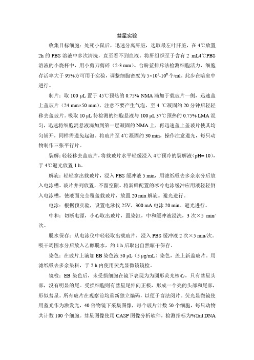
彗星实验收集目标细胞:处死小鼠后,迅速分离肝脏,选取最左叶肝脏,在4℃放置2h的PBS溶液中多次清洗,直至看不到血液。
将肝组织至于含有2 mL4℃PBS 溶液的小烧杯中,用小剪刀剪碎(2-3 mm)。
台盼蓝排斥法检测细胞活力,细胞存活率大于95%方可用于实验,调整细胞密度为5×10⁵-106个/ml。
此步在暗室中进行。
制片:取100 μL置于45℃预热的0.75% NMA滴加于载玻片一侧,迅速盖上盖玻片(24 mm×50 mm),注意不要产生气泡,至4 ℃凝固约20分钟后轻轻移去盖玻片。
吸取10 μL待检测的细胞悬液与100 μL 37℃预热的0.75% LMA混匀,迅速将细胞混悬液滴加到第一层凝固的NMA上,再迅速盖上盖玻片使其均匀铺开,同样需避免起泡。
将玻片至4℃凝固约30 min,操作注意避光,每只动物制作三张平行片。
裂解:轻轻移去盖玻片,将载玻片水平轻缓浸入4℃预冷的裂解液(pH= 10),于4℃避光放置1 h。
解旋:轻轻拿出载玻片,浸入PBS缓冲液5 min,用滤纸吸去多余水分后放入电泳槽。
玻片并列放置,不留空隙。
将新鲜配置的冰冷电泳缓冲应用液轻轻倒入电泳槽,使液面完全覆盖载玻片,放置20 min解旋。
避光进行。
电泳:根据预实验,设置电泳仪25V、300 mA电泳20 min。
避光进行。
中和:切断电源,小心取出玻片,置染缸,中和缓冲液浸洗,3次×5 min/次。
脱水保存:从电泳仪中轻轻取出载玻片,浸入PBS缓冲液2次×5 min/次。
吸干周围水分后放入乙醇脱水,约1 h后取出自然晾干保存。
染色:在玻片上滴加EB染色液50 μL(5 μg/mL)染色,盖上新盖玻片。
用滤纸吸去多余染料,于2 h内使用荧光显微镜镜检。
镜检:EB染色后,未受损细胞在镜下表现为为圆形荧光核心,只有彗星头部,没有明显的尾。
受损细胞则有彗星尾伸向正极,形成一个亮的头部和尾部,形似彗星。
彗星实验(Comet assay),又称单细胞凝胶电泳(Single cell gel electrophoresis,SCGE),各种理化因子作用细胞后引起的DNA链的断裂可用该方法检测[1~3],并在统计学基础上对损伤程度做出评估[4]。
本实验对Singh 等[5]建立的碱性彗星实验的一些步骤作了改良。
用超净工作台上的紫外消毒灯[可发射波长为254 nm的紫外线(Ultraviolet,UV),属于UVC波段范围]作为DNA损伤的诱导因子[7~9],诱导K562细胞DNA损伤,用改良彗星实验检测损伤程度,验证改良的实验系统是否可靠,同时筛选并评价DNA损伤的分析指标。
1 材料与方法1.1 细胞K562细胞,来源于第四军医大学免疫学教研室,37 ℃、5% CO2培养箱中培养,取对数期细胞进行实验。
1.2 紫外线照射装置紫外消毒灯(ZSZ-20型,20 W,天津市紫晶特种光源有限公司)。
1.3 主要试剂和仪器培养基:10%新生牛血清(杭州四季青公司),90%RPMI-1640培养液(Hyclone公司);双抗(青、链霉素,100 UI/ml);TritonX-100(Genview分装);二甲基亚砜、肌苷酸钠(Sigma分装);低熔点琼脂糖(FMC 分装);常熔点琼脂糖(Spanish分装)。
其余生化试剂均为分析纯。
电泳仪:由西北大学物理系提供;电泳槽:DYC33A型(北京市六一仪器厂);显微镜:Leica DM LB 2 (Leica 公司);彗星图象分析软件:CASP软件(casp-1.2.2,/index.php下载);CO2培养箱:BB16HF型(上海力申科学仪器有限公司);环地牌紫外辐照计(北京师范大学光电仪器厂)。
1.4 实验分组及UV处理收集对数生长的K562细胞,台盼蓝染色计数,细胞活力大于95%,用Hank's(pH7.4)调整细胞密度至1×10⁵/ml,接种于塑料培养皿中(ф=35 mm,2 ml/plate),然后进行紫外线照射(0.3 mW/cm2)。
彗星试验一、实验原理正常细胞核是由DNA和蛋白质构成,DNA的双螺旋以核小体的组蛋白为核心形成超螺旋结构,再进一步形成高级结构,形成染色质,染色质的缠绕形成细胞核。
彗星实验又称单细胞凝胶电泳实验,它能有效地检测并定量分析细胞中DNA单,双链缺口损伤的程度。
当各种内源性和外源性DNA损伤因子诱发细胞DNA链断裂时,其超螺旋结构受到破坏,在细胞裂解液作用下,细胞膜、核膜等膜结构受到破坏,细胞内的蛋白质、RNA 以及其他成分均扩散到细胞裂解液中,而核DNA由于分子量太大只能留在原位。
在中性条件下,DNA片段可进入凝胶发生迁移,而在碱性电解质的作用下,DNA发生解螺旋,损伤的DNA断链及片段被释放出来。
由于这些DNA的分子量小且碱变性为单链,所以在电泳过程中带负电荷的DNA会离开核DNA 向正极迁移形成“彗星”状图像,而未受损伤的DNA部分保持球形。
DNA受损越严重,产生的断链和断片越多,长度也越小,在相同的电泳条件下迁移的DNA量就愈多,迁移的距离就愈长。
通过测定DNA迁移部分的光密度或迁移长度就可以测定单个细胞DNA损伤程度,从而确定受试物的作用剂量与DNA损伤效应的关系。
毒鼠强的中毒机制尚不清楚,但有文献显示,毒鼠强的致惊厥作用可能是拮抗γ-氨基丁酸(γ-GABA)的作用,造成脑细胞凋亡。
因此,本实验通过检测不同剂量的毒鼠强对大鼠脑组织细胞DNA的损伤情况,探讨细胞DNA损伤与毒鼠强的毒作用间的关系。
二、试验目的:检测毒鼠强与细胞DNA损伤的关系。
三、试剂与器材(一)试验细胞来源:大鼠脑细胞(大鼠采用SD大鼠)(二)试剂1、毒鼠强纯品(98.5%,公安部物证鉴定中心提供)2、无钙、镁磷酸盐缓冲液(PBS缓冲液)将Nacl4.0g,KH2PO40.1g,KCL0.2g,Na2HPO40.58g,Na2HPO4·12H2O.45g,溶于500ml蒸馏水中。
3、琼脂糖用无钙、镁磷酸盐缓冲液配置4、细胞裂解液Nacl73.05g,Na2EDTA·2H2O18.6g,Tris(三羟甲基氨基甲烷)0.61g,肌氨酸钠5g,双蒸水400ml,先加入一些NaOH在磁力搅拌器上加热溶解。
PH9143|DNA损伤试剂盒(彗星电泳法)DNA Damage Assay kitCatalog No:PH9143Size:☐20T Store at4℃简介DNA在自由基(如·OH)的攻击下容易发生损伤,即脱氧戊糖遭到破环,磷酸二脂键的断裂,碱基的破环或脱落,便可进一步产生单链断裂或双链断裂。
将细胞固定于低熔点琼脂糖中,取少量细胞涂在载玻片上,用碱高盐溶液破坏细胞膜,再用碱溶液使DNA分子解旋。
将载玻片置于电泳液中,在电场的作用下,DNA分子向阳极移动。
如果DNA损伤严重,碎片多,则电泳速度快。
未受损伤的DNA大分子则由于细胞膜的阻隔,滞留在原处。
用PI染色或银染,可观察到DNA受损的细胞形同彗星现象,作定性分析。
也可用相关的软件作定量分析。
组分Components20Assay StorageLysis Bufffer100mL4℃DMSO8mL4℃正常熔点琼脂糖NMA30mg4℃低熔点琼脂糖LMA30mg4℃Propidium Iodide(PL)400ul4℃避光自备试剂和仪器低速离心机、水平电泳仪、荧光显微镜、37℃和45℃恒温水浴箱、载玻片、盖玻片、平皿、微量移液器、1.5ml Microube、0.4mmol/LTris-HCl(pH7.5)缓冲液、PBS使用参考1、细胞用冰冷的PBS洗一次,离心收集,用PBS重悬使其密度为1x106个/mL;2、铺胶:下述各浓度琼脂糖凝胶均用PBS配制:第1层凝胶的制备:将载玻片的磨砂面向上,45℃预热,将预热45℃的100μL的0.5%正常熔点琼脂糖(NMA)铺于载玻片上,盖上干净的盖玻片,再置4℃下10min使NMA凝固。
第2层凝胶的制备:将10μL细胞(约104个)和75μL的0.7%低溶点琼脂糖LMA(在37℃下水浴加热至少20min使之完全溶化)混合均匀。
然后,轻轻揭去盖玻片,迅速将含细胞的LMA滴到第1层琼脂糖上,立即盖上另一干净盖玻片,置4℃10min使第2层LMA凝固。
Comet Assay 彗星分析系统软件功能:彗星试验-单细胞凝胶电泳Single cell gel electrophoresis是一种快捷、高灵敏度的检测DNA受损的方法.Comet Assay 专业彗星试验图像分析软件,提供专业快速的测量方法。
测量指标包括头半径,尾长度,积分光密度,头/尾DNA含量比例,尾矩,Olive 矩等多种参数。
具有自动或手动调节分割彗星头部和尾部功能同时提供对双染色系统的分析系统可广泛用于环境毒理学中对DNA具有损伤作用的分析:如紫外光、电离辐射、氧自由基等因素造成的DNA损伤。
以及细胞凋亡,抗癌药物的药物毒理学研究,遗传毒理研究和肿瘤治疗的跟踪监测等单细胞凝胶电泳(彗星试验):原理、应用和局限性彗星试验:原理、应用和局限性1. 前言过去十年中,彗星试验或单细胞凝胶电泳(SCGE)已成为一个评定DNA损伤的标准方法,广泛应用于遗传毒性试验、人类生物监测和分子流行病学、生态毒理学以及DNA损伤修复的基础研究,且因其简单、灵敏、多功能、快速、经济实用而赢得了广大科研工作者的青睐,和其有关的文献数目逐年上升。
在应用彗星试验时不会过多去考虑它如何运作和它提供何种信息,因为它在论证DNA损伤中的成功已足以证明它的价值。
这一点实在太可惜,因为它不仅能告诉我们细胞内存在的损伤的类型,而且能告诉我们损伤的程度。
尽管彗星试验是检测DN A断裂的基本方法,但由于对特异性核酸内切酶损伤的引入使其可以检测紫外线诱导的嘧啶二聚体,氧化碱基和烷基化损伤。
2.检测原理和检测指标2.1 背景二十世纪七十年代,Peter Cook和其同事们制定了一种用非离子去污剂使细胞溶解来研究核结构的方法。
这种处理除去胞膜、胞质和核质,并使核小体破裂(几乎所有的组蛋白均被浓盐提取),剩下的就是由核基质或RNA、蛋白质组成的支架以及DNA所构成的类核,其DNA的双螺旋以核小体的组蛋白为核心形成负超螺旋结构。
超螺旋的存在使DNA不能自由旋转,Cook 等提出一个模型,DNA间断的附加于基质,有效的形成一系列环状、而不是线性分子。
Lotte Bjergbæk (ed.), DNA Repair Protocols, Methods in Molecular Biology, vol. 920,DOI 10.1007/978-1-61779-998-3_6, © Springer Science+Business Media New York 2012C hapter 6T he Comet Assay: A Sensitive Genotoxicity Testfor the Detection of DNA Damage and RepairGünter S peit and A ndreas R othfussA bstractT he comet assay (single-cell gel electrophoresis) is a simple and sensitive method for studying DNA damage and repair. In this microgel electrophoresis technique, a small number of cells suspended in a thin agarose gel on a microscope slide is lysed, electrophoresed, and stained with a flu orescent DNA-binding dye. Cells with increased DNA damage display increased migration of chromosomal DNA from the nucleus towards the anode, which resembles the shape of a comet. The assay has manifold applications in fundamental research for DNA damage and repair, in genotoxicity testing of novel chemicals and pharmaceuticals, environmental biomonitoring, and human population monitoring. This chapter describes a standard protocol of the alkaline comet assay and points to some useful modi fic ations.K ey words:A lkaline comet assay ,A lkali-labile sites ,B iomonitoring ,C rosslinks ,D NA-strand breaks ,E xcision repair ,G enotoxicity testing ,S ingle-cell gel electrophoresisT he comet assay (single-cell gel electrophoresis) is a useful techniquefor studying DNA damage and repair with manifold applications.In this microgel electrophoresis technique, a small number of cellssuspended in a thin agarose gel on a microscope slide is lysed, electro-phoresed, and stained with a flu orescent DNA-binding dye. Cellswith increased DNA damage display increased migration of chro-mosomal DNA upon electrophoresis from the nucleus towards theanode, which resembles the shape of a comet (Fig. 1). In its alka-line version, which is mainly used, DNA-strand breaks and alkali-labile sites become apparent, and the extent of DNA migrationcorrelates with the amount of DNA damage in the cell. The cometassay combines the simplicity of biochemical techniques for detectingDNA single-strand breaks and/or alkali-labile sites with the singlecell approach typical of cytogenetic assays. The advantages of the 1.I ntroduction7980G. Speit and A. Rothfusscomet assay include its simple and rapid performance, its sensitivity for detecting DNA damage, the analysis of data at the level of the individual cell, the use of extremely small cell samples, and the usability of virtually any eukaryote cell population. Apart from image analysis, which greatly facilitates and enhances the possibili-ties of comet measurements, the cost of performing the assay is extremely low. The comet assay has already been used in many studies to assess DNA damage and repair induced by various agentsin a variety of cells in vitro and in vivo ( 1) . The test has widespread applications in genotoxicity testing in vitro and in vivo ( 2–5 ) , DNA damage and repair studies ( 6) , environmental biomonitoring ( 7,8 ) , human population monitoring ( 9, 10 ) , reproductive toxi-cology ( 11) , and radiobiology ( 12 ) .T he alkaline version (pH >13) of the comet assay introduced by Singh and coworkers ( 13) detects a broad spectrum of DNA lesions, that is, DNA single- and double-strand breaks and alkali-labile sites. Modi fi e d versions of the assay introduced by Olive ( 14) involved lysis in alkaline buffer followed by electrophoresis at either neutral or mild alkaline (pH 12.1) conditions to detect DNA double-strand breaks or single-strand breaks, respectively. However, since the majority of genotoxic agents induce much more single-strand breaks and alkali-labile sites than double-strand breaks, the alkaline version (pH >13) of the comet assay has been identi fi e d to show the highest sensitivity for detecting induced DNA damage and hasbeen recommended for genotoxicity testing (2 ) . Importantimprovements of the test procedure were introduced by Klaude F ig. 1. P hotomicrographs of human lymphocytes in the comet assay. ( a ) Untreated cell (control). ( b ) Cell exhibiting increased DNA migration after mutagen treatment.816 The Comet Assay: A Sensitive Genotoxicity Test for the Detection…and coworkers ( 15 ) . The use of agarose-precoated slides in combination with drying of gels and fi x ation of the comets led to a further simpli fi c ation and a much better handling of the test. The comet assay is especially suited for studies with a high number of samples since it can be performed in a high-throughput fashion and analysis of slides can be automated ( 16– 18 ) .A broad spectrum of DNA-damaging agents increases DNA migration in the comet assay such as ionizing radiation, hydrogenperoxide and other radical-forming chemicals, alkylating agents, polycyclic aromatic hydrocarbons (PHAs) and other adduct-forming chemicals, radiomimetic chemicals, various metals, or UV-irradiation. In principle, the alkaline version of the comet assay detects all kinds of directly induced DNA single-strand breaks and any lesion that can be transformed into a single-strand break under alkaline conditions (i.e., alkali-labile sites).I n addition to directly induced strand breakage, processes which introduce single-strand nicks in the DNA, such as incision during excision repair processes, are also detectable. In some cases (e.g., UV, PAHs) the contribution of excision repair to the induced DNA effects in the comet assay seems to be of major importance( 19) . Some speci fi c classes of DNA base damage can be detected with the comet assay in conjunction with lesion-speci fi c endonu-cleases ( 6) . These enzymes, applied to the slides for a short time after lysis, nick DNA at sites of speci fi c base alterations and the resulting single-strand breaks can be quanti fi e d in the comet assay. Using this modi fi c ation of the comet assay, oxidized DNA bases have been detected with high sensitivity with the help of endonu-clease III, formamidopyrimidine-DNA-glycosylase (FPG) or cell extracts in in vitro tests and in samples obtained from humanstudies( 6, 20– 22 ) .C rosslinks (DNA-DNA or DNA-protein) as induced by chemicals, such as nitrogen mustard, cisplatin, cyclophosphamide or formaldehyde may cause problems in the standard protocol. The induction of crosslinks reduces the ability of the DNA to migrate in the agarose gel by stabilizing chromosomal DNA( 23,24 ) . Crosslinks can be detected by adjusting the duration of unwinding and/or electrophoresis to such an extent that control cells exhibit signi fi c ant DNA migration. A lower extent in DNA migration in treated samples compared to untreated controls wouldthen indicate an induction of crosslinks ( 25) . Another possibility is to induce DNA migration with a second strand-breaking agent (e.g., ionizing radiation, methyl methanesulfonate) after exposure towards the (assumed) crosslinking agent and performing the comet assay immediately thereafter. A crosslinking effect is then determined as reduced migration in comparison with the effect ofthe strand-breaking agent alone ( 23,24 ) . Post-treatment of samples with Proteinase K allows to distinguish betweenDNA-DNA and DNA-protein cross-links ( 24) . 1.1.D etection of DNADamage82G. Speit and A. RothfussA widely used approach for determining DNA repair is to monitor a time-dependent removal of lesions (i.e., the decrease in DNAmigration) after treatment with a DNA-damaging agent. The comet assay has been successfully used to follow the rejoining of strand breaks induced by ionizing radiation or reactive oxygenspecies ( 26,27 ) as well as the repair of various kinds of DNA damage induced by chemical mutagens ( 28,29 ) . A useful extension of repair studies includes the additional use of lesion-speci fi cenzymes (6 ) , or cell extracts ( 30, 31 ) . Thereby, the repair of speci fi c types of DNA lesions can be followed and, due to its high sensitiv-ity, this approach enables the analysis of very low (“physiological”)levels of DNA damage ( 32) . A common alternative approach is the use of repair inhibitors or repair-de fi c ient cells. Incubation of cells with inhibitors of DNA- (repair-) synthesis, such as hydroxyurea, cytosine arabinoside, or aphidicolin leads to an accumulation ofincomplete repair sites as DNA breaks ( 19,33 ) . Mutant cell lines either with a speci fi c defect in a repair pathway (e.g., xeroderma pigmentosum) or with a hypersensitivity towards speci fi c DNA damaging agents (e.g., various mutant rodent cell lines) are well suited to elucidate DNA repair pathways and the biological conse-quences of disturbed DNA repair or to evaluate the repair compe-tence of cells ( 19,34– 36 ) . While the standard version of the comet assay provides information on DNA damage and repair in the whole genome of a cell, the introduction of a combination of the comet assay with fl u orescence in situ hybridization (FISH) addition-ally allows to measure DNA damage and repair in speci fi c genomicregions ( 37–39 ) .T he purpose of this protocol is to provide information on the application of the alkaline comet assay for the investigation of DNA damage and repair in mammalian cells in vitro. For establishing the method, we recommend to start with experiments using blood samples and the induction of DNA damage by a standard mutagen (e.g., methyl methane sulfonate, MMS). The method described here is based on a protocol established by R. Tice according to theoriginal work of Singh et al. (13 ) and includes the modi fi c ations introduced by Klaude and coworkers ( 15) . An outline of the pro-tocol is diagrammed in Fig.2 .1. M icroscope slides (with frosted end).2.C overslips (24 × 60 mm). 3. N ormal melting-point agarose.4. L ow melting point (LMP) agarose.5. H orizontal gel electrophoresis unit.1.2.M easuring DNA Repair 2.M aterials836 The Comet Assay: A Sensitive Genotoxicity Test for the Detection… 6. F luorescence microscope equipped with an excitation fi l ter of515–560 nm and a barrier fi l ter of 590 nm.7.P hosphate-buffered saline (PBS) (without Ca 2+ and Mg 2+ ). 8.L ysing solution (1 L): 2.5 M NaCl, 100 mM EDTA, 10 mM Tris (set pH to 10.0 with ~7 g solid NaOH). Store at room temperature. Final lysing solution (100 mL, made fresh): add 1 mL of Triton X-100 and 10 mL of DMSO to 89 mL of lysingsolution, and then refrigerate (4 °C) for 60 min before use.9.E lectrophoresis buffer: 300 mM NaOH/1 mM EDTA. Prepare from stock solutions of 10 N NaOH (200 g/500 mL distilled H 2 O ), 200 mM EDTA (14.89 g/200 mL of dH 2 O , pH 10.0). Store at room temperature. For 1× buffer, mix 45 mL NaOH,7.5 mL of EDTA, and add water to 1,500 mL (total volume needed depends on gel box capacity). Mix well and store at 4 °C. Make fresh before each run.10.N eutralization buffer: 0.4 M Tris–HCl, pH 7.5. Store at room temperature.11. E thidium bromide staining solution: 10× stock: 200 μ g /mL.Store at room temperature. For 1× stock (20 μ g /mL), mix1 mL with 9 mL dH2 O and fi l ter. C aution : Ethidium bromide is a mutagen. Handle with care.StainingNeutralisationF ig.2.S cheme for the performance of the comet assay.84G. Speit and A. Rothfuss1. C lean slides with ethanol before use. Wear gloves.2. S cratch slides with a diamond pen, drawing a line width-wise approximately 5 mm from the end of the slide to improve the adhesion of the agarose.3. F or the bottom layer, prepare 1.5 % normal melting agarose (300 mg in 20 mL of PBS) and boil until the agarose is com-pletely melted. Dip the slides brie fl y into hot (>60 °C) agarose. The agarose should reach to and cover half of the frosted part of the slide to ensure that the agarose will stick properly. Wipe off the agarose from the bottom side of the slide and lay the slide horizontally. This step has to be performed quickly to ensure a good distribution of agarose. Dry slides overnight at room temperature. Slides can be stored for several weeks.4. P repare 0.5 % LMP agarose (100 mg in 20 mL of PBS). Microwave or heat until near boiling and the agarose dissolves. Place the LMP agarose in a 37 °C water bath to cool.5. A dd 120 μ L of LMP agarose (37 °C) mixed with 5,000–50,000 cells (see Subheading 3.2 ) in ~5–10 μ L (do not use more than 10 μ L ). Add coverslip, and place the slide in a refrigerator for ~2 min (until the agarose layer hardens). Using ~10,000 cells results in ~1 cell per microscope fi e ld (250× magni fi c ation). From this step until the end of electrophoresis, direct light irradiation should be avoided to prevent additional DNA damage.6. G ently slip off the coverslip and slowly lower slide into cold, freshly made lysing solution. Protect from light, and place at 4 °C for a minimum of 1 h. Slides may be stored for extended periods of time in cold lysing solution (but generally not lon-ger than 4 week). If precipitation of the lysing solution is observed, slides should be rinsed carefully with distilled water before electrophoresis. 1. W hole blood: Mix ~5 μ L whole blood with 120 μ L of LMP agarose, and layer onto the slide.2. I solated lymphocytes: Add 4 mL of whole blood to a tube with4 mL prewarmed (37 °C) Ficoll. Centrifuge for 25 min at ~320 × g . Carefully remove the lymphocytes and resuspend them in 8 mL RPMI 1640 medium. Centrifuge again for 10 min at ~180 × g . Remove the supernatant and repeat the washing step. Incubate the cells for 30 min at 37 °C. Centrifugefor 10 min at ~130 × g , discard the supernatant and resuspend the pellet in 375 μ L of RPMI 1640 medium. Count the cells and adjust to 1,500 cells/ μ L . Mix 10 μ L of the suspension with 120 μ L LMP agarose and layer onto the slide.3.M ethods (S eeN otes 1and 2)3.1.P reparationof Slides3.2. P reparation of Cells( S ee N otes 3 and 4)856 The Comet Assay: A Sensitive Genotoxicity Test for the Detection… 3. C ell cultures. (a) M onolayer cultures: Gently trypsinize the cells (for approx. 2 min with 0.15 % trypsin, stop by adding serum or com-plete cell culture medium) to yield approximately 1 × 10 6 cells/mL. Add 10 μ L of cell suspension to 120 μ L LMP agarose, and layer onto the slide. (b) S uspension cultures: Add ~15,000 cells in 10 μ L (or smaller volume) to 120 μ L of LMP agarose and layer onto the slide. 1. A fter at least 1 h at 4 °C, gently remove the slides from the lysing solution. 2. P lace the slides in the gel box near the anode (+) end, posi-tioning them as close together as possible. 3. F ill the buffer reservoirs with electrophoresis buffer (4 °C) until the slides are completely covered (avoid bubbles over the agarose). Perform the electrophoresis in an ice bath (4 °C). 4. L et slides sit in the alkaline buffer for 20–60 min to allow unwinding of the DNA and the expression of alkali-labile damage. For most experiments with cultured cells, 20 min are recommended. 5. T urn on power supply to 25 V (~0.8–1.5 V/cm, depending on gel box size) and adjust current to 300 mA by slowly raising or lowering the buffer level. Depending on the purpose of the study and on the extent of migration in control samples, allow the electrophoresis to run for 20–40 min. For most experi-ments, 20 min is recommended. 6. T urn off the power. Gently lift the slides from the buffer and place on a staining tray. Coat the slides with drops of neutralization buffer, and let sit for at least 5 min. Repeat two more times. 7. D rain the slides, rinse carefully with distilled water, and let them dry (inclined) at room temperature. Slides can be stored for a longer time before staining. To stain, rinse the slides brie fl y in distilled water, add 30 μ L 1× ethidium bromide staining solution, and cover with a coverslip. Antifade can be used to prevent slides from drying or fading out if necessary, i.e., when automated analysis is used. S lides should be stained one by one and evaluated immedi-ately. It is possible to rinse stained (evaluated) slides in distilled water, remove the coverslip, let the slides dry and stain them at a later time point for reevaluation. F or visualization of DNA damage, observations are made of ethid-ium bromide-stained DNA at 250× (or 400×) magni fi c ation usinga fl u orescence microscope. Generally, 100 randomly selected cells3.3.E lectrophoresisand Staining ( S eeN otes 5–7)3.4.E valuation of DNAEffects (S ee N ote 8)86G. Speit and A. Rothfussper sample are analyzed. In principle, evaluation can be done infour different ways:1. I mage analysis systems are used to quantify DNA damage.Parameters such as tail intensity (percentage DNA in the tail),tail moment, and tail length are commonly used. It is impor-tant to note that the some parameters (e.g., tail moment) maybe calculated differently among image analysis systems. For thepurpose of inter-laboratory comparison of DNA damageparameters, tail intensity is probably the most suited.2. A utomated systems have been established, which search for com-ets and carry out the analysis with minimal human intervention.3. C ells are scored visually according to tail size into fiv e classes(from undamaged, 0, to maximally damaged, 4). Thus thetotal score for 100 comets can range from 0 (all undamaged)to 400 (all maximally damaged).4. C ells are analyzed using a calibrated scale in the ocular lens ofthe microscope. For each cell, the image length (diameter of thenucleus plus migrated DNA) is measured in microns, and themean is calculated. Alternatively, the length of the comet (orcomet tail) can be measured on a photomicrograph. These mea-surements are very laborious and may only give limited informa-tion because the tail length saturates at higher levels of damage.F or the statistical analysis of comet assay data, a variety of para-metric and nonparametric statistical methods are used. The mostappropriate means of statistical analysis depends on the kind ofstudy and has to take into account the various sources of assay vari-ability. For a powerful statistical analysis of in vitro test data, appro-priate replication and repeat experiments have to be performed (2,40, 41). For example, the median DNA migration of 50 cells persample and the mean of 2–3 samples per data point may be deter-mined. Also, the mean from repeat experiments can be determined.The use of the median should be preferred over the average since anormal size distribution is usually not observed. Analyses are mainlybased on changes in group mean response but attention shouldalso be paid to the distribution among cells which often providesadditional important information. Recommendations for appropriatestatistical analyses of comet assay data have been published (40, 41).4.N otes1. M any technical variables have been used including the concen-tration and amount of LMP agarose, the composition of thelysing solution and the lysis time, the alkaline unwinding, theelectrophoresis buffer, electrophoretic conditions, and6 The Comet Assay: A Sensitive Genotoxicity Test for the Detection…87DNA-speci fic dyes for staining. Some of these variables mayaffect the sensitivity of the test. To allow for a comparisonobtained in different laboratories and for a critical evaluationof data, it is absolutely necessary to clearly describe the tech-nical details of the method employed.2. T he simplicity of the comet assay combined with the need ofonly low number of cells per sample enables the conduct ofin vitro studies with high ef fic iency. Therefore, the comet assaycan be used in a high-throughput fashion (17). Furthermore,the introduction of automated image analysis systems forcomet assay slides further can speed up test performance (18).3. M any other cell types have been used and it is an advantage ofthe comet assay that virtually any eukaryote cell population isamenable to analysis. The comet assay is particularly suited forthe investigation of organ- or tissue-speci fic genotoxic effectsin vivo (2–5), the only requirement being the preparation ofan intact single cell suspension.4. F or the demonstration of a positive effect, mix 200 μLheparinized whole blood with 50 μL of a 2.5 × 10 −4M methylmethanesulfonate (MMS) solution ( fin al concentration:5 × 10 −5M), incubate for 1 h at 37 °C and then use 10 μL forthe test.5. F or each cell type, the method should be adjusted scienti fic allyto obtain valid and reproducible results. It is important to de fin ethe optimal time for alkaline treatment and electrophoresis. It isrecommended that the conditions must be such that the DNAfrom the control cells exhibit, on the average, some migration.This effect ensures sensitivity and enables an evaluation ofintralaboratory experiment-to-experiment variability (5).6. T he temperature during alkaline treatment and electrophoresissigni fic antly in flu ences the amount of DNA migration (42). It isnecessary to establish stable and reproducible conditions and itmay be useful to use a cooled electrophoresis unit or to place theelectrophoresis unit in a jar fil led with ice or in a cooled room.7. I f speci fic types of base damage should be determined by usinglesion-speci fic endonucleases or cell extracts, the standard proto-col has to be modi fie d in the following way: after at least 1 h at4 °C, gently remove slides from the lysing solution and washthree times in enzyme buffer. Drain slides and cover with 200 μLof either buffer or enzyme in buffer. Seal with a coverslip andincubate for 30 min at 37 °C. Remove coverslip, rinse slides withPBS and place them on the electrophoretic box (6, 20–22).8. I t is strongly recommended to include some measure of cyto-toxicity into any study since increased DNA migration may alsooccur due to non-genotoxic cell killing. However, such an effectmay depend on the cell type used. While no increased DNAmigration had been observed in human leukocytes (43)or cell88G. Speit and A. Rothfusslines such as V79 (43, 44)and L5178Y (45), TK-6 cells showedincreased DNA migration after treatment with non-genotoxiccytotoxins when viability in treated cultures fell below 75 % (46).Therefore, acute cytotoxic effects should be determined byTrypan blue exclusion measurements or flu orochrome-mediatedviability tests. Furthermore, individual dead or dying cells maybe identi fie d by their speci fic microscopical image, i.e., necroticor apoptotic cells may result in comets with small or nonexis-tent head and large, diffuse tails (47). These cells are commonlycalled “hedgehogs,” “ghost cells,” “clouds,” or “non-detect-able cell nuclei (NDCN).” Such cells have been detected aftertreatment with cytotoxic, non-genotoxic agents (44, 46, 48).However, since these microscopic images are also seen aftertreatment with high doses of radiation or high concentrationsof strong mutagens, such comets are not uniquely diagnosticfor apoptosis/necrosis (49, 50). For the evaluation of geno-toxic effects, it is recommended to record these cells but toexclude them from image analysis under the principle that theyrepresent dead cells.R eferences1. D hawan A, Bajpayee M, Parmar D (2009)Comet assay: a reliable tool for the assessment of DNA damage in different models. Cell Biol Toxicol 25:5–322. T ice RR, Agurell E, Anderson D, Burlinson B,Hartmann A, Kobayashi H, Miyamae Y, Rojas E, Ryu J-C, Sasaki YF (2000) The single cell gel/comet assay: guidelines for in vitro and in vivo genetic toxicology testing. Environ Mol Mutagen 35:206–2213. H artmann A, Agurell E, Beevers C, Brendler-Schwaab S, Burlinson B, Clay P, Collins A, Smith A, Speit G, Thybaud V, Tice RR (2003) Recommendations for conducting the in vivo alkaline Comet assay. Mutagenesis 18:45–51 4. B rendler-Schwaab S, Hartmann A, Pfuhler S,Speit G (2005) The in vivo comet assay: use and status in genotoxicity testing. Mutagenesis 20:245–2545. B urlinson B, Tice RR, Speit G, Brendler-Schwaab SY, Collins AR et al (2007) Fourth International Workshop on Genotoxicity Testing: results of the in vivo Comet assay workgroup. Mutat Res 627:31–356. C ollins AR (2009) Investigating oxidativeDNA damage and its repair using the comet assay. Mutat Res 681:24–327. J ah AN (2008) Ecotoxicological applicationsand signi fic ance of the comet assay. Mutagenesis 23:207–2218. F renzilli G, Nigro M, Lyons BP (2009) TheComet assay for the evaluation of genotoxic impact in aquatic environments. Mutat Res 681:80–929. D usinska M, Collins AR (2008) The cometassay in human biomonitoring: gene-environ-ment interactions. Mutagenesis 23:191–205 10. V alverde M, Rojas E (2009) Environmentaland occupational biomonitoring using the Comet assay. Mutat Res 681:93–10911. B aumgartner A, Cemeli E, Anderson D (2009)The comet assay in male reproductive toxicol-ogy. Cell Biol Toxicol 25:81–9812. O live PL (2009) Impact of the comet assay inradiobiology. Mutat Res 681:13–2313. S ingh NP, McCoy MT, Tice RR, Schneider EL(1988) A simple technique for quanti fic ation oflow levels of DNA damage in individual cells.Exp Cell Res 175:184–19114. O live PL (1989) Cell proliferation as a require-ment for development of contact effect in Chinese hamster V79 spheroids. Radiat Res 117:79–9215. K laude M, Erikson S, Nygren J, Ahnström G(1996) The comet assay: mechanisms and tech-nical considerations. Mutat Res 363:89–96 16. F rieauff W, Hartmann A, Suter W (2001)Automatic analysis of slides processed in the comet assay. Mutagenesis 16:133–13789 6 The Comet Assay: A Sensitive Genotoxicity Test for the Detection…17. W ood DK, Weingeist DM, Bhatia SN,Engelward BP (2010) Single cell trapping andDNA damage analysis using microwell arrays.Proc Natl Acad Sci U S A 107:10008–10013 18. S tang A, Brendamour M, Schunck C, Witte I(2010) Automated analysis of DNA damage inthe high-throughput version of the comet assay.Mutat Res 698:1–519. S peit G, Hartmann A (1995) The contributionof excision repair to the DNA-effects seen inthe alkaline single cell gel test (comet assay).Mutagenesis 10:555–55920. C ollins AR, Duthie SJ, Dobson VL (1993)Direct enzymic detection of endogenous oxi-dative base damage in human lymphocyte DNA. Carcinogenesis 14:1733–173521. D ennog C, Hartmann A, Frey G, Speit G(1996) Detection of DNA damage after hyper-baric oxygen (HBO) therapy. Mutagenesis 11:605–60922. S peit G, Schütz P, Bonzheim I, Trenz K,Hoffmann H (2004) Sensitivity of the FPG protein towards alkylation damage in the cometassay. Toxicol Lett 146:151–15823. P fuhler S, Wolf HU (1996) Detection of DNA-crosslinking agents with the alkaline comet assay. Environ Mol Mutagen 27:196–20124. M erk O, Speit G (1999) Detection of cross-links with the comet assay in relationship togenotoxicity and cytotoxicity. Environ Mol Mutagen 33:167–17225. F uscoe JC, Afshari AJ, George MH, DeAngeloAB, Tice RR, Salman T, Allen JW (1996) Invivo genotoxicity of dichloroacetic acid: evalu-ation with the mouse peripheral blood micro-nucleus assay and the single cell gel assay.Environ Mol Mutagen 27:1–926. C ollins AR, Ai-guo A, Duthie SJ (1995) Thekinetics of repair of oxidative DNA damage (strand breaks and oxidised pyrimidines) in human cells. Mutat Res 336:69–7727. C ollins AR, Dusinska M, Horvathova E, MunroE, Savio M, Stetina R (2001) Inter-individualdifferences in repair of DNA base oxidation,measured in vitro with the comet assay.Mutagenesis 16:297–30128. H artmann A, Speit G (1996) The effect ofarsenic and cadmium on the persistence of mutagen-induced DNA lesions in human cells.Environ Mol Mutagen 27:98–10429. H artmann A, Speit G (1995) Genotoxic effectsof chemicals in the single cell gel (SCG) testwith human blood cells in relation to the induc-tion of sister chromatid exchanges (SCE).Mutat Res 346:49–5630. L angie S, Knaapen AD, Brauers K, van BerloD, van Schooten F-J, Godschalk WL (2006)Development and validation of a modi fie dcomet assay to phenotypically assess nucleotideexcision repair. Mutagenesis 21:153–15831. G aivao I, Piasek A, Brevik A, ShaposhnikovS, Collins AR (2009) Comet assay-based methods for measuring DNA repair in vitro;estimates of inter- and intra-individual varia-tion. Cell Biol Toxicol 25:45–5232. C ollins AR, Harrington V, Drew J, Melvin R(2003) Nutritional modulation of DNA repairin a human intervention study. Carcinogenesis24:511–51533. G edik CM, Ewen SWB, Collins AR (1992)Single-cell gel electrophoresis applied to the analysis of UV-C damage and its repair in human cells. Int J Radiat Biol 62:313–320 34. G reen MHL, Lowe JE, Harcourt SA, AkinluyiP, Rowe T, Cole J, Anstey AV, Arlett CF (1992)UV-C sensitivity of unstimulated and stimu-lated human lymphocytes from normal and xeroderma pigmentosum donors in the cometassay: a potential diagnostic technique. MutatRes 273:137–14435. H elbig R, Speit G (1997) DNA effects in repair-de fic ient V79 Chinese hamster cells studied withthe comet assay. Mutat Res 377:279–28636. T ebbs RS, Flannery ML, Meneses JJ, HartmannA, Tucker JD, Thompson LH, Cleaver JE, Pedersen RA (1999) Requirement for the Xrcc1DNA base excision repair gene during early mouse development. Dev Biol 208:513–529 37. S haposhnikov S, Frengen E, Collins AR (2009)Increasing resolution of the comet assay usingflu orescent in situ hybridization—a review.Mutagenesis 24:383–38938. G lei M, Hovhannisyan G, Pool-Zobel BL(2009) Use of Comet-FISH in the study of DNA damage and repair: review. Mutat Res 681:33–4339. S pivak G, Cox RA, Hanawalt PC (2009) Newapplications of the Comet assay: Comet-FISHand transcription-coupled DNA repair. Mutat Res 681:44–5040. L ovell DP, Omori T (2008) Statistical issues inthe use of the comet assay. Mutagenesis 23:171–18241. W iklund SJ, Agurell E (2003) Aspects of designand statistical analysis in the Comet assay.Mutagenesis 18:167–17542. S peit G, Trenz K, Schütz P, Rothfuss A, MerkO (1999) The in flu ence of temperature duringalkaline treatment and electrophoresis on results obtained with the comet assay. ToxicolLett 110:73–7843. H artmann A, Speit G (1997) The contributionof cytotoxicity to effects seen in the alkaline comet assay. Toxicol Lett 90:183–188。