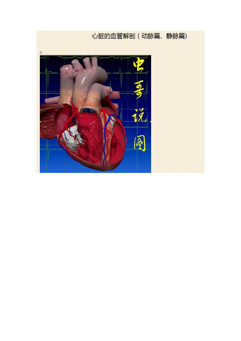
心脏解剖图
- 格式:ppt
- 大小:1.31 MB
- 文档页数:15


心脏3D动画正前方看心脏和室间隔的效果切开右心耳,往外翻,耳朵里面交错纵横的这些结构叫梳状肌(像上图梳子)心耳位于心房上方,血流本来就慢,当房颤时血流速度更慢,再加上左右心耳这些纵横交错的梳状肌结构,所以很容易在这里形成血栓所以房颤超过48小时要抗凝治疗3周才能复律。
(一是防止新的血栓形成,二是要让已经形成血栓机化)。
另外,即使房颤复律为窦性,左右心耳一般不能马上恢复节律收缩,仍处于顿抑状态,叫心房顿抑,所以仍要抗凝4周。
这就是房颤抗凝前3后4 的由来。
下面我们把右心室游离壁也切掉,把三尖瓣、乳头肌透明化,就暴露出清爽的右房右室内部结构看这里,这是一个倒放的漏斗,在右心室这个漏斗负责收集右心室流出道的血液汇向肺动脉,由于这个漏斗形似圆锥体,故又叫动脉圆锥部。
何谓流出道、流入道?看模拟一个血流:蓝色血流代表流入道它位置较低,绿色代表流出道它位置较高右心室有个Y字型扁平肌肉隆起非常醒目,分三个部分c为隔带,a为隔带前脚,b 为隔带后脚,隔带向下延伸为隔缘肉柱,是心脏最大一个肉柱,我们所说的右束支就走在里面,隔缘肉柱的末端和前乳头肌连在一起。
室上嵴和界脊:把心脏抽象的切成两部分室间隔分法一:经典的解剖学划分方法:以这个丫字作为天然的势力划分范围,分为:窦部——(窦部上面有内侧乳头肌)、小梁化部——(小梁化部上面有肌小梁和肉柱)、漏斗部——(漏斗部上有面圆锥乳头肌供三尖瓣隔瓣附着)。
分法二:经典病理学分法分膜部、肌部。
没有肌肉就叫膜部,有肌肉就是肌部,膜部势力范围很小,是心室肌最薄的地方,是室间隔缺损最好发的地方。
分法三:经典供血分法。
把室间隔分为前缘和后缘,也就是我们常说的前室间隔和后室间隔,有时又叫它室间隔上部和室间隔下部,前室间隔由前降支分出的穿隔支供血,后室间隔由后降支供血,所以这种分法也适用于心梗定位。
分法四:分室间隔上部、室间隔中部、室间隔下部,就是把上面两分法改为3分法。
分法五:超声切面分法:分上段、中段、下段,就是在左室长轴切面切断,切线刚好经过间隔上部A点、中部B点、下部C点,它是一个平面的概念。

心脏的解剖结构及生理(含彩图)-心脏位置图XXX of the HeartThe heart is a pump that propels blood flow in the entire circulatory system。
It is located in the mediastinum。
behind the sternum and the second to sixth rib cartilage。
and in front of the XXX。
with two-thirds of the heart on the left side of the chest cavity and one-third on the right side.The heart is composed of cardiac muscle cells。
valves。
and four chambers。
The left ventricle mainly forms the heart apex。
while the heart base consists of the great vessels and veins。
The four chambers of the heart are the left atrium。
left ventricle。
right atrium。
and right ventricle。
The tricuspid valve is een the right atrium and ventricle。
while the mitral valve is een the left atrium and XXX is een the right XXX。
and the aortic valve is een the left XXX ventricular septa separate the left and right sides of the heart。