胰腺囊性病变
- 格式:ppt
- 大小:7.39 MB
- 文档页数:37
胰腺囊性病变的病理学基础及影像学诊断周诚;杨正汉;谭晔【期刊名称】《中国医学计算机成像杂志》【年(卷),期】2002(008)005【摘要】胰腺和胰周囊性病变可包括从良性到恶性的不同疾病.由于其病理基础不同,影像学表现上存在一定的差异.本文从病理基础和影像学表现两个方面讨论常见的胰腺囊性病变.先天性囊肿为胰腺导管发育异常所致,囊肿内有完整内皮,影像学上符合薄壁的单纯囊肿表现.潴留性囊肿是由于胰腺导管阻塞所致,主要见于肿瘤患者,在潴留囊肿前方可见实性肿块.外伤性假囊肿主要内容物为血肿,胰腺炎假囊肿内为坏死组织、炎性渗出、血液和胰酶,胰周结核主要是淋巴结的干酪性坏死.胰腺癌的囊性变为肿瘤中心的坏死、液化.浆液性囊腺瘤为良性过程,以多发小囊为主要类型.黏液性囊腺瘤为潜在恶性的病变,分叶状囊实性肿块为其特点,在影像学上与黏液性囊腺癌不易鉴别.假囊肿特别是胰腺炎假囊肿和囊腺瘤之间的鉴别是最重要的,重症胰腺炎的临床病史有助于鉴别.【总页数】6页(P348-353)【作者】周诚;杨正汉;谭晔【作者单位】100730,卫生部北京医院,北京大学第五临床医学院放射科;100730,卫生部北京医院,北京大学第五临床医学院放射科;100730,卫生部北京医院,北京大学第五临床医学院放射科【正文语种】中文【中图分类】R36【相关文献】1.胰腺囊性病变的影像学诊断及鉴别诊断 [J], 余鸿鸣;张庆雷;朱斌;李丹燕2.胰腺囊性病变的影像学诊断现状 [J], 鲍润贤;孙鼎元3.胰腺假性囊肿的病理学基础及影像学诊断 [J], 段舒怀;胡治红4.胰腺假性囊肿的病理学基础及影像学诊断 [J], 段舒怀胡治红5.胰腺囊性病变的影像学诊断及鉴别诊断 [J], 李相宁因版权原因,仅展示原文概要,查看原文内容请购买。
Publicationdate November 3, 2012Cystic pancreatic lesions are increasingly identified due to the widespread use of CT and.Most of these cysts are incidental findings and are benign or low-grade neoplasms.The characterization and management of these cysts is a dilemma since there is a significant overlap in the morphology of benign and premalignant lesions.MR is the imaging study of choice to both characterize and follow these pancreatic cysts.In many cases these lesions remain undetermined and guidelines are needed for follow up and management.by Marc Engelbrecht, Jennifer Bradshaw and Robin Smithuis发表于2012年11月3日胰腺囊性病变广泛应用于CT和MR大多数囊性病变是被偶然发现的,多为良性或低度恶性肿瘤。
由于在良性和癌前病变的形态极其相似,因此对于这些囊性病变的处理处于两难的困境。
选择和遵循这些胰腺囊肿的影像学研究。
在许多情况下,一些病变仍然未制定相关指南来进行跟踪和处理。
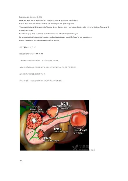
查看原图分类胰腺囊性病变可分为以下几组:假性囊肿常见的囊性肿瘤:IPMN -导管内乳头状黏液性肿瘤SCN-浆液性囊腺瘤MCN -粘液性囊腺瘤少见的囊性肿瘤:SPEN (实性假乳头状上皮性肿瘤)肿瘤的囊性变:神经内分泌癌相关疾病:•胰腺炎•囊腺瘤•肿瘤系统的方法当检测出胰腺有囊性病变的时候,第一步首先怀疑是否为假性囊肿或囊性肿瘤。


国际医学放射学杂志IntJMedRadiol2020Nov 鸦43穴6雪胰腺囊性病变的影像表现与临床特点(下)徐建国1唐光健2彭泰松1赵丽丽1于萍1任龙飞1许志高1【摘要】胰腺囊性病变(PCL)是胰腺上皮和间质组织发生囊腔病变的一大类疾病,以胰腺内囊性包块为主要特征,具有不同的病因、临床和组织病理学特点。
本文的前两部分介绍了胰腺炎症相关囊性病变(包括胰腺假性囊肿与胰腺包裹性坏死)与胰腺真性囊肿(包括孤立性胰腺上皮囊肿、von Hippel-Lindau 病、多囊肾和囊性纤维化)以及常见的胰腺浆液性囊腺瘤、胰腺黏液性囊腺瘤和胰腺实性假乳头状瘤的影像表现与临床特点。
本篇为最后一部分,就常见的胰腺导管内乳头状黏液瘤与胰腺少见囊性肿瘤予以介绍和分析,以期为临床诊断与治疗提供重要依据。
【关键词】胰腺囊性病变;体层摄影术,X 线计算机;磁共振成像;导管内乳头状黏液瘤;囊性肿瘤中图分类号:R576;R445.2;R445.3文献标志码:APancreatic cystic lesions:imaging findings and clinical features (Part 3)XU Jianguo 1,TANG Guangjian 2,PENGTaisong 1,ZHAO Lili 1,YU Ping 1,REN Longfei 1,XU Zhigao 1.1Department of Radiology,Third People ’s Hospital of Datong City,Datong 037008,China;2Department of Radiology,Peking University First Hospital【Abstract 】Pancreatic cystic lesions (PCLs)are a broad spectrum of diseases with cystic lesions in pancreaticepithelial and interstitial tissues,characterized by cystic mass,with various etiology,clinical and histopathological characteristics.The first two parts of the article introduces the imaging manifestations and clinical features of 1)pancreatitis related cystic lesionincluding pancreatic pseudocyst and pancreatic encapsulated necrosis,2)pancreatic true cysts including isolated pancreatic epithelial cyst,von Hippel Lindau disease,polycystic kidney and cystic fibrosis,and3)the common cystic tumors of the pancreas including serous cystadenoma,mucinous cystadenoma and solid pseudopapilloma of the pancreas.In the last part of this issue,we introduce another common cystic tumor of pancreas (intraductal papillary mucinous neoplasm of the pancreas)and rare cystic tumors of the pancreas,in order to provide an important basis for clinical diagnosis and treatment of pancreatic cystic lesions.【Keywords 】Cystic disease of pancreas;Tomography,X -ray computed;Magnetic resonance imaging;Intraductalpapillary mucinous neoplasm;Cystic tumorIntJMedRadiol,2020,43(6):716-720作者单位:1大同市第三人民医院医学影像科,大同037008;2北京大学第一医院放射科通信作者:唐光健,E-mail:***************.com DOI:10.19300/j.2020.J18513图文讲座3.4胰腺导管内乳头状黏液瘤胰腺导管内乳头状黏液瘤(intraductal papillary mucinous neoplasm,IPMN )实际上并不是囊性肿瘤,由于肿瘤分泌黏液,引起胰腺导管扩张,大体病理与影像表现为伴有囊的病变,故在胰腺囊性病变内一并讨论。