Positive and negative transcriptional regulation of aromatase
- 格式:pdf
- 大小:174.13 KB
- 文档页数:7

生物钟如何决定睡眠的时间?东南大学韩俊海课题组发现了一个关键分子调控黎明时的觉醒订阅号APExBIO研究意义睡眠的时间受生物钟(circadian clock)的严格控制,但是,生物钟如何决定睡眠的时间在很大程度上还不清楚。
在脊椎动物和无脊椎动物中,睡眠和觉醒的状态由起搏器神经元(pacemaker neurons)的电活动调节,而抑制性GABA能输入负责起搏器神经元的昼夜节律调节和抑制。
之前有研究显示生物钟输出分子WAKE在黄昏时上调GABA A受体水平以促进睡眠,但是黎明时的觉醒是如何发生的呢?来自南京东南大学发育与疾病相关基因教育部重点实验室的韩俊海教授课题组以果蝇作为研究模型,发现了一个关键分子——Fbxl4,其表达可以节律性地降解GABA A受体来增加起搏器神经元的活动,以促进黎明时的觉醒。
研究结果在线发表在Cell子刊《Current Biology》。
睡眠是从蠕虫到人类进化上保守且必要的行为。
睡眠由两个独立的过程严格控制:一是生物钟(circadian clock)决定睡眠时间,二是稳态机制控制睡眠的量和深度。
生物钟包含一个负转录反馈回路(negative transcriptional feedback loop),使大多数动物的生理和行为与日常的环境振荡(environmental oscillations)同步。
睡眠的时间可以被认为是生物钟的输出。
有几种分子,如褪黑激素,前动力蛋白2和WAKE已被确定为调节睡眠时间的生物钟输出分子。
生物钟也调节起搏器神经元(pacemaker neurons)的电活动,其调节睡眠和觉醒(wakefulness)状态。
在脊椎动物和无脊椎动物中,生物钟驱动钠和钾电导的反相振荡,以控制起搏器神经元中膜电位的每日循环。
相反,起搏器神经元的电活动被抑制性GABA能输入(inhibitory GABAergic inputs)抑制,GABA能输入是哺乳动物中枢神经系统(CNS)和果蝇唤醒促进大腹侧外侧神经元(lLNvs)中的抑制性信号传导的主要来源。

水产专业英语词汇水产业:Fishery淡水养殖:Fresh water aquaculture 海水养殖:Mariculture 南美白对虾:Pacific white shrimp 斑节对虾:tiger shrimp 拟穴青蟹:mud crab淡水小龙虾:cray fish蛤:clam螺丝:snail牡蛎:oyster双壳类:bivalve腹足类:gastropod海星:sea star海胆:sea urchin海带:kelp鲈鱼:bass加州鲈:Large mouth bass大菱鲆:founder石斑鱼:grouper鲍鱼:abalone鲤鱼:carp草鱼:grass carp青鱼:sapphire鳙鱼:bighead carp鲢鱼:chub鲶鱼:catfish罗非鱼:tilapia鲟鱼:sturgen淡水龟:tortoise海龟:turtle鳖:softshell turtle浮游生物:plankton浮游植物:phytoplankton浮游动物:zooplankton硅藻:diatom甲藻:Dinoflagellate轮虫:rotifer丰年虫:artemia桡足类:copepod枝角类:Cladoceran无节幼体:Nauplii细菌:bacterium弧菌:vibrosis真菌:fungi病毒:virus寄生虫:parasite食物转换率:FCR(feed conversion ratio)维生素:vitamin 蛋白质:protein脂类:lipidAquaculture水产养殖Genetics 遗传学Genomics 基因组学Teleosts 硬骨鱼类Shellfish 贝类Shrimp 虾Physiologic 生理的Mechanisms 机制Maturity 成熟Bodyweight 体重Ovary 卵巢Testis 精巢Dimorphism 二态性Hormone 激素disulfide bonds 二硫键mediate 介导receptor 受体reproduction 繁殖immunity 免疫endocrine 内分泌的promotion 促进clone 克隆signal transduction 信号转导Transformation 转化Transfection 转染Infection 感Characterize 鉴定Gene 基因Pool 池塘Photoperiod 光周期water temperature 水温reproductive manipulation 繁殖调控egg 卵sperm 精子semen 精液female 雌鱼male 雄fertilization 受精volume 体积hatch 孵化gentle aeration 微充气larvae 仔鱼triplicate 重复3次dose 剂量Gonad 性腺RNA extraction RNA提取Ethanol 酒精Sex 性别Blood 血Brain 脑Eye 眼Gill 鳃Kidney 肾Intestine 肠Liver 肝Muscle 肌肉Pituitary 垂体Skin 皮肤Spleen 脾脏Stomach 胃Microscope 显微镜Aliquot 份Activate 激活Digestion 消化Protocol 实验计划Primer 引物Polymerase 聚合酶Reverse transcription 反转录Sequence 序列Fragment 片段Amplification 扩增Template 模板Cycle 循环Intron 内含子Exon 外显子Vertebrate 脊椎动物Kit 试剂盒Instruction 用法说明Agarose gel 琼脂糖胶Band 带Propagate 繁殖amino acid 氨基酸alignment 比对phylogenetic trees进化树marker 标记Quantitative 定量的Stage时期Procedure 程序Software 软件Concentration 浓度Motility 活力encoding region 编码区signal 信号residue 残基flounder 牙鲆common carp 鲤鱼grass carp 草鱼tissue 组织population 群体sex ratio 性比negative 负的positive 正的control 对照grow 生长production 生产molecular 分子的stock 群体sex determination mechanisms性别决定机制superfemale 超雌个体progeny 后代feeding 喂养manipulation 管理environment 环境study 研究species 种类catfish 鲶鱼microsatellite 微卫星elucidate 阐明further investigations 进一步的研究significantly 显著地spawn 产卵expression 表达up-regulate 上调fold 倍gynogenesis 雌核发育homozygosity 纯合性meiotic减数的irradiate灭活heterologous 异源的cold shock冷休克diploid 二倍体的morphology形态homogamete同配marine 海洋flatfish比目鱼coastal areas沿海地区adult成体individual个体investment投资chromosome染色体approach方法monosexual单性的second meiotic division第二次减数分裂induce诱导mitogynogenesis卵裂雌核发育the first mitotic division第一有丝分裂homologous同源的development发育paternal父母的offspring子代Nowadays当前Cryopreserve冷冻Technique技术Practical实际的Feasible可行的Neomal伪雄鱼Theoretical理论的genetic diversity遗传多样性sex control性别控制lab实验室sex differentiation性分化heterozygosity杂合度recombination重组locus位点Additionally另外地Increment增量cultivated population养殖群体base pairs碱基对feasibility可行性parameter参数survival rate成活率gynogenetic population雌核发育群体thaw解冻fisheries水产trial实验experiment实验storage保存liquid nitrogen液氮waterbath水浴appropriate适宜的condition条件haploid单倍体diploid二倍体hybrid杂交子treatment处理incubator培养容器percentage百分率embryo胚胎fertilization rate受精率survival rate成活率data数据batch批randomly随机地sample取样classic经典的mixture混合物enzyme酶Ligation连接Restriction限制性的Dilute稀释pre-amplification预扩automatic自动的denaturation变性anneal退火elongation延伸polyacrylamide gel聚丙烯酰胺胶silver staining银染molecular weight分子量allele等位基因index指数Initiation起始Duration持续Putative推定的Polymorphism多态性linkage disequilibrium连锁不平衡artificial人工的diploidization二倍化optimization优化proportion比例sex reversal性逆转crossover交换incorporation整合eel鳗鲡summary摘要abstract摘要discussion 讨论references参考文献material and method材料和方法result结果title题目manuscript草稿revise修改review评审funding资助genetic sexing遗传性别鉴定Acknowledgments致谢Mechanism机制Article文章Foundation基础Methylation甲基化Aromatase芳香化酶Promoter启动子Androgen雄激素Estrogen雌激素Masculinization雄性化inverse relationship负相关suppress抑制genotypic sex determination遗传性别决定temperature-dependent sex determination温度性别决定thermosensitive period温度敏感期non-mammalian vertebrates非哺乳类脊椎动物sex steroid性类固醇激素hypothesize假定pattern模式pathway通路epigenetic表观遗传的nuclear细胞核transcription factors转录因子biosynthesis生物合成mutual共同的dinucleotides二核苷酸transcription start site转录起始位点overall总体上frequencies频率phenotype表型genotype基因型housekeeping gene持家基因exogenous外源的temperature-dependent温度依赖的sex-dependent性别依赖的rear养殖culture养殖stimulate刺激block阻止bioinformatic生物信息的putative推定的co-transfection共转染transcriptional activators转录激活因子luciferase reporter assay荧光素酶报告基因检测opening reading frame开放阅读框similarity相似度development发育demethylation去甲基化process过程sex differentiation性分化silence沉默DNAmethyltransferases DNA甲基转移酶Identify鉴定、发现Influence影响Consistent一致的Nutrition营养Disease疾病Ingredient成分body size体长polygenic多基因的monosex populations单性群体determine确定match匹配consequence结果migration迁移discern辨别hypermethylate超甲基化phenomenon现象conserved保守的threshold阈值scheme计划Fertlizating rate and survial rate are the important parameters of revaluatingThe quality of eggs受精率和成活率是评价卵的重要参数For improving the culturing production ,feeding and managent is very important饲养的管理对于提高养殖产量非常重要的The first step of breeding project design is the selection of the base population基础群的选择是育种计划设计的第一步The tilapia have a unique breeding characteristics: Male territory estoblisher and female mouth brooders罗非鱼具有奇特的繁殖习性:雄性领地占领者和雌性口腔孵化者The half-smooth tongue sole showed significand growth difference半滑舌鳎两性之间生长差异显著I have ten pools providing for culturing tilapia我有十个池塘养殖罗非鱼Reproductive manipulation is of importance for gonad maturity繁殖调控对鱼类的性腺成熟时非常重要的The hatching stage of sole eggs is 3 days舌鳎卵需要三天才能孵化Nowadays there are a lot of of nile tilapia femilies all over the world目前世界上有很多罗非鱼的品系。

第一节概述围绕基因表达过程中发生的各种各样的调节方式都通称为基因表达调控(gene regulation或gene control)。
几个基本概念1、顺式作用元件和反式作用因子:基因活性的调控主要通过反式作用因子(通常是蛋白质)与顺式作用元件(通常在DNA 上)相互作用而实现。
顺式作用元件是指对基因表达有调节活性的DNA序列,其活性只影响与其自身同处在一个DNA分子上的基因;同时,这种DNA序列通常不编码蛋白质,多位于基因旁侧或内含子中,如启动子和终止子,都是典型的顺式作用元件。
反式作用因子是能调节与它们接触的基因的表达的各种扩散分子(通常是蛋白质),如RNA聚合酶、转录因子。
2、结构基因和调节基因:结构基因(structural gene)是编码蛋白质或RNA的基因。
细菌的结构基因一般成簇排列,多个结构基因受单一启动子共同控制,使整套基因或都表达或都不表达。
调节基因(regulator gene)是编码合成那些参与其他基因表达调控的RNA或蛋白质的特异DNA 序列。
调节基因编码的调节物质通过与DNA上的特定位点结合控制转录是调控的关键。
比如:它能使结构基因在需要某种酶时就合成某种酶,不需要时,则停止合成,它对不同染色体上的结构基因有调节作用。
调节物与DNA特定位点的相互作用能以正调控的方式(启动或增强基因表达活性)调节靶基因,也能以负调控的方式(关闭或降低基因表达活性)调节靶基因。
DNA位点通常位于受调节基因的上游,但也有例外.3、操纵基因和阻遏蛋白操纵基因(operator)是操纵子中的控制基因,在操纵子上一般与启动子相邻,通常处于开放状态,使RNA聚合酶能够通过并作用于启动子启动转录。
但当它与调节基因所编码的阻遏蛋白结合时,就从开放状态逐渐转变为关闭状态,使转录过程不能发生。
阻遏蛋白(aporepressor)是负调控系统中由调节基因编码的调节蛋白,它本身或与辅阻遏物(corepressor)一起结合于操纵基因,阻遏操纵子结构基因的转录。


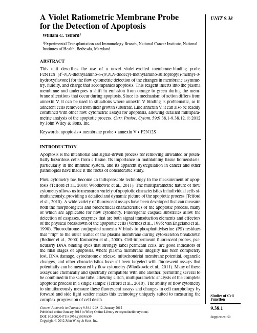
UNIT9.38 A Violet Ratiometric Membrane Probefor the Detection of ApoptosisWilliam G.Telford11Experimental Transplantation and Immunology Branch,National Cancer Institute,NationalInstitutes of Health,Bethesda,MarylandABSTRACTThis unit describes the use of a novel violet-excited membrane-binding probeF2N12S[4 -N,N-diethylamino-6-(N,N,N-dodecyl-methylamino-sulfopropyl)-methyl-3-hydroxyflavone]for theflow cytometric detection of the changes in membrane asymme-try,fluidity,and charge that accompanies apoptosis.This reagent inserts into the plasmamembrane and undergoes a shift in emission from orange to green during the mem-brane alterations that occur during apoptosis.Since its mechanism of action differs fromannexin V,it can be used in situations where annexin V binding is problematic,as inadherent cells removed from their growth substrate.Like annexin V,it can also be readilycombined with otherflow cytometric assays for apoptosis,allowing detailed multipara-metric analysis of the apoptotic process.Curr.Protoc.Cytom.59:9.38.1-9.38.12.C 2012by John Wiley&Sons,Inc.Keywords:apoptosis r membrane probe r annexin V r F2N12SINTRODUCTIONApoptosis is the intentional and signal-driven process for removing unwanted or poten-tially hazardous cells from a tissue.Its importance in maintaining tissue homeostasis,particularly in the immune system,and its apparent dysregulation in cancer and otherpathologies have made it the focus of considerable study.Flow cytometry has become an indispensable technology in the measurement of apop-tosis(Telford et al.,2010;Wlodkowic et al.,2011).The multiparametric nature offlowcytometry allows us to measure a variety of apoptotic characteristics in individual cells si-multaneously,providing a detailed and dynamic picture of the apoptotic process(Telfordet al.,2010).A wide variety offluorescent assays have been developed that can measureboth the morphological and biochemical characteristics of the apoptotic process,manyof which are applicable forflow cytometry.Fluorogenic caspase substrates allow thedetection of caspases,enzymes that are both signal transduction elements and effectorsof the physical breakdown of the apoptotic cells(Vermes et al.,1995;van Engeland et al.,1998).Fluorochrome-conjugated annexin V binds to phosphatidylserine(PS)residuesthat“flip”to the outer leaflet of the plasma membrane during cytoskeleton breakdown(Bedner et al.,2000;Komoriya et al.,2000).Cell-impermeantfluorescent probes,par-ticularly DNA binding dyes that strongly label permeant cells,are good indicators ofthefinal stages of apoptosis,where plasma membrane integrity has been completelylost.DNA damage,cytochrome c release,mitochondrial membrane potential,organellechanges,and other characteristics have all been targeted withfluorescent assays thatpotentially can be measured byflow cytometry(Wlodkowic et al.,2011).Many of theseassays are chemically and spectrally compatible with one another,permitting several tobe combined in the same tube,allowing a rich,multiparametric analysis of the completeapoptotic process in a single sample(Telford et al.,2010).The ability offlow cytometryto simultaneously measure thesefluorescent assays and changes in cell morphology byforward and side light scatter makes this technology uniquely suited to measuring the complex progression of cell death.Current Protocols in Cytometry9.38.1-9.38.12,January2012Published online January2012in Wiley Online Library().DOI:10.1002/0471142956.cy0938s59Copyright C 2012John Wiley&Sons,Inc.Studies of Cell Function9.38.1 Supplement59AVioletRatiometric Membrane Probefor the Detectionof Apoptosis 9.38.2Supplement 59Current Protocols in CytometryCell membrane alterations and eventual breakdown are key characteristics used in the routine measurement of cell death.Alterations in membrane symmetry and the “flipping”of PS residues to the outer leaflet of the plasma membrane are relatively “early”mor-phological changes in apoptosis,occurring prior to complete breakdown of membrane permeability and cell structure.These alterations are believed to serve as a recognition signal for phagocytic cells tasked to remove apoptotic cells,and thus occur prior to the most terminal stages of complete membrane breakdown (Bedner et al.,2000,Komoriya et al.,2000).As a result,they have proven useful targets for measuring the onset of apop-tosis.The PS binding protein annexin V has proven to be a tremendously useful reagent for measuring this process;it can be conjugated to a variety of fluorescent probes,and readily labels apoptotic cells from a variety of species and tissues (Bedner et al.,2000;Komoriya et al.,2000).The measurement of membrane asymmetry based on specific binding to a single lipid does impose some technique restrictions,however.Analysis of adherent cells by flow cytometry requires their removal from their growth substrate,usually by enzymatic means with trypsin,by chelating divalent cations in the cell solu-tion with EDTA,or by mechanical dissociation (i.e.,scraping).All of these operations can have a detrimental effect on PS asymmetry in the plasma membrane.Trypsin and mechanical scraping can cause PS “flipping”even in healthy cells.Annexin V required the presence of calcium and magnesium to bind PS residues,making EDTA usage prob-lematic.Some cell types,including megakaryocytes,some myeloid cell lineages,and platelets normally possess high levels of PS on their cell surfaces as well.To address these limitations,other fluorescent techniques for measuring membrane al-terations in cell death have been pursued.One such avenue has been the development of fluorescent “designer”molecules that can intercalate into the plasma membrane,and demonstrate altered spectral characteristics based on membrane symmetry.An example of such a membrane binding probe,designated F2N12S (4 -N ,N -diethylamino-6-(N ,N ,N -dodecyl-methylamino-sulfopropyl)-methyl-3-hydroxyflavone),readily binds to plasma membranes (Shynkar et al.,2007;Das et al.,2008;Klymchenko et al.,2009;Kułakowska et al.,2010;Oncul et al.,2010;Roche et al.,2010;Hope-Roberts et al.,2011).When excited with a violet laser diode now found on many flow cytometers,it undergoes a spectral shift from orange to green fluorescence as cells transition from viable to apoptotic (Shynkar et al.,2007;Oncul et al.,2010;Hope-Roberts et al.,2011).This shift correlates roughly with the appearance of annexin V binding in apoptotic cells,suggesting that this spectral shift does indeed correlate with loss of membrane symmetry.Like annexin V ,F2N12S can detect alterations in membrane structure that occur in intermediate stage cell death.It does not specifically measure phosphatidylserine orientation,however,but rather undergoes a spectral shift based on changes in membrane symmetry,fluidity,and charge that occur during cell death and other processes (Das et al.,2008;Klymchenko et al.,2009;Kułakowska et al.,2010;Roche et al.,2010).Therefore,it can be used with adherent cells following trypsinization or mechanical removal from a growth substrate.Like annexin V ,this assay can be readily combined with other apoptosis assays utilizing unfixed cells,including annexin V itself.BASICPROTOCOL 1LABELING WITH F2N12S AND DNA DYE ALONE Both suspension and adherent cells can be analyzed using F2N12S.Unlike many live cell assays for apoptosis,the presence of protein during the F2N12S labeling can reduce labeling level,adversely affecting the assay.Protocols can be modified depending on differences in cell type.Studies of Cell Function 9.38.3Current Protocols in Cytometry Supplement 59F2N12S is minimally excited by 488-nm or red lasers,and can therefore be readilycombined with one of many DNA dyes for multiparametric analysis of apoptosis with nocompensation.F2N12S should be minimally combined with one DNA dye to distinguishearly-and late-stage apoptotic cells,and to detect artifacts.Choice of dye will be dictated both by the spectral properties of F2N12S and otherprobes to be included,such as annexin V conjugates and caspase substrates.As a minimalprocedure,F2N12S can be combined with a single DNA-binding dye,such as propidiumiodide (PI)and 7-aminoactinomycin D (7-AAD)dyes;however,other DNA dyes can beused.For example,red-excited DNA dyes with varying degrees of cell permeability canalso be used with F2N12S.SYTOX Red and TO-PRO-3(Invitrogen Life Technologies)can both be excited with red HeNe or diode lasers and are compatible with F2N12S.UV-or violet-excited DNA dyes like SYTOX Blue,DAPI,and the Hoechst dyes cannot beused,as their spectral emission will overlap with F2N12S.F2N12S is also compatible with most of the fluorogenic enzyme substrates used to detectcaspase activity in live cells.The so-called FLICA substrates (available from a varietyof sources)are all based on fluorochromes similar to fluorescein,and can be co-labeledwith F2N12S.F2N12S labeling requires unfixed cells,and is therefore not compatiblewith antibodies against cleaved caspases that requires cell permeabilization.F2N12S canalso be combined with mitochondrial membrane potential probes,including DiIC1(5)and rhodamine 123.As above,it cannot use used with TUNEL or other assays requiringcell fixation and permeabilization.Positive and negative controls for apoptosis should be present in the assay both atinstrument set up and again to verify the reagent function.A negative control is usuallyuntreated cells (although overgrown or otherwise stressed cell lines can have an apoptoticcomponent even in the absence of a specific inducer).A positive control can be anexperimental sample,or can be induced using a drug.Staurosporin,actinomycin D,andcamptothecin are drugs that disrupt calcium flux,transcription,and type II topoisomeraseactivity,respectively,and are examples of apoptosis inducers that work over a wide varietyof cell types.They are frequently used to produce a positive control to verify the operationof apoptosis assays.This unit describes the use of this reagent,and modifications to itsuse allowing it to be combined with other fluorescent apoptosis reagents.MaterialsF2N12S reagent (Invitrogen Life Technologies,cat.no.A35137):sold as a solutionand is stored in anhydrous conditions at minus 20◦C;it can withstand multiplefreeze/thaw cycles without harmDNA dyes including:Propidium iodide (PI;see recipe)7-aminoactinomycin D (7-AAD;see recipe)Cells (this reagent has been tested for a variety of cell lines and primary cell types;EL4mouse lymphoma cells have been used in the examples shown)Standard Hank’s balanced salt solution (HBSS)containing calcium and magnesiumand no phenol red makes a good incubation buffer for F2N12S (this buffershould not be confused with wash buffer below,which can be used for stepspreceding F2N12S labeling)Fluorescent alignment verification microsphere standard,to verify violet laseralignment:Single intensity Rainbow Ultra beads (Spherotech)or AlignFlow UVbeads (Invitrogen)are well-excited with violet lasers and are good for alignmentverification;rainbow 8-peak (Spherotech)or InSpeck Blue (Invitrogen)multi-intensity bead cocktails are also good for monitoring laser alignment,since degradation in low-signal resolution can be observedA VioletRatiometric Membrane Probefor the Detection of Apoptosis 9.38.4Supplement 59Current Protocols in CytometryFlow cytometer equipped with a violet laser diode or other violet laser (e.g.,F2N12S can be readily carried out on BD Bioscience LSR II,LSR Fortessa,FACSVerse,Influx,and FACSAria series cytometers,Beckman-Coulter Gallios and Astrios,Stratedigm S series,Miltenyi Biotec MACSQuant,Partec CyFlow series,the Life Technologies/Applied Biosystems Attune,and Cytek Development modified BD instruments with a violet laser)1.Thaw the F2N12S reagent (supplied at 100μl in DMSO)prior to starting the experiment.Prepare all DNA dye solutions in advance.2.Harvest the cells to be labeled,and resuspend in HBSS with no added protein at roughly 0.5to 1million cells per ml,in a volume of 1ml.3.Add 1μl of the F2N12S reagent per 1ml of cells.4.Immediately add the DNA binding dye.For PI,add 2μl of a 1mg/ml stock solution per ml.For 7-AAD,add 5μl of a 1mg/ml stock solution per ml.5.Incubate 5min at room temperature.6.Analyze by flow cytometry within 30min of labeling.ALTERNATEPROTOCOL 1LABELING WITH F2N12S AND MULTIPLE APOPTOSIS REAGENTS F2N12S can be readily combined with caspase detection,annexin V binding,and DNA dye permeability for a powerful multiparametric apoptosis assay.The main limitation will be the sophistication of the instrument available for analysis.The potential combinations are numerous,and should be carefully planned prior to the beling with many of these reagents can be carried out simultaneously,although some have different volume,temperature and wash characteristics.For instruments with blue-green 488-nm and violet lasers,a sample protocol could be:Fluorescein caspase substrate (488-nm laser,FITC detector)PE-conjugated annexin V (488-nm laser,PE detector)7-aminoactinomycin D (488-nm laser,PE-Cy5detector)F2N12S (violet laser,green,and orange detectors)For instruments with 488-nm,red and violet lasers,a sample protocol could be:Fluorescein caspase substrate (488-nm laser,FITC detector)7-aminoactinomycin D (488-nm laser,PE-Cy5detector)APC-or Alexa Fluor 647-conjugated annexin V (red laser,APC detector)F2N12S (violet laser,green and orange detectors)Note that both of these combinations are spectrally compatible both with each other and with the intended analysis instrument.Ensure these compatibilities before proceeding.ALTERNATEPROTOCOL 2LABELING WITH F2N12S,ANNEXIN V ,AND DNA DYES In this alternate protocol,we initially label with a fluorochrome-conjugated annexin V followed by F2N12S and a DNA dye.Additional Materials (also see Basic Protocol 1)Wash buffer Annexin V (F2N12S can be readily combined with almost any annexin V conjugates that are not UV or violet excited,with probes such as Alexa Fluor 405and Pacific Blue annexin V being excluded;FITC,PE,PE-Cy5,APC,andStudies of Cell Function 9.38.5Current Protocols in Cytometry Supplement 59Cy5or Alexa Fluor 647are all commercially available and are compatible withF2N12S)CentrifugeNOTE:A wash buffer composed of colorless RPMI-1640with 2%fetal bovine serummakes a good generic wash and incubation buffer for labeling steps preceding F2N12Sincubation.This buffer contains calcium and magnesium and is compatible with annexinV labeling.It should be washed out with the same standard HBSS containing no proteinused for F2N12S prior to F2N12S labeling.1.Harvest the cells to be labeled,and resuspend in complete medium or wash buffer atroughly 0.5to 1million cells per ml,in a volume of 200μl.2.Add the annexin V reagent to the cells.Incubate for 10min at room temperature.Concentrations typically range from 1:20to 1:100for solutions between 0.2and 1mg/ml,and should be titered beforehand.3.Add 2to 3ml HBSS with no protein to each tube and centrifuge 5min at 400×g ,room temperature.The volume of wash buffer should be the reasonable maximum that the tube can hold.Ifusing 1.5-ml conical tubes,use 1ml;if using 12×75–mm sample tubes,use 3ml.4.Add 1μl of the F2N12S reagent.Then immediately add the DNA binding dye.For7-AAD,add 5μl of a 1mg/ml stock solution per ml.Incubate for an additional 5minat room temperature.5.Analyze by flow cytometry within 30min of labeling.ALTERNATE PROTOCOL 3LABELING WITH F2N12S,CASPASE SUBSTRATES,ANNEXIN V ,AND DNADYESIn this alternate protocol,we add a fourth apoptosis reagent to the annexinV/F2N12S/DNA dye,namely a fluorogenic caspase substrate.This results in apowerful assay for multiple apoptotic characteristics.Make certain your fluorescentprobes are spectrally compatible with each other and your intended instrument beforebeginning.Additional Materials (also see Basic Protocol 1)Fluorogenic caspase substrates including:PhiPhiLux (Oncoimmunin)NucView 488(Biotium)CellEvent (Invitrogen Life Technologies)1.Harvest the cells to be labeled,and resuspend in complete medium or wash buffer atroughly 0.5to 1million cells per ml,in a volume appropriate for the planned caspaseassay.For both reagents,incubate 45min at 37◦C.For FLICA,cells are typically resuspended at 0.5ml,and the final FLICA stock reagentadded 10μl (see the manufacturer’s instructions).For PhiPhiLux,the supernatant shouldbe completely decanted,leaving the cells in minimal volume,and the cells resuspendedin 50μl of the substrate.The NucView and CellEvent reagents typically require shorterincubations.2.For the last 10min of the caspase substrate incubation,add the annexin V .Again,concentrations for annexin V typically range from 1:20to 1:100for solutionsbetween 0.2and 1mg/ml,and should be titered beforehand.AVioletRatiometric Membrane Probefor the Detectionof Apoptosis 9.38.6Supplement 59Current Protocols in Cytometry3.Finish the incubation at 37◦C,and add 1to 3ml HBSS to each tube,depending on the tube size (1ml for 1.5-ml conical tubes,3ml for 12×75–mm sample tubes).4.Centrifuge 5min at 400×g ,room temperature,decant,and resuspend cells in 1ml HBSS.5.Add 1μl of the F2N12S reagent.Simultaneously add the DNA binding dye,and incubate for an additional 5min at room temperature.6.Analyze by flow cytometry within 30min of labeling.BASICPROTOCOL 2FLOW CYTOMETER SETUP,DATA ACQUISITION,AND ANALYSIS The instrument should have two PMT detectors aligned to a violet laser source.The same filter set appropriate for blue and green fluorochromes can be used,i.e.,a 530/30nm for the green fluorescence component,and 575/26or 585/42nm filter for the orange com-ponent.A 550-to 560-nm long or shortpass dichroic is required to separate the signals,depending on the instrument.Optical layouts for common commercial instruments are shown in Figure 9.38.1.For high-end instruments equipped for quantum-dot detection,the detectors with filters for Qdot 525and 565or 585can be used.Flow cytometers with violet lasers are typically equipped with a blue bandpass filter,i.e.,450/50nm;this filter will not work for F2N12S.The filter configuration of your cytometer should be carefully examined and changed if necessary.Violet laser diodes are the most common source of violet laser light for flow cytometry,and are now frequently found on newer instrumentation.The minimum violet laser power installed on commercial instrumentation is usually 20to 50mW;this power level is sufficient for cuvette cytometers.Higher power violet lasers (50to 100mW)are becoming standard equipment on multilaser cytometers;however,this will probably not significantly improve F2N12S sensitivity.If analysis and/or sorting are carried out on a jet-in-air instrument,higher power levels (at least 50mW)are essential.The use of both positive and negative controls is essential for correct setup.Figure 9.38.1Optical configurations for several commercial instruments,including the BD Bio-sciences LSR II,LSR Fortessa,and FACSAria series (left)and the Beckman-Coulter Gallios (right).Note the use of a longpass dichroic for the BD instruments,and a shortpass dichroic for the Beckman-Coulter.In all cases,any blue filters in the configuration were removed or not used.Studies of Cell Function 9.38.7Current Protocols in Cytometry Supplement 59MaterialsPositive and negative controlsFluorogenic caspase substrates including:PhiPhiLux (Oncoimmunin)NucView 488(Biotium)CellEvent (Invitrogen Life Technologies)Annexin V (F2N12S can be readily combined with almost any annexin Vconjugates that are not UV or violet excited,with probes such as Alexa Fluor405and Pacific Blue annexin V being excluded;FITC,PE,PE-Cy5,APC,andCy5or Alexa Fluor 647are all commercially available and are compatible withF2N12S)Flow cytometer equipped with a violet laser diode or other violet laser (e.g.,F2N12S can be readily carried out on BD Bioscience LSR II,LSR Fortessa,FACSVerse,Influx,and FACSAria series cytometers,Beckman-Coulter Galliosand Astrios,Stratedigm S series,Miltenyi Biotec MACSQuant,Partec CyFlowseries,the Life Technologies/Applied Biosystems1.Make sure the instrument has been properly configured for detecting F2N12S asdescribed above (Fig.9.38.1).Instrument alignment (especially after changing filters and/or dichroics)should be verifiedusing an alignment microsphere suspension.2.Set the violet laser green and orange detectors to linear scaling.Construct a two-parameter dot plot for forward versus side scatter,and a second two-parameter dotplot with F2N12S orange fluorescence on the x axis and F2N12S green fluorescenceon the y axis (Fig.9.38.2).3.Run the negative control.Adjust the detector voltage so that this population is onscale for orange fluorescence,but relatively low for green.Viable cells should have a higher level of orange fluorescence and a lower level ofgreen;this should appear as a diagonal cell distribution weighted toward the orange(Fig.9.38.2).4.Run the positive control.Apoptotic cells should have a higher level of green fluorescence and a lower level oforange;this should appear as a diagonal cell distribution weighted toward the green.5.Verify the fluorescence setting for other assays in the sample,including caspasesubstrates,annexin V ,etc.No compensation should be required between F2N12S and other probes;however,com-pensation may be required between some of the supplementary probes,i.e.,FLICA andPE-conjugated annexin V ,for example (Fig.9.28.3).6.Gates can now be drawn for the F2N12S viable and apoptotic populations(Fig.9.38.2).For some cell types,the apoptotic cells transition rapidly from the high orange/lowgreen state;for others,they may transit through an intermediate that is low for bothfluorescent parameters.If desired,the two fluorescent parameters can be depicted as asingle parameter by creating a virtual parameter of the ratio between green and orangefluorescence.Most third-party flow cytometry software packages can produce this virtualparameter.Once the correct settings have been determined for F2N12S,it can be analyzed simul-taneously with any other included apoptosis reagents.For example,the apoptotic cells,as identified by F2N12S,can be gated and displayed for annexin V binding and caspaseactivity.A Violet Ratiometric Membrane Probe for the Detection of Apoptosis9.38.8Supplement 59Current Protocols in Cytometry forward scatter viablescatter-viable scatterapoptoticscatter apoptotic F2N12S orange 585/42 nmscatter-viableviable 64K 64K 128K 192K 256K P a c i f i c B l u e -A P a c i f i c B l u e -AP a c i f i c B l u e -A 64K 128K 192K 256K 64K128K192K 256K128K 192K 256K forward scatteractinomycin D 5 g/ml 4 hr64K 64K 128K 192K 256K 64K128K192K 256K 128K 33.1681.95192K 256K Pacific Orange-A 64K 128K 192K 256K Pacific Orange-A 64K 128K 192K 256K Pacific Orange-A64K 128K 192K 256K64K 128K192K 256K Pacific Orange-A64K 128K 192K 256K64K 128K 192K 256K Pacific Orange-A 64K 128K 192K 256K Pacific Orange-A64K 128K 192K 256KFigure 9.38.2F2N12S spectral shift from orange to green fluorescence during apoptosis.EL4cells were either untreated or induced with actinomycin D for 4hr,and labeled with F2N12S.Untreated cells showed a higher ratio of orange fluorescence compared to green (top row).Induced cells,even when gated for “viability”based on forward and side scatter,started to transition to the predominantly green form (middle right dot plots).Cells in an advanced state of apoptosis have a much higher ratio of green fluorescence to orange (bottom right dot plots).For the color version of this figure go to /protocol/cy0938.Studies of Cell Function 9.38.9Current Protocols in Cytometry Supplement 59P a ci fi c B l ue -A 7-AAD no treatment10110210310464K 128K192K256K64K 128K 192K 256KF2N12S orange 585/42 nmP aci f icBlue-A 7-AAD Pacific Orange-A actinomycin D 5 g/ml 4 hr510210310410543264K128K192K256K64K 128K 192K 256K P a cifi c Bl ue-A F2N12S orange 585/42 nm64K128K192K 256K 64K 128K 192K 256KFigure 9.38.3Simultaneous labeling with F2N12S,annexin V ,and 7-AAD.EL4cells were either untreated or induced with actinomycin D for 4hr,and labeled with APC-conjugated annexin V ,7-AAD,and F2N12S.Untreated cells again showed a higher ratio of orange fluorescence compared to green (top row).Induced cells gated for annexin V and 7-AAD negativity started to transition to the predominantly green form (middle right dot plot).Cells positive for annexin V and 7-AAD show a much higher ratio of green fluorescence to orange (bottom right dot plot).For the color version of this figure go to /protocol/cy0938.REAGENTS AND SOLUTIONSUse deionized,distilled water in all recipes and protocol steps.For common stock solutions,see APPENDIX 2A ;for suppliers,see SUPPLIERS APPENDIX .Propidium iodide (PI)This can be prepared as a 1mg/ml stock solution in water,and stored up to 4months at 4◦C.It should be added to samples at a final concentration of 2μg/ml (1:500stock).It is excited at 488nm and typically detected in the orange or red detector of most instruments (575to 585nm or >650nm).A VioletRatiometric Membrane Probefor the Detectionof Apoptosis 9.38.10Supplement 59Current Protocols in Cytometry 7-aminoactinomycin D (7-AAD)This DNA binding dye is supplied as a lyophilized solid,and dissolved in 95%ethanol or DMSO at 1mg/ml.Stock solutions should be stored up to 3month at −20◦ing ethanol as a diluent prevents freezing of the stock and can speed up sample preparation.7-AAD is added to samples at 5μg/ml final concentration (1:200stock).It is excited at 488nm and detected in the long red detector of most instruments (>650nm).7-AAD is slightly more cell-permeable than PI,and may detect apoptotic cells at a slightly earlier stage.A modified lower molecular weight form of 7-AAD called SYTOX AADvanced is available from Invitrogen Life Technologies,and can be substituted for 7-AAD at the same concentration.It is also provided in lyophilized form and must be dissolved in DMSO.COMMENTARY Anticipated Results In the case of EL4cells induced with the drug actinomycin D,shown in Figures 9.38.2,9.38.3,and 9.38.4,the uninduced cells show predominantly orange fluorescence.When the cells are induced,the scatter “viable”popu-lation (as determined by forward versus side scatter)already shows some shift toward the green (Fig.9.38.2).The advanced apoptotic cells are predominantly green.Interestingly,a state often appears where the cells lose both orange and green fluorescence during induc-tion;this may be a transition state between the two fluorescence populations.When the cells are simultaneously labeled with other probes,the timing of the F2N12S transition becomes apparent.In Figure 9.38.3,cells were also labeled with APC-conjugated annexin V and 7-AAD.The F2N12S transition appears to occur at approximately the same time as annexin V binding,but prior to loss of membrane permeability using 7-AAD.If a caspase 3fluorogenic substrate is included in the experiment,the F2N12S transition oc-curs after caspase 3activation in this EL4cell model (Fig.9.38.4).In all cases,cells can ei-ther be gated for other characteristics prior to F2N12S,or vice versa.Background Information F2N12S was a membrane-binding fluores-cent probe originally designed to measure membrane hydration in plasma membranes,and has been used for this and other stud-ies in membrane fluid dynamics in both in-tact cells and isolated membranes.This probe was subsequently found to distinguish viable from apoptotic cells,via a mechanism related to a combination of changes in membrane asymmetry,increased membrane fluidics,and charge.While not originally designed for flow cytometry,this reagent can take advantage of widespread incorporation of inexpensive vio-let laser diodes into benchtop cytometers.Critical Parameters and Troubleshooting This reagent is relatively easy to use,and should present few problems if properly ap-plied.Problems with F2N12S labeling are mainly connected with the incubation buffer,which should be protein free.HBSS contain-ing calcium and magnesium works well for this purpose,since it is more enriched and bet-ter buffered than PBS.Calcium and magne-sium are necessary for annexin V binding,if it is included in the assay.If the cells or cell lines used have not been previously characterized,it should be ensured that they are undergoing apoptosis.Some cell lines are resistant to many apoptotic stimuli,and the absence of apoptosis following appar-ent induction may be the result of this.This emphasizes the importance of combining mul-tiple apoptotic assays into a single tube;in addition to providing much more information about the apoptotic process,it also provides multiple internal verifications that apoptosis is indeed occurring.It is also important to remember that apoptosis can be highly vari-able from cell type to cell type.Instances of apoptosis occurring via caspase-independent pathways have been demonstrated,for exam-ple,and DNA fragmentation (once thought to be a relatively universal indicator of receptor-mediated cell death)has been shown to be ab-sent in some cell types.Multiple,simultaneous assays will provide verification that apoptosis is truly occurring.Good negative and positive controls are critical both to verify the assay is working,and to assist in the set up of the instrument.The proper detection optics is critical for this technique.Many flow cytometers with。
视黄酸在免疫调节中的研究进展王慧;陈永;管剑龙【摘要】This article reviews the research progress on vitamin A in nonspecific and specific immune function,so as to arouse the attention to vitamin A in food supplement and drug use of patients with relevant diseses in clinical practice.%本文对维生素A在机体固有免疫和特异性免疫方面的研究进展进行综述,以引起临床对于维生素A在相关疾病患者食物、药物方面的使用的重视.【期刊名称】《实用药物与临床》【年(卷),期】2017(020)009【总页数】4页(P1102-1105)【关键词】视黄酸;免疫耐受;自身免疫;综述【作者】王慧;陈永;管剑龙【作者单位】上海市杨浦区市东医院,上海200438;复旦大学附属华东医院免疫风湿科,上海200040;复旦大学附属华东医院免疫风湿科,上海200040【正文语种】中文维生素A(Vitamin A,Vit A)通过其衍生物视黄酸(Retinoic acid,RA)在胚胎发育,决定细胞、组织分化中起关键作用。
儿童Vit A缺乏与视觉缺陷密切相关。
Vit A缺乏也增加感染性疾病的患病几率[1],这显示了Vit A在免疫功能中的地位。
RA在促进免疫耐受方面有调节作用,在效应免疫反应中有广泛作用。
Guo等[2]研究发现,肿瘤(B16.OVA黑色素瘤细胞系)微环境中的RA是周围组织的5倍,是肿瘤特异性CD8+ T细胞增殖、聚集的信号通路之一,介导抗肿瘤免疫。
Pino-Lagos 等[3]研究发现,RA直接调控CD4+ T细胞分化,调节炎症反应。
近年来,国外对Vit A及其活性物质RA在免疫/炎症反应中的研究较多,国内尚未引起重视。
Controlled cell proliferation is a predominant theme in normal embryonic and post-embryonic development, and, in many instances, cell-type specification and cell proliferation are intimately coupled. Several secreted intercellular signaling proteins that behave as morphogens during pattern formation are also implicated in the regulation of the cell cycle. Hedgehogs (Hhs) are one such class of morphogens that regulate an enormous variety of developmental events in the fly and vertebrate embryo and plays a central role in several cancers.The vertebrate Hh family is represented by at least three members: Dhh (Desert Hh), Ihh (Indian Hh) and Shh (Sonic Hh), two Patched homologs, Ptc1 (Patched-1) and Ptc2 (Patched-2); and three homologs of Ci (Cubitus interruptus, a 155 kDa cytoplasmic zinc finger protein), Gli1, Gli2 and Gli3 (Ref.1). Shh is the most extensively characterized vertebrate homolog, and is involved in morphogenesis of several organs including the eye, hair and lungs. It acts as both a short-range, contact-dependent factor and as along-range, diffusible morphogen. Shh genes are highly conserved and have been identified within a variety of species, including human, mouse, frog, fish, and chicken. In the human embryo, Shh is expressed in the notochord, the floorplate of the neural tube, the gut, and in the developing limbs. Dhh and Ihh play more restricted roles: Dhh acts in the regulation of spermatogenesis and organization of the perineurium, which ensheaths peripheral nerves, and Ihh in coordinating proliferation and maturation of chondrocytes during development of the endochondral skeleton. Hh signals act as morphogens to induce distinct cell fates at specific concentration thresholds. In Drosophila, Hh patterns the segment, wing, leg, eye, and regions of the fly brain either directly, or through the recruitment of other signaling factors such as Dpp (Decapentaplegic) and Wg (Wingless) (Ref.2).The Hh-signaling pathway comprises three main components: the Hh ligand; a transmembrane receptor circuit composed of the negative regulator Ptc plus an activator, Smo (Smoothened) a GPCR (G-Protein Coupled Receptor); and finally a cytoplasmic complex that regulates the Ci or Gli family of transcriptional effectors. Additional pathway components are thought to modulate the activity or subcellular distribution of these molecules. There is positive and negative feedback at the transcriptional level as the Gli1 and Ptc1 genes are direct transcriptional targets of activation of the pathway (Ref.3). Ptc, a twelve-pass membrane protein binds Hh ligand, and in the absence of ligand, Ptc interacts with and inhibits Smo, a seven-pass membrane protein. This repression culminates in a transcription factor, Ci (Ci75) in Drosophila and Gli in vertebrates acting as a transcriptional repressor. When Hh binds Ptc, its interactions with Smo are altered such that Smo is no longer inhibited. This leads to Ci/Gli protein entering the nucleus and acting as a transcriptional activator for the same genes it represses when Ptc is free to interact with and inhibit Smo. The determination of diverse cell fates by Shh signaling occurs by regulating the combination of Gli genes expressed in a cell. The transcriptional effects of Hh signaling are directed to particular target genes by the specificity of the Ci zinc fingers in DNA sequence recognition (Ref.4). The processing and nuclear import of Ci is regulated via a complex of Ci with the cytoplasmic members of the Hh signaling pathway, Cos2 (Costal-2; Cos-FlyBase), Fused (Fu) and SUFU (Suppressor of Fused). Cos2 tethers the Ci-containing complex to the microtubules. On Hh signaling, the complex is released from microtubules and full-length Ci enters the nucleus (Ref.5). Kinases including GSK3Beta (Glycogen Synthase Kinase-3Beta), Slimb and PKA (Protein Kinase-A) oppose activation of the Shh pathway by regulating the stability of intermediate signaling transcription factors of Hh pathway. SUFU interacts directly with Ci proteins, repressing Hh signaling. In the absence of Hh signal, Cos2 and SUFU binding to Ci prevent Ci activation and retain it in the cytoplasm. Most of Ci is available for cleavage in a process which is dependent upon its phosphorylation by the PKA and which involves Cos2and Slimb. Uncleaved, full-length Ci is actively exported from the nucleus. Upon Hh reception, Fu is activated and acts on Cos2 and SUFU, alleviating their negative effect on Ci. As a result, Ci cleavage is reduced, Ci155 nuclear import overcomes its export and Ci is activated. Ci activation requires Cos2 and Fu to antagonize SUFU negative effect. Activated nuclear Ci interacts with the CBP (CREB Binding Protein) to fully activate the transcription of Hh target genes (Ref.6).Since their isolation, members of the Hh family of intercellular signaling proteins have been recognized as key mediators of many fundamental processes in embryonic development. Their activities are central to the growth, patterning, and morphogenesis of many different regions within the body plans of vertebrates and insects, and most likely other invertebrates (Ref.7). Inactivation of Shh or components in its signal transduction pathway, such as Gli2 and Gli3, gives rise to various degrees of lung and foregut malformations, with fusion of lung lobes, hypoplasia and esophageal atresia or stenosis (Ref.1). Further, misregulation of Hh signaling in humans is associated with congenital malformations of the CNS (Central Nervous System, spina bifida, holoprosencephaly type 3, hpe3), head (cleft palate), and limb (syn- and polydactyly) and with a predisposition for developing a variety of tumors of the skin (basal cell carcinoma) and CNS (medulloblastoma, glioblastoma) (Ref.8). Hh signal transduction has been the focus of intense research over the past decade due to the central role it plays in development and its emerging biomedical relevance in areas ranging from regenerative medicine to oncology (Ref.3).。
Journal of Steroid Biochemistry &Molecular Biology 95(2005)17–23Positive and negative transcriptional regulation of aromataseexpression in human breast cancer tissue ଝShiuan Chen ∗,Jingjing Ye,Ikuko Kijima,Yoshiyuki Kinoshita,Dujin ZhouDepartment of Surgical Research,Beckman Research Institute,City of Hope National Medical Center,Duarte,CA 91006,USAAbstractBy performing primer-specific RT-PCR analyses,three laboratories including ours have found that exons I.3and PII are the two major exon Is present in aromatase mRNAs isolated from breast tumors.These results suggest that promoters I.3and II are the major promoters directing aromatase expression in breast tumors.The characterization of transcription factors that interact with the two elements near promoters I.3and II,i.e.,S1and CREaro,helps us better understand the mechanism of the switch of promoter usage between normal breast tissue and cancer tissue.The positions of the two regulatory regions were mapped by DNase I footprinting and DNA deletion analyses.We applied the yeast one-hybrid approach to screen a human breast tissue hybrid cDNA expression library for genes encoding the proteins binding to these regions.Our results suggest that in normal breast tissue,the function of promoters I.3and II is suppressed through the binding of EAR-2,COUP-TFI,and RAR ␥to S1,and through the binding of Snail/Slug proteins to their binding site that quenches the CREaro activity.In cancer tissue,the expression levels of EAR-2,COUP-TF1,EAR ␥,Snail,and Slug decrease,and aromatase expression is then up-regulated through the binding of ERR ␣to S1and the binding of CREB1or related factors to CREaro.In a separate study,we found that estrogen could up-regulate aromatase expression in breast cancer cells by a non-genomic action of ER ␣via cross-talk with growth factor-mediated pathways.Our preliminary results suggest that protein kinase C delta participates in this ER ␣-growth factor mediated regulation.To further understand the regulatory mechanism,we have recently initiated an in vivo footprinting analysis of the −260/+76bp region of promoter I.3.The experiments were conducted with both MCF-7and MDA-MB-231breast cancer cells.Our results revealed several footprinted sites.Five regions (sites 1–5)were then selected for functional analysis through DNA site-directed mutagenesis experiments.This analysis has also confirmed the promoter I.3TATA site and Snail/Slug binding site.These mutants showed higher luciferase activity when compared to the wild-type,indicating that the proteins binding to these sites were acting as repressors.By reviewing findings from our laboratory and other laboratories,a detailed mechanism for the transcriptional regulation of aromatase expression in breast cancer tissue is summarized and discussed.©2005Elsevier Ltd.All rights reserved.Keywords:Transcriptional regulation;Cancer tissue;Promoter I.3;Aromatase;Breast cancerAbbreviations:COUP-TF1,chicken ovalbumin upstream promoter-transcription factor I;COX2,cyclooxygenase-2;CREaro,cAMP responsive element in aromatase gene;CREB1,cAMP responsive element binding pro-tein 1;EAR-2,v-erb A related gene-2;EGF,epidermal growth factor;ERR ␣,estrogen-related receptor;ER ␣,estrogen receptor ␣;GRIP1,glucocorticoid receptor-interacting protein 1;LRH-1,liver receptor homologue-1;MAP kinase,mitogen-activated protein kinase;MTA3,metastasis-associated pro-tein 3;PI3kinase,phosphatidylinositol 3-kinase;PKC-␦,protein kinase C-␦;PPAR ␥,peroxisome proliferator-activated receptor ␥;RAR ␥,retinoic acid receptor ␥ଝPresented at the VIIth International Aromatase Conference:AROMATASE 2004,Edinburgh,Scotland,UK,6–8September 2004.∗Corresponding author.Present address:Department of Surgical Re-search,Beckman Research Institute of the City of Hope,1500East Duarte Road,Duarte,CA 91010,USA.Tel.:+16263598111x63454;fax:+16263018972.E-mail address:schen@ (S.Chen). 1.IntroductionDuring the past 5years,several endocrine therapy stud-ies have been carried out to compare aromatase inhibitors [anastrozole (Arimidex),letrozole or exemestane]with ta-moxifen [1–5].In all of these studies,aromatase inhibitors were demonstrated to be superior to tamoxifen with regard to disease progression,incidences of locoregional and dis-tant relapses,and contralateral breast cancers.While results from recent clinical trials are exciting,several issues about the use of aromatase inhibitors against breast cancer have been recognized.We have learned from the Arimidex,tamox-ifen,alone or in combination (ATAC)trial [1],aromatase in-hibitors were found to be better tolerated than tamoxifen and were associated with lower incidences of endometrial can-0960-0760/$–see front matter ©2005Elsevier Ltd.All rights reserved.doi:10.1016/j.jsbmb.2005.04.00218S.Chen et al./Journal of Steroid Biochemistry&Molecular Biology95(2005)17–23cer,vaginal bleeding and discharge,cerebrovascular events, venous thromboembolic events,and hotflushes.However, musculoskeletal disorders and fractures associated with os-teoporosis were significantly less frequent in patients receiv-ing tamoxifen.Therefore,to prevent the side effects of total body depletion of estrogen by aromatase inhibitors,there is a need for the development of a treatment strategy targeting the suppression of estrogen formation in breast tumors.Aromatase is expressed in a tissue-specific manner.This enzyme is mainly expressed in the ovaries of premenopausal women.A very high level of aromatase is expressed in pla-centa of pregnant women.In postmenopausal women and men,adipose tissue and skin cells are the major sources of estrogen production.However,the aromatase activity in these tissues is significantly lower than that in the ovaries of premenopausal women,and the level of circulating es-trogen is much lower in postmenopausal women and in men than in premenopausal and pregnant women.Since aro-matase is the enzyme responsible for the synthesis of estro-gens,and estrogens play a major role in the development of breast cancer,abnormal expression of aromatase in breast cancer cells,and/or surrounding adipose stromal cells,es-pecially in postmenopausal women,may have a significant influence in tumor maintenance and growth in breast cancer patients.Immunocytochemical analysis from our laboratoryfirst identified the presence of aromatase in both breast cancer ep-ithelial and stromal cells[6].Ourfindings have recently been confirmed by immunocytochemical analysis using newly de-veloped anti-aromatase monoclonal antibodies,#677and#F2 (presented by Dr.H.Sasano at the Aromatase2004meeting). Dr.Sasano has pointed out that by evaluating43cases of breast cancer specimens,aromatase is detected in both breast cancer epithelial and stromal cells.Furthermore,it is found that there is a good correlation between the biochemical activ-ity of aromatase and the immunopositivity of carcinoma cells with the antibody#677.Therefore,it can be stated that aro-matase is expressed in breast cancer tissue(both cancer and surrounding adipose stromal cells),probably at a higher level than normal breast tissue,as demonstrated by enzyme activity measurement and ing co-culture experiments,our laboratory has previously demonstrated that tumor aromatase can stimulate breast tumor growth in both an autocrine and a paracrine manner[7].2.Transcriptional regulation of aromatase expression in breast tumorsA complex mechanism is involved in the control of human aromatase expression.At least10exon Is/promoters have been reported[8].By performing primer-specific RT-PCR analyses,our laboratory and two other laboratories[9–11] have found that exons I.3and PII are the two major exon Is present in aromatase mRNAs isolated from breast tumors. These results suggest that promoters I.3and II are the major promoters directing aromatase expression in breast cancer and surrounding stromal cells andfibroblasts.Thesefindings indicate that there is a switch of the regulatory mechanism of aromatase expression from normal breast tissue to cancer tissue.It is known that adipose stromal cells andfibroblasts isolated from non-cancerous tissue have mainly exon I.4con-taining aromatase mRNA[12,13].Our approaches to determine the molecular mechanism of transcriptional regulation of aromatase expression in breast tumors include:detection of the regulatory elements in promoters I.3and II by DNase I footprinting and DNA deletion/mutation analysis,identification of cis-transcription factors using yeast one-hybrid analysis by screening a human breast tissue hybrid cDNA expression library,veri-fication of the interaction of the factors with the regulatory elements by DNA mobility shift analysis,assessment of the regulatory effects of the factors by mammalian cell transfection experiments,and confirmation of the presence of transcription factors in breast cancer cells and tissues by RT-PCR analysis.These steps would assure us about the roles of the identified transcription factors in the modulation of aromatase expression under physiological condition.A study from our laboratory has identified a negative regulatory element that is situated between promoters I.3 and II,which down-regulates the action of these promot-ers.This negative regulatory element is thought to be a si-lencer element(S1)because it acts in an orientation-and promoter-independent manner[14].The position of S1(5 -CCAAGGTCAGAAATGCTGCAATTCAAGCCA-3 )was mapped by DNase I footprinting and DNA deletion analyses. We applied the yeast one-hybrid approach to screen a hu-man breast tissue hybrid cDNA expression library for genes encoding the proteins binding to the silencer region.Most proteins identified using this approach belong to the nuclear receptor superfamily.Fifty percent of the positive clones en-code for ERR␣,and other positive clones include EAR-2, EAR-3(COUP-TF1),RAR␥,and p120E4F[15].The inter-action of ERR␣,EAR-2,COUP-TFI,and RAR␥with S1was confirmed by DNA mobility shift analysis[15].In contrast to thefindings that ERR␣behaves as a positive regulatory factor, the other three nuclear receptors were found,by mammalian cell transfection experiments,to act as negative regulatory factors by binding to S1[16].Furthermore,the negative ac-tion of the three nuclear receptors could override the positive effect of ERR␣.RT-PCR analysis of11cell lines and55hu-man breast tumor specimens has shown that these nuclear receptors are expressed in human breast tissue.Since EAR-2,COUP-TFI,and RAR␥are expressed at high levels,it is likely that S1is a negative regulatory element that suppresses aromatase promoters I.3and II in normal breast tissue.In can-cer tissue,S1may function as a positive element since ERR␣is expressed,while EAR-2and RAR␥are only present in a small number of tumor specimens.This hypothesis is sup-ported by thefinding that there is a weak inverse correlation between the expression of COUP-TFI and that of aromatase in breast tumor tissue.These results demonstrate that nuclearS.Chen et al./Journal of Steroid Biochemistry&Molecular Biology95(2005)17–2319receptors play important roles in modulating aromatase ex-pression in human breast tissue.ERR␣(ERR␣-1in the earlier publication)is an orphan receptor without known endogenous ligands.Since ERR␣plays an important role in regulating aromatase expression, its ligands may be useful in modulating aromatase expres-sion.Experiments were performed to understand the molec-ular basis of the constitutive activity of this receptor[17]and to search for its ligands[18,19].Briefly,our results suggest that in ERR␣,the phenyl side chain of Phe-232(referred to as Phe-329in reference[17])is situated in the ligand-binding pocket that places the AF-2domain in an active conforma-tion.Therefore,this receptor is active without the require-ment of ligands.In addition,our studies have revealed that toxaphene and chlordane(two organochlorine pestides)[18] and isoflavone phytoestrogens(such as genistein)[19]are antagonists and agonists of ERR␣,respectively.Thus,these compounds can modulate aromatase expression by interact-ing with ERR␣.During our identification of the CREaro element(i.e., cAMP responsive element in the aromatase gene)[20]by DNA deletion analysis,we have learned that there is a nega-tive regulatory region positioned upstream from the CREaro. Using the yeast one-hybrid approach to screen a human breast tissue hybrid cDNA expression library in the absence of cAMP,we found that the zinc-finger transcriptional factor Snail interacted with a regulatory region near promoter I.3 of the human aromatase gene[21].DNA mobility shift as-says and mutation analyses using recombinant Snail protein expressed in Escherichia coli has revealed that this protein interacts with a segment,5 -CTGATGAAGT-3 ,which is be-tween66and76bp upstream from the transcriptional start site of promoter ing mammalian cell transfection ex-periments,Snail was found to act as a repressor of promoter I.3activity.To demonstrate the inhibitory activity against aromatase expression,a stable Snail expressing MDA-MB-231breast cancer cell line was generated,and the aromatase RNA messages in the Snail transfected cell line were found to be30%of those in the vector transfected cell line.RT-PCR analysis on RNAs isolated from12cell lines has confirmed that Snail is expressed at a higher level in normal breast ep-ithelial cell and stromalfibroblast cell lines than in breast cancer cell lines.In addition,Snail mRNA was detected in only16out of55breast cancer specimens.On the other hand, aromatase mRNA was detected in54out of the55speci-mens.Our results indicate that Snail acts as a repressor that down-regulates the expression of aromatase in normal breast tissue by suppressing the function of promoter I.3.A reduc-tion of the expression of Snail in breast cancer tissue further suggests a cancer-protective role for this protein in normal breast tissue.In this yeast one-hybrid screening experiment, we have also identified the protein Slug(another member of the Snail family)that binds to this region and acts as a repressor[22].This is thefirst time that Slug and Snail pro-teins have been identified in human breast tissue.When the yeast one-hybrid screening was performed in thepresenceScheme1.of100M of dibutyl cAMP,CREB1protein was identified. The interaction of CREB1was demonstrated by DNA mo-bility shift experiments[22].Based on the results obtained from the functional characterization of CREaro and from the examination of their binding proteins,we hypothesize that in normal breast tissue,the function of promoters I.3and II can be suppressed through the binding of Snail/Slug proteins to their binding site that quenches the CREaro activity.In cancer tissue,the expression levels of Snail/Slug decrease, and aromatase expression is then up regulated through the binding of CREB1or related factors to CREaro.By reviewing our results,it is thought that in normal breast tissue,the function of promoters I.3and II is suppressed through the binding of EAR-2,COUP-TFI,and RAR␥to S1,and through the binding of Snail/Slug proteins to their binding site,resulting in quencheing the CREaro activity.In cancer tissue,the expression levels of EAR-2,COUP-TF1, EAR␥,Snail,and Slug decrease,and aromatase expression is then up-regulated through the binding of ERR␣to S1and the binding of CREB or related factors to CREaro(Scheme1).Results from our laboratory and other laboratories reveal that cAMP plays a critical role in up-regulating the expression of aromatase,increasing estrogen biosynthesis in breast can-cer tissue.Several factors may be involved in increasing the level of cAMP in breast cancer tissue.For example,Zhao et al.[23]suggested that prostaglandin PGE2synthesized in breast cancer cells induces cAMP response.Furthermore,estrogen is capable of increasing cAMP production in breast cancer cells by stimulating adenylate cyclase[24].These observa-tions suggest a paracrine loop between estrogen production (by aromatase)and cAMP synthesis in breast cancer tissue.Dr.S.Bulun’s laboratory has reported a similar positive and negative regulatory mechanism to explain the interac-tion of SF-1and COUP-TFI with S1in endometrium[25]. In addition,Dr.E.Simpson’s laboratory recently reported a similar mechanism in pre-adipocytes[26].LRH-1was shown to bind to S1,leading to an increase of the promoter II ac-tivity.When the cultured human pre-adipocytes were differ-entiated into mature adipocytes,a time-dependent induction of PPAR␥and a rapid loss of the expression of LRH-1and aromatase were observed.20S.Chen et al./Journal of Steroid Biochemistry&Molecular Biology95(2005)17–233.Induction of aromatase expression in breastcancer cells through a non-genomic action of ER␣In this study,we have found that estrogen,the product of aromatase,can up-regulate its expression.ER transient transfection experiments were performed using the SK-BR-3breast cancer cell line,which is ER-negative and expresses aromatase[27].When SK-BR-3cells were transfected with the expression plasmid pCI-ER␣,but not pCI-ER,aro-matase activity was elevated by17-estradiol(E2)in a dose-dependent manner.The E2induction could be enhanced by co-transfection with the coactivator GRIP1,and suppressed by antiestrogens such as tamoxifen and ICI182,ing aromatase gene exon Is-specific RT-PCR,the level of pro-moter I.1-driven transcripts was found to be elevated in E2-treated ER␣-transfected cells.This suggested that E2induced aromatase expression through the up-regulation of promoter ing DNA deletion analysis of the5 -flanking region of promoter I.1,the section between−300and−280bp up-stream from exon I.1was identified to be important for me-diating E2induction.However,a direct binding of ER␣to this20bp-region could not be demonstrated.It was found that E2induction could be suppressed by the MEK inhibitor, PD98059,and the EGF receptor tyrosine kinase inhibtor, PD153035.A significant induction of aromatase expression was also detected in ER positive-MCF-7breast cancer cells after transfection with pCI-ER␣following by E2treatment. Furthermore,after ER␣transfection and E2treatment,the aromatase activity in Her-2over expressed MCF-7cells was drastically higher than that of the wild-type MCF-7cells.In addition,aromatase induction in MCF-7cells could also be suppressed by PD153035.These results suggest that E2up-regulates aromatase expression by a non-genomic action of ER␣via cross-talk with growth factor-mediated pathways. While the usage of promoter I.1in aromatase expression in breast cancer tissue is not clearly defined,it is utilized for aro-matase expression in breast cancer cell lines such as SK-BR-3.In addition,ourfinding may explain the results generated from a recent phase III randomized trial[3],the aromatase inhibitor letrozole was found to be a more effective neoad-juvant endocrine therapy than tamoxifen for ErbB-1and/or ErbB-2positive and ER positive breast cancer.We hypothe-size that in ErbB-1/ErbB-2positive and ER␣positive breast cancer cells,aromatase activity may be induced following the non-genomic mechanism described here.Therefore,by removing estrogen,aromatase inhibitor treatment would be an effective way to suppress the cancer growth.We are ex-cited about thisfinding that serves as a model to study the cross-talk between hormone-mediated pathways and growth factor-mediated pathways.Keshamouni et al.[28]have previously reported that E2 can induce a slow but persistent activation of MAP kinase in MCF-7cells.Results generated from these investigators in-dicate that E2-induced MAP kinase activation is mediated by a Heregulin/Her-2/PKC-␦/Ras pathway.To further assess the molecular mechanism of the E2/ER␣-induced aromatase ex-pression in breast cancer cells,we have examined the effects of PKC inhibitors on the aromatase expression in SK-BR-3cells.As shown in Fig.1,the E2/ER␣-induced aromatase activity/expression was effectively suppressed by5M of rottlerin(a PKC-␦specific inhibitor)or5M of bisindolyl-maleimide HCl(a pan-PKC inhibitor),but not by10nM of GO6979(an inhibitor of calcium-dependent PKC sioforms). These results support the hypothesis that PKC-␦participates in E2/ER␣-induced aromatase expression in breast cancer cells.The SK-BR-3cells were transfected with0.5g of pSG5-hER␣.After a5h incubation,the medium was changed to McCoy’s5A containing10%charcoal dextran treated FBS. Cells were treated with10nM E2and PKC inhibitor.After a 24h incubation,the cells were assayed for aromatase activity.4.Characterization of the promoter I.3region by in vivo footprinting analysisTo confirm our results generated from DNase I foot-printing analysis and to search additional regulatory sites in the promoter I.3region,our laboratory recently carried out an in vivo footprinting analysis[29]of a334-bpsegment Fig.1.Effect of various PKC inhibitors on E2/ER␣-induced aromatase expression in SK-BR-3cells.S.Chen et al./Journal of Steroid Biochemistry&Molecular Biology95(2005)17–2321of promoter I.3(−260/+76bp region).The experiments were conducted with both MCF-7and MDA-MB-231breast cancer cells.For the in vivo samples,MCF-7and MDA-MB-231cells were treated with dimethyl sulfate(DMS) before DNA extraction.For the in vitro samples,MCF-7and MDA-MB-231cells were lysed and the DNA was extracted. This was followed by DMS treatment.For DNA sequencing, Maxam–Gilbert analysis was performed on MCF-7cells.All of the DNA samples were precipitated once again and then treated with piperidine for cleaving the methylated guanine by DMS.Three overlapping primers,with sequences from the aromatase promoter I.3region,were synthesized for the analysis.Ligation-mediated PCR(LM-PCR)was used for the exponential amplification of DNA fragments that were generated from the treatment of DMS and piperidine.The third primer was IRD(infraredfluorochrome dye)-labeled for visualization.Electrophoresis and scanning were per-formed in a Li-C or DNA sequencer.If the footprints were seen in the in vivo samples,but not in the in vitro samples,the footprinted regions were believed to associate with protein binding that protects DNA from DMS treatment in vivo.Our results revealed several footprinted sites in the aromatase promoter I.3region.Similar results were generated from the studies when they were performed using either the MCF-7or the MDA-MB-231cells.In addition to the confirmation of the promoter I.3TATA site and Snail/Slug binding site,five regions(sites1–5)were then selected for functional analysis: sites1–3are located at the upstream of promoter I.3while sites4and5are located at the downstream of promoter I.3. The human aromatase promoter I.3−260/+74region was inserted into the multiple cloning site(MCS)of the pGL3-promoter vector(wild-type).The pGL3-promoter vector contained both the luciferase reporter gene and the SV40promoter,which are located downstream of the MCS.The level of the promoter activity was assessed by the luciferase activity present in the transfected cells(MCF-7and HepG2 cells).We preparedfive separate mutants(M1–M5)that were each mutated on only one of itsfive sites.The mutant’s label indicates which site number was mutated.For example, the site1mutant(M1)contained a changed sequence in its first site only.The constructs were transiently transfected into MCF-7and HepG2cells using Lipofectin.After48h of incubation following the transfection,the cells were lysed. Aliquots of lysate were used for the assay of luciferase.The relative luciferase activity was obtained by normalization with protein concentrations.These mutants showed higher luciferase activity when compared to the wild-type,indi-cating that the proteins binding to these sites were acting as repressors.Similar results were obtained for experiments that were performed with either MCF-7or HepG2.Our preliminary results show that the footprinted sites identified by our in vivo footprinting analysis play regulatory roles in modulating the activity of aromatase promoter I.3.5.A complex mechanism to modulate aromatase expression in human breast cancer tissueBy reviewingfindings from our laboratory and other labo-ratories,a complex regulatory mechanism for the aromatase expression in human breast cancer tissue is summarized here. As shown in Scheme2,ERR␣is expressed at higher level in the breast cancer tissue and is proposed to be a positive regulatory factor that up-regulates promoter I.3and II[15]. Through the binding to ER␣,E2has been reported to up-regulate the expression of MTA3which suppresses theac-Scheme2.22S.Chen et al./Journal of Steroid Biochemistry&Molecular Biology95(2005)17–23tivity of Snail/Slug[30].These two zinc-finger proteins have been shown to down-regulate the activity of promoter1.3. By suppressing the expression of Snail/Slug through MTA3, E2/ER␣indirectly enhances the activity of promoter1.3. On the other hand,through cross-talk with ErbB1/ErbB2, E2/ER␣can up-regulate PKC-␦/MAP kinase and promoter I.1of the aromatase gene.Furthermore,ER␣’s and Snail’s function can be modulated by the PI3kinase/Akt pathway [31].Therefore,the ligands of ER␣and ERR␣,e.g.,phy-toestrogens,can regulate aromatase expression.As indicated above,results from our laboratory and other laboratories re-veal that cAMP plays a critical role in up-regulating pro-moters I.3and II,increasing estrogen biosynthesis in breast cancer tissue.Several factors may be involved in increas-ing the level of cAMP in breast cancer tissue.For exam-ple,prostaglandin PGE2synthesized in breast cancer cells induces cAMP response[23].Resent research from Dr. Brueggemeier’s laboratory has demonstrated that by sup-pressing prostaglandin formation,COX2inhibitors reduce aromatase activity/expression in a dose-dependent manner.6.ConclusionSuppression of in situ estrogen biosynthesis can be achieved by the prevention of aromatase expression in breast tumors or by the inhibition of aromatase activity.RT-PCR analysis of exon I usage of breast tissue reveals that there is a switch in the aromatase promoter usage in normal tissue ver-sus cancereous tissue,i.e.,from a glucocorticoid-stimulated promoter I.4to more efficient cAMP-stimulated promoters I.3and II.During the last several years,our laboratory and other laboratories have made significant progress in char-acterizing several important regulatory elements that may affect aromatase expression in breast cancer cells.We have learned that the mechanism for the transcriptional regulation of aromatase expression in breast cancer tissue is complex. With the advancement of experimental approaches and molecular tools,new regulatory pathways are identified.Re-sults from our laboratory and other laboratories demonstrate that growth factor pathways,such as ErbB1/ErbB2/IGF1, can modulate the expression of aromatase and plays a role in the determination of the responsiveness of breast cancer toward the treatment of anti-estrogens/aromatase inhibitors. An understanding of the regulatory mechanism for the expression of aromatase in breast tumors will provide us with information concerning the role of aromatase in tumors and,potentially,the development of a modality to treat estrogen-dependent breast cancer by controlling the estrogen biosynthesis in estrogen-producing cells. AcknowledgementSupported by the National Institutes of Health grant CA-44735.References[1]The ATAC(Arimidex Tamoxifen alone or in Combination)Trialists’Group,Anastrozole alone or in combination with tamoxifen versus tamoxifen alone for adjuvant treatment of postmenopausal women with early breast cancer:first results of the ATAC randomised trial, Lancet359(2002)2131–2139.[2]E.P.Winer,C.Hudis,H.J.Burstein,R.T.Chlebowski,J.N.Ingle,S.B.Edge,E.P.Mamounas,J.Gralow,L.J.Goldstein,K.I.Pritchard,S.Braun,M.A.Cobleigh,nger,J.Perotti,T.J.Powles,T.J.Whe-lan,G.P.Browman,American Society of Clinical Oncology technol-ogy assessment on the use of aromatase inhibitors as adjuvant ther-apy for women with hormone receptor-positive breast cancer:status report2002,J.Clin.Oncol.20(2002)3317–3327.[3]M.J.Ellis,A.Coop,B.Singh,L.Mauriac,A.Llombert-Cussac,F.Janicke,ler,D.B.Evans,M.Dugan,C.Brady,E.Quebe-Fehling,M.Borgs,Letrozole is more effective neoadjuvant endocrine therapy than tamoxifen for ErbB-1-and/or ErbB-2-positive,estrogen receptor-positive primary breast cancer:evidence from a phase III randomized trial,J.Clin.Oncol.19(2001)3808–3816.[4]P.E.Goss,J.N.Ingle,S.Martino,N.J.Robert,H.B.Muss,M.J.Piccart,M.Castiglione,D.Tu,L.E.Shepherd,K.I.Pritchard,R.B.Livingston,N.E.Davidson,L.Norton, E.A.Perez,J.S.Abrams, P.Therasse,M.J.Palmer,J.L.Pater,A randomized trial of letro-zole in postmenopausal women afterfive years of tamoxifen ther-apy for early-stage breast cancer,New Engl.J.Med.349(2003) 1793–1802.[5]R.C.Coombes,E.Hall,L.J.Gibson,R.Paridaens,J.Jassem,T.De-lozier,S.E.Jones,I.Alvarez,G.Bertelli,O.Ortmann,A.S.Coates,E.Bajetta,D.Dodwell,R.E.Coleman,L.J.Fallowfield,E.Mick-iewicz,J.Andersen,P.E.Lonning,G.Cocconi,A.Stewart,N.Stuart,C.F.Snowdon,M.Carpentieri,G.Massimini,J.M.Bliss,A random-ized trial of exemestane after two to three years of tamoxifen therapy in postmenopausal women with primary breast cancer,New Engl.J.Med.350(2004)1081–1092.[6]J.M.Esteban,Z.Warsi,M.Haniu,P.F.Hall,J.E.Shively,S.Chen,Detection of intratumoral aromatase in breast carcinomas,an im-munohistochemical study with clinico-pathologic correlation,J.Am.Pathol.140(1992)337–343.[7]X.Z.Sun,D.Zhou,S.Chen,Autocrine and paracrine actions ofbreast tumor aromatase.A three-dimensional cell culture study in-volving aromatase transfected MCF-7and T-47D cells,J.Steroid Biochem.Mol.Biol.63(1997)29–36.[8]S.E.Bulun,S.Sebastian,K.Takayama,T.Suzuki,H.Sasano,M.Shozu,The human CYP19(aromatase P450)gene:update on phys-iologic roles and genomic organization of promoters,J.Steroid Biochem.Mol.Biol.86(2003)219–224.[9]C.Zhou,D.Zhou,J.Esteban,J.Murai,P.K.Siiteri,S.Wilczyn-ski,S.Chen,Aromatase gene expression and its exon I usage in human breast tumors.Detection of aromatase messenger RNA by reverse transcription-polymerase chain reaction(RT-PCR),J.Steroid Biochem.Mol.Biol.59(1996)163–171.[10]S.E.Bulun,L.S.Noble,K.Takayama,M.D.Michael,V.Agarwal,C.Fisher,Y.Zhao,M.M.Hinshelwood,Y.Ito,E.R.Simpson,Endocrine disorders associated with inappropriately high aromatase expression, J.Steroid Biochem.Mol.Biol.61(1997)133–139.[11]N.Harada,A unique aromatase(P-450arom)mRNA formed by al-ternative use of tissue-specific exons1in human skinfibroblasts, mun.189(1993)1001–1007.[12]M.S.Mahendroo,C.R.Mendelson,E.R.Simpson,Tissue-specificand hormonally controlled alternative promoters regulate aromatase cytochrome P450gene expression in human adipose tissue,J.Biol.Chem.268(1993)19463–19470.[13]Y.Zhao,C.R.Mendelson,E.R.Simpson,Characterization of the se-quences of the human CYP19(aromatase)gene that mediate regula-tion by glucocorticoids in adipose stromal cells and fetal hepatocytes, Mol.Endocrinol.9(1995)340–349.。