脱髓鞘性假瘤
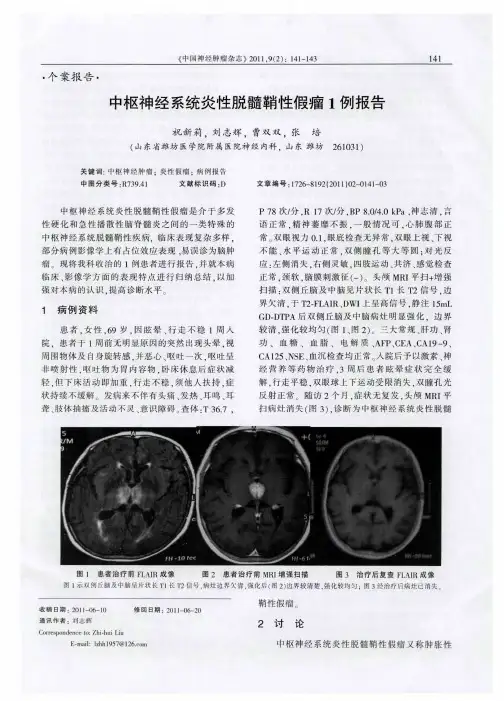
- 格式:pptx
- 大小:4.74 MB
- 文档页数:11


作者单位:430070 武汉,广州军区武汉总医院放射科作者简介:邱怀明(1975-),男,湖北麻城人,硕士,住院医师,主要从事介入治疗工作。
・中枢神经影像学・脑脱髓鞘性假瘤的C T及M RI诊断邱怀明,高保安,曾晓华,金德勤【摘要】 目的:分析脑脱髓鞘性假瘤的CT及MRI表现,提高对该疾病的影像诊断水平。
方法:结合文献报道,回顾性分析3例经手术病理证实的脑脱髓鞘性假瘤的CT及MRI表现。
结果:所有病例均表现为脑内单发肿块样病变,CT平扫多呈低密度改变,MRI检查多表现为长T1、长T2信号,增强扫描呈弥漫性或环形强化,病灶周围水肿及占位效应相对较轻。
结论:脑脱髓鞘性假瘤的CT及MRI表现缺乏特征性,但全面认识该疾病的临床特征、影像表现和病理改变,可提高对该疾病的诊断水平。
【关键词】 脱髓鞘疾病;体层摄影术,X线计算机;磁共振成像【中图分类号】R814.42;R744.5 【文献标识码】A 【文章编号】100020313(2005)022*******CT and MRI of Demyelinating Pseudotumor of the B rain Q IU Huai2ming,GAO Bao2an,ZEN G Xiao2hua,et al.Department of Radiology,PL A Wuhan G eneral Hospital,Wuhan430070,P.R.China【Abstract】 Objective:To analyze the CT and MRI manifestations of demyelinating p seudotumor of the brain and to in2 crease the diagnosis accuracy.Methods:The CT and MRI imaging features of three pathologically proved cases of demyelina2 ting pseudotumor of the brain were retrospectively analyzed.R esults:All cases appeared as a solitary mass without adjacent accompanying lesions.On plain CT scans,the majority of lesions presented as low density;on plain MRI scans,the lesions manifested as homogenous low signal on T1WI and high signal on T2WI.The majority showed diffuse or annular enhance2 ment.Peritumoral edema and space occupying2effect were found relatively mild.Conclusion:The CT and MRI manifestations of demyelinating p seudotumor of the brain are short of characteristic.But good understanding of clinical,radiologic and pathologic manifestations can increase the diagnosis accuracy of this disease.【K ey w ords】 Demyelinating diseases;Tomography,X2ray computed;Magnetic resonance imaging 中枢神经系统脱髓鞘性假瘤是介于多发性硬化和急性播散性脑脊髓炎之间的一类特殊的中枢神经系统脱髓鞘性疾病,临床罕见,常误诊为脑肿瘤。

《中枢神经系统瘤样脱髓鞘病变诊治指南》要点瘤样脱髓鞘病变(tumefactive demylinatinglesions,TDLs),既往也称瘤样炎性脱髓鞘病(TIDD),或脱髓鞘假瘤(DPT),是中枢神经系统(CNS)一种相对特殊类型的免疫介导的炎性脱髓鞘病变,绝大多数为脑内病变,脊髓TDLs鲜有报道。
影像所见病变体积较大,多伴周边水肿,且具有占位效应,或/和MRI增强影像改变,易与脑肿瘤相混淆,因此得名。
尽管脑活检是诊断TDLs的金标准,但有其局限性。
目前,对TDLs诊断仍主要依靠临床与影像特点,国内外尚缺乏TDLs相关诊断标准或专家共识,部分患者误诊“肿瘤”而行手术切除或伽玛刀治疗情况。
近年来,国内TDLs临床研究进展迅速,诊断经验日趋成熟。
1 临床特点1.1 发病特点TDLs的发病率及患病率等流行病学资料缺如。
急性或亚急性起病居多,少数慢性起病,鲜有前驱感染症候,个别发病前有疫苗接种及感冒受凉史。
男女患者比例基本相当,各年龄段均可发病,以中青年为多。
1.2 自然病程近年来,国内外临床研究发现,大多数TDLs为单次病程,少数可向复发-缓解型MS(RRMS)转化,或再次以TDLs形式复发,极少数可与视神经脊髓炎谱系疾病(NMOSD)重叠。
1.3 临床症候绝大多数患者TDLs脑内受累,少数脊髓也可受累。
与脑胶质瘤相比,多数TDLs临床症候相对较显著,少数亦可表现为影像病灶大、临床症候相对较轻的特点,与胶质瘤类似。
TDLs以头痛、言语不清、肢体力弱起病多见。
部分患者早期可仅表现为记忆力下降、反应迟钝、淡漠等精神认知障碍症候,易被患者及家属忽视。
随病情进展,症状可逐渐增多或加重,也可有视力下降。
TDLs的临床症候主要取决于病变累及的部位及范围,活动期症状可逐渐增多或加重,但很少仅表现癫痫发作(在脑胶质瘤中多见)。
当TDLs病变较弥漫或多发时,可影响认知功能,部分出现尿便障碍。
TDLs以白质受累为主,还可累及皮层及皮层下白质。
炎症脱髓鞘假瘤病变都有哪些症状
炎症脱髓鞘假瘤病变都有哪些症状,这是很多患者特别想了解的,因为这种疾病对于患者的身体危害还是比较严重的,所以为了尽快的让自己得到有效的治疗康复,就想具体了解一下炎症脱髓鞘假瘤病变都有哪些症状,下面内容就做了具体介绍,你可以尽快了解。
1、头痛,脱髓鞘疾病中出现头痛的机率不高,但脱髓鞘病人也有头痛发作。
正常的神经纤维,感觉冲动发生于神经末梢和细胞体,运动冲动发生于细胞体;病变的神经纤维,冲动可发生于轴突的中部而向周围和中枢传导,这种异位冲动可以由易患性增高,对机械刺激非常敏感所致,也可是自发的紧随着同一纤维的正常冲动后发生或某个刺激在病变部位引起反复兴奋,可造成疼痛。
2、脱髓鞘的同时伴有淋巴细胞、浆细胞、多形核白细胞的浸润,形成严重的炎性反应。
3、多发性硬化是因为自身免疫障碍而导致的中枢神经系统的脱髓鞘性疾病。
多在20--40岁之间发病,而在10岁以下和50岁以上发病者很少,起病可急可缓,表现为:易激动、强哭、强笑、记忆力减退、构音障碍或语音轻重不一、视力障碍、感觉减退或感觉异常、肢体活动不利或瘫痪、小便障碍、阳痿等。
专家提示:脱髓鞘病变病情发展具波动性,一次发作后症状可自行缓解或经治疗后缓解,经过一段时间可再复发。
然而,每次复发可遗留一定程度的功能缺损,总趋势是病情逐渐恶化。
因此,希望广大患者能够准确认识脱髓鞘病变的症状,做到疾病的早发现,早治疗,以确保自身的生活质量!
炎症脱髓鞘假瘤病变都有哪些症状,相信很多的患者已经通过以上的介绍,有了全面的认识和了解,所以为了不让这种疾病对自己身体构成严重伤害,在了解以上介绍的症状有哪些以后,要尽快的通过正确的治疗方法,让自己的这种疾病得到康复。
瘤样炎性脱髓鞘病(tumor-like inflammatory demyelinating diseases, TIDD)是中枢神经系统(central nervous system, CNS)的一种较为特殊的脱髓鞘病,是CNS炎性假瘤中的一种。
近年来,TIDD受到越来越多的关注,但多为个案或数例的报道。
对其临床及影像特点观察研究比较浅显。
随着近年影像技术发展及脑活检术的应用,发现该病罹患人数并不在少数,特别是其临床、影像等方面与脑肿瘤有诸多相似之处, 临床中二者易于混淆、误诊或误治。
即使是神经内科、神经外科及影像科的资深专家有时对TIDD及脑肿瘤的鉴别也有一定困难。
有的TIDD因误诊肿瘤而行手术病灶切除,有的因误诊而直接行放疗或γ刀治疗,部分患者因此而遗留功能障碍,甚至发生放射性脑病。
因此,有必要提高对TIDD的诊断与鉴别诊断水平,以满足临床工作的需要。
为此,结合我们已经积累的50多例TIDD患者(其中经病理证实的有26例)的临床、影像、病理方面的特点,以及我们积累的不同脑肿瘤(病理证实的)方面的临床经验,并参考国内外文献,对TIDD与脑肿瘤进行详细的对比剖析,提供相应的临床、实验室指标、影像特点,以供研究神经系统疾病相关的专科医师在TIDD的诊断与鉴别诊断中进行参考。
海军总医院神经内科戚晓昆一、TIDD的概念及误诊原因CNS炎性脱髓鞘病变为神经系统免疫介导性疾病,包括多发性硬化(multiple sclerosis, MS)、视神经脊髓炎(neuromyelitis optica, NMO)、播散性脑脊髓炎等。
其中一种CNS脱髓鞘病其影像学具有占位效应,而临床上表现为病灶大而症候少,酷似脑肿瘤,被称之为瘤样炎性脱髓鞘病(TIDD) ,或肿瘤性脱髓鞘病(tumefactive demyelinating lesions,TDLs),亦或称脱髓鞘假瘤(demyelinating pseudotumor, DPT)。