脑组织供血及脑血管解剖
- 格式:ppt
- 大小:2.65 MB
- 文档页数:35

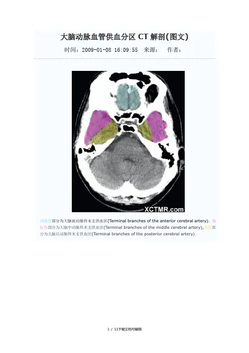
大脑动脉血管供血分区CT解剖(图文)时间:2009-01-08 16:09:55 来源:作者:浅蓝色部分为大脑前动脉终末支供血区(Terminal branches of the anterior cerebral artery),粉红色部分为大脑中动脉终末支供血区(Terminal branches of the middle cerebral artery),黄色部分为大脑后动脉终末支供血区(Terminal branches of the posterior cerebral artery)浅蓝色部分为大脑前动脉终末支供血区(Terminal branches of the anterior cerebral artery),粉红色部分为大脑中动脉终末支供血区(Terminal branches of the middle cerebral artery),黄色部分为大脑后动脉终末支供血区(Terminal branches of the posterior cerebral artery),绿色部分为脉络膜前动脉供血区(Anterior choroidal artery),浅蓝色部分为大脑前动脉终末支供血区(Terminal branches of the anterior cerebral artery),粉红色部分为大脑中动脉终末支供血区(Terminal branches of the middle cerebral artery),黄色部分为大脑后动脉终末支供血区(Terminal branches of the posterior cerebral artery),绿色部分为脉络膜前动脉供血区(Anterior choroidal artery),褐色部分为大脑前动脉深穿支供血区(Penetrating branches of the anterior cerebral artery),枣红色部分为大脑后动脉深穿支及后交通动脉供血区(Penetrating branches of the posterior cerebral artery and posterior communicating artery),浅蓝色部分为大脑前动脉终末支供血区(Terminal branches of the anterior cerebral artery),粉红色部分为大脑中动脉终末支供血区(Terminal branches of the middle cerebral artery),黄色部分为大脑后动脉终末支供血区(Terminal branches of the posterior cerebral artery),绿色部分为脉络膜前动脉供血区(Anterior choroidal artery),褐色部分为大脑前动脉深穿支供血区(Penetrating branches of the anterior cerebral artery),枣红色部分为大脑后动脉深穿支及后交通动脉供血区(Penetrating branches of the posterior cerebral artery and posterior communicating artery),亮红色部分为大脑中动脉深穿支供血(Penetrating branches of the middle cerebral artery)浅蓝色部分为大脑前动脉终末支供血区(Terminal branches of the anterior cerebral artery),粉红色部分为大脑中动脉终末支供血区(Terminal branches of the middle cerebral artery),黄色部分为大脑后动脉终末支供血区(Terminal branches of the posterior cerebral artery),绿色部分为脉络膜前动脉供血区(Anterior choroidal artery),枣红色部分为大脑后动脉深穿支及后交通动脉供血区(Penetrating branches of the posterior cerebral artery and posterior communicating artery),亮红色部分为大脑中动脉深穿支供血(Penetrating branches of the middle cerebral artery)浅蓝色部分为大脑前动脉终末支供血区(Terminal branches of the anterior cerebral artery),粉红色部分为大脑中动脉终末支供血区(Terminal branches of the middle cerebral artery),黄色部分为大脑后动脉终末支供血区(Terminal branches of the posterior cerebral artery),枣红色部分为大脑后动脉深穿支及后交通动脉供血区(Penetrating branches of the posterior cerebral artery and posterior communicating artery),亮红色部分为大脑中动脉深穿支供血(Penetrating branches of the middle cerebral artery)浅蓝色部分为大脑前动脉终末支供血区(Terminal branches of the anterior cerebral artery),粉红色部分为大脑中动脉终末支供血区(Terminal branches of the middle cerebral artery),黄色部分为大脑后动脉终末支供血区(Terminal branches of the posterior cerebral artery),浅蓝色部分为大脑前动脉终末支供血区(Terminal branches of the anterior cerebral artery),粉红色部分为大脑中动脉终末支供血区(Terminal branches of the middle cerebral artery),黄色部分为大脑后动脉终末支供血区(Terminal branches of the posterior cerebral artery),浅蓝色部分为大脑前动脉终末支供血区(Terminal branches of the anterior cerebral artery),粉红色部分为大脑中动脉终末支供血区(Terminal branches of the middle cerebral artery),黄色部分为大脑后动脉终末支供血区(Terminal branches of the posterior cerebral artery),浅蓝色部分为大脑前动脉终末支供血区(Terminal branches of the anterior cerebral artery),粉红色部分为大脑中动脉终末支供血区(Terminal branches of the middle cerebral artery),浅蓝色部分为大脑前动脉终末支供血区(Terminal branches of the anterior cerebral artery)(学习的目的是增长知识,提高能力,相信一分耕耘一分收获,努力就一定可以获得应有的回报)11 / 11下载文档可编辑。


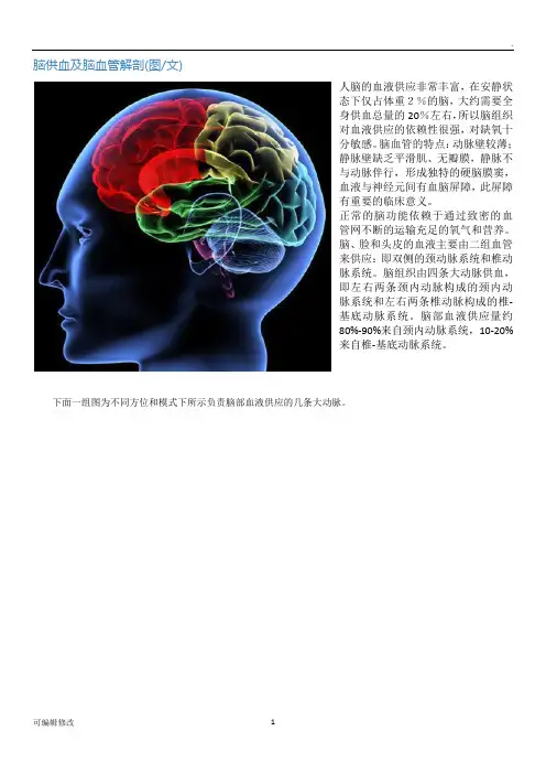
脑供血及脑血管解剖(图/文)人脑的血液供应非常丰富,在安静状态下仅占体重2%的脑,大约需要全身供血总量的20%左右,所以脑组织对血液供应的依赖性很强,对缺氧十分敏感。
脑血管的特点:动脉壁较薄;静脉壁缺乏平滑肌、无瓣膜,静脉不与动脉伴行,形成独特的硬脑膜窦,血液与神经元间有血脑屏障,此屏障有重要的临床意义。
正常的脑功能依赖于通过致密的血管网不断的运输充足的氧气和营养。
脑、脸和头皮的血液主要由二组血管来供应:即双侧的颈动脉系统和椎动脉系统。
脑组织由四条大动脉供血,即左右两条颈内动脉构成的颈内动脉系统和左右两条椎动脉构成的椎-基底动脉系统。
脑部血液供应量约80%-90%来自颈内动脉系统,10-20%来自椎-基底动脉系统。
下面一组图为不同方位和模式下所示负责脑部血液供应的几条大动脉。
颈总动脉于第四颈椎相当于甲状软骨上缘处分为颈内A和颈外A两个分支,其中颈外动脉负责脸部和头皮的血液供应,颈内动脉分出后沿颈部向上直至颅底,经颈动脉管进入海绵窦,紧靠海棉窦内侧壁,穿出海棉窦行至蝶骨的前床突内侧,开始分支(颈内A按行程分为四段:即颈段、颈内动脉管段、海棉窦段和脑段,临床上将后两段合称为“虹吸部”),其颅外的颈段无任何分支,颈内动脉管段先后分出颈鼓A和翼管A两个小支,海棉窦段先后分出海棉窦支、垂体支和脑膜支,脑段在前床突内侧处分出眼动脉,在视交叉外侧正对前穿质处分成大脑前动脉(ACA)和最大终末支的大脑中动脉(MCA)两个主要终末支。
供应除部分颞叶和枕叶之外的大脑前3/5的血液,即又称为前循环系统。
椎-基底动脉供应脊髓上部、大脑的后2/5(枕叶、颞叶的一部分、丘脑后大半部和丘脑下部的小部分)、脑干和小脑的血液,故又称为后循环系统。
两侧大脑前动脉通过前交通动脉相连,颈内动脉的末端通过后交通动脉和大脑后动脉相连,于是围绕脚间窝形成一完整的血管环即大脑动脉环(Willis动脉环)。
Willis动脉环是一种代偿的潜在装置。