盆部与会阴
- 格式:doc
- 大小:135.00 KB
- 文档页数:5
盆部与会阴一、境界与分区盆部1、定义:盆部指界线以下,盆膈以上,属于小骨盆的部分。
向上与腹腔相通,向下借盆膈与会阴部的软组织相邻。
界线由骶岬、髂翼、弓状线、耻骨梳、耻骨嵴和耻骨联合上缘围成。
2、组成骨骼:小骨盆(男性骨盆窄、入口小、耻骨下角为70-75度、小骨盆腔为漏斗状,女性骨盆宽,入口大,耻骨下角为90-100度、小骨盆腔为圆桶状P275表格)盆部肌:闭孔内肌、梨状肌、肛提肌和尾骨肌盆膈:由盆膈上、下筋膜及其间的肛提肌和尾骨肌共同构成。
盆腔内的脏器1、男性盆部器官①输尿管盆部和壁内部:两侧输尿管越过髂总血管的前方,入盆后即为输尿管的盆部,它在腹膜外沿盆壁行向前下内,经输精管的后下方交叉后,进入膀胱底,续为输尿管的壁内部。
②膀胱:为贮尿的肌性囊,可分为:尖、体、底、颈四部分。
其形态和位置随尿液的充盈程度而变化。
膀胱底内面,两侧输尿管口与尿道内口之间的三角形区域称膀胱三角。
输尿管斜穿膀胱壁形成输尿管口上方的纵行皱襞,称输尿管皱襞。
两输尿管口之间有隆起的粘膜横襞,称输尿管间襞。
★重点1、膀胱的位置,毗邻及机能状态膀胱的位置,随年龄及盈虚状态而不同,空虚时呈锥体状,位于盆腔前部,可分尖、体、底、颈四部。
充盈时可升至耻骨联合上缘以上,此时腹膜返折处亦随之上移,膀胱前外侧壁则直接邻贴腹前壁。
临床上常利用这种解剖关系,在耻骨联合上缘上进行膀胱穿刺或做手术切口,可不伤及腹膜。
儿童的膀胱位置较高。
空虚的膀胱,前方与耻骨联合相邻,其间为耻骨后隙,膀胱下外侧面邻肛提肌,闭孔内肌及其筋膜,其间充满疏松CT等,称膀胱旁组织,内有输尿管盆部,男性还有输精管壶腹穿行。
膀胱后方借直肠膀胱隔与精囊、输精管壶腹及其后方的直肠相邻,女性则借膀胱阴道隔与子宫颈及阴道前壁相邻,膀胱上面覆盖腹膜,并与肠袢相邻,女性还与子宫相邻。
膀胱的后下部即膀胱颈,下接尿道。
男性邻贴前列腺,女性与尿生殖膈相邻。
★膀胱三角(名解)膀胱空虚时,其内粘膜面呈现许多皱襞,唯其底部有一三角形的平滑区,称膀胱三角,两侧角即左、右输管口,两口之间有呈横向隆起的输尿管间襞,三角的前下角为尿道内口,膀胱三角是结核与结石、肿瘤的好发部位。
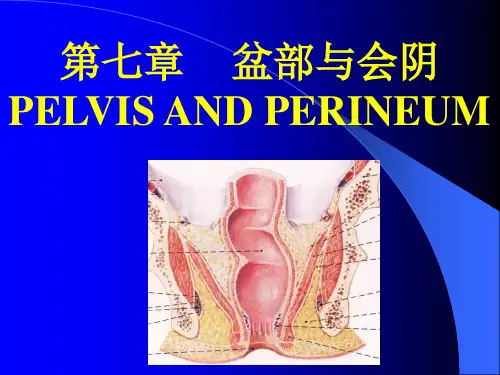
盆部和会阴盆部和会阴位于躯干的下部,以骨盆为支架。
盆部由盆壁、盆底和盆腔内的脏器组成。
盆 腔向上经骨盆上口与腹腔相通,向下借盆膈与会阴相邻。
会阴是指盆膈以下封闭骨盆下口的 全部软组织。
骨性标志及体表投影 从腹前正中线下端向外,依次可触得耻骨联合上缘、耻骨嵴、耻骨结 节、腹股沟韧带、髂前上棘、髂嵴。
在后方,可触得髂后上棘、坐骨结节和骶角。
左、右骶 角之间是骶管裂孔 sacral hiatus,骶角为骶管麻醉穿刺的标志。
两侧髂嵴最高点连线通过第四 腰椎棘突。
两侧髂后上棘连线平第二骶椎棘突。
第一节 盆 部【解剖方法】1.检查盆部腹膜及其形成的结构 腹膜从腹前壁向下覆盖膀胱上壁、侧壁和后壁的一部 分。
向后在男性覆盖输精管壶腹及精囊上端;在女性则覆盖子宫大部分、输卵管、卵巢以及 阴道后壁的最上部,然后皆反折至直肠,覆盖直肠中1/3 的前面及直肠上 1/3 的前面和两侧, 向上与腹后壁腹膜相续。
男性腹膜在膀胱与直肠之间,形成膀胱直肠陷凹;女性则在膀胱与子宫间形成膀胱子宫 陷凹,在子宫与直肠间形成直肠子宫陷凹(又称 Douglas 窝),以手指探查该窝的深度。
腹膜在子宫的前、后面反折,并且向两侧伸延至盆侧壁,形成双层的腹膜皱襞,称子宫 阔韧带(图 235,6)。
阔韧带上缘游离,内含输卵管,韧带的基底宽阔,与盆底前后的腹膜续 连,卵巢位于阔韧带后面,以卵巢系膜与阔韧带后层相连。
观察腹膜从骨盆上口髂总动脉的分叉处延伸至卵巢上端形成的卵巢悬韧带(又称骨盆漏 斗韧带)(图 235)。
韧带内有结缔组织和平滑肌,并有卵巢动、静脉经过,韧带的后内侧与输 尿管邻近,待后解剖时予以注意这种关系。
将子宫底、体往前推,显示有腹膜皱襞自子宫颈呈弓形向后,绕过直肠两侧至骶骨前面,称 直肠子宫襞,内藏子宫骶韧带(图 237)。
把子宫复位,再检查由子宫角向前至腹环进腹股沟管的子宫圆韧带。
2.探查盆腔的脏器 在女性盆部,将腹膜自盆壁往内翻至直肠及子宫外侧,然后修洁由 腹后壁下行的输尿管,往下追踪至膀胱,沿途可见它循盆壁、卵巢后缘转向前内行经阔韧带 下方,在距子宫颈外侧约 2cm处,有子宫动脉跨过其前上方(图 235)。


局部解剖学资料(盆部与会阴)盆部与会阴一、名词解释1.会阴:perineum有广义和狭义之分。
广义会阴是指盆膈以下封闭骨盆下口的全部软组织的总称,其境界略呈菱形,即位于前方耻骨联合下缘、两侧坐骨结节、后方尾骨尖四者之间的区域。
此区域又可借两侧坐骨结节之间的连线分为前方的尿生殖三角(尿生殖区)和后方的肛门三角(肛区)。
狭义的会阴在男性系指阴囊根与肛门之间的软组织,女性则指阴道前庭后端与肛门之间的软组织,又称产科会阴。
2.盆膈:pelvic diaphragm 位于小骨盆下口的肛提肌和尾骨肌共同构成盆底肌,盆底肌的上、下面分别覆盖有盆膈上筋膜和盆膈下筋膜,三者共同构成盆膈。
在两侧肛提肌前内侧缘之间留有一狭窄的裂隙,称盆膈裂孔,被下方的尿生殖膈封闭,男性有尿道通过,女性有尿道和阴道通过。
盆隔的后部有肛管通过。
盆膈有支持和固定盆腔内脏器的作用,并可与腹肌、膈肌一起协同增加腹压。
3.前列腺鞘:fascial sheath of prostate盆膈上筋膜向上返折,移行为盆筋膜的脏层,包裹于盆内脏器的周围,其中包裹前列腺外面的部分形成的前列腺鞘,具有支持和固定前列腺的作用。
前列腺鞘与前列腺囊之间有静脉丛。
4.阴部管(Alcock管):pudendal canal 又称Alcock管,位于坐骨直肠窝的外侧壁,坐骨结节上方约3cm处,是闭孔筋膜形成的裂隙。
管内有阴部内动脉、阴部内静脉和阴部神经通过。
5.浅会阴筋膜:superficial fascia of perineum 又称Colles 筋膜。
该筋膜向前方和两侧分别附着于耻骨弓和坐骨结节;后方终止于尿生殖膈后缘(相当于两侧坐骨结节连线处),并与尿生殖膈上、下筋膜相愈着,在正中线上还与男性尿道球中隔、会阴中心腱愈着;前上方与阴囊肉膜、浅阴茎筋膜和腹前壁的Scarpa筋膜相延续。
6.会阴浅隙:superficial perineal space 又称会阴浅袋,位于浅会阴筋膜与尿生殖膈下筋膜之间,该间隙内有会阴肌浅层、阴部神经、阴部内动脉的末支及其伴行的静脉。
第五章盆部与会阴一、概述了解盆部和会阴部的境界、分区和体表标志。
[07级临五]单选:14、关于盆部的表面解剖正确的是(左右髂棘最高点过第四腰椎棘突)(137)37. Which one is not the important surface landmark (E)A. pubic crest.B. pubic arch.C. ischial tuberosity.D. apex of coccyx.E. promontory of the sacrum.二、盆部1.了解骨盆的整体观及盆壁肌。
掌握盆膈的构成。
1. The bony pelvis (A) A. is formed by the two hip bones, sacrum, coccyx and their joints.B. can be divided into abdominal and pelvic part by the terminal line.C. is enclosed at the inferior pelvic aperture by perineum.D. is wider, shorter and more funnel-shaped in female than in male.E. pelvic outlet is formed by the terminal line.[07级临五]多选:3、盆底肌包括()【填空】盆膈的肌肉包括肛提肌和尾骨肌。
肛提肌包括前列腺提肌(男)、耻骨阴道肌(女)、耻骨直肠肌、耻尾肌和髂尾肌4部分。
2. The levator ani does not include (A) A. coccygeus. B. levator prostate. C. puborectalis. D. pubococcygeus. E. iliococcygeus.【盆膈】又称盆底,它由肛提肌及覆盖其上下面的筋膜构成[07级临七]单选:21、盆膈的组成/[06级临五]选择:36、盆隔构成3. The pelvic diaphragm (E) A. encloses the whole inferior aperture of the pelvis. B. is located in the anal triangle.C. consists of levator ani, sphincter ani externus, superior and inferior fascia of pelvic diaphragm.D. has a hiatus of pelvic diaphragm for digestive and urogenital canal.E. can support the pelvic viscera.5. The hiatus of the pelvic diaphragm (A) A. is a cleft between the anteromedial margin of pelvic diaphragm.B. is filled with the urogenital diaphragm.C. is enclosed by superior and inferior fascia of urogenital diaphragm.D. is the main channel connecting the pelvic spaces and perineal space.E. is penetrated by the membranous part of the urethra in male.2.了解盆筋膜的配布、盆筋膜间隙的名称和位置。
51. The parietal pelvic fascia on the surface of the obturator internus forms (BCD)A. obtrurator membrane.B. obturator fascia.C. obturator canal.D. tendinous arch of levator ani.E. pudendal canal.52. The visceral pelvic fascia forms (BC)A. pudendal canal.B. prostatic sheath.C. cardinal ligament of uterus.D. superior fascia of pelvic diaphragm.E. retrorectal space.53. Pelvic fascial spaces include (CD)A. deep perineal space.B. superficial perineal spaceC. retropubic space.D. retrorectal space. .E. ischioanal fossa.55. Peritoneum in pelvis forms (BDE)A. the uterosacral ligament.B. the rectouterine pouch.C. the round ligament of uterus.D. the mesovarium.E. the vesicouterine pouch.65. Rectovesical or rectovaginal septum (ABCD) A. is between the rectum and urinary bladder or vagina. B. is in coronary plane.C. contacts upwards to peritoneum.D. ends downwards at the pelvic diaphragm.E. contains prostate in male.耻骨后隙和直肠后隙的位置及临床意义?【答案】耻骨后隙位于耻骨联合后面与膀胱下外侧面之间,内有大量疏松结缔组织。
临床上可经此间隙施行耻骨上切口的腹膜外膀胱手术。
直肠后隙位于骶前筋膜与直肠筋膜之间,内含疏松结缔组织和静脉丛,向上与腹膜后隙相延续。
可经此间隙行腹膜后隙充气造影术,以助诊断;由于腹膜后隙和直肠后隙相通,因而感染化脓时脓液可相互扩散。
6. The retropubic space (B) A. is located between the urinary bladder and rectum or uterus.B. is usually used for extra-peritoneal operation of urinarry bladder.C. is rich in venous plexus and unsuitable for organ separation.D. is suitable for exudation absorption.E. doesn’t communicating with other pelvic spaces7. The retrorectal space (E) A. surround the inferior part of the ampulla of rectum. B. is the space between the sacrum and presacral fascia.C. is rich in venous plexus and unsuitable for rectus separation.D. communicates downwards with the ischioanal fossa.E. communicates upwards with the retroperitoneal space .3.了解盆部的血管、神经和淋巴。
掌握髂内动脉的主要分支;掌握子宫动脉与输尿管的位置关系。
[07级临七]单选:22、髂内动脉壁支(142)[06级临五]选择:11、髂内动脉的分支(142)38. Which is wrong about the internal iliac artery (D)A. It arises from the common artery.B. It is a short trunk descending into the lesser pelvis.C. The ureter runs anterior to it.D. Send out the inferior epigastric a.E. The internal iliac vein accompanies the artery on its medial side.39. The internal iliac artery doesn’t send (D)A. superior gluteal artery.B. obturator artery.C. internal pudendal artery.D. superior rectal artery.E. inferior vesical artery.50. The internal pudendal artery (D) A. Arises from the anterior trunk of the internal iliac artery.B. Passes through the lesser sciatic foramen to enter the gluteal region.C. Runs along the lateral surface of the obturator internus.D. Supplies the perineum.E. Runs below the pudendal nerve in the pudendal canal.13.Female ureter crosses AA. above uterine arteryB. under uterine arteryC. in front of uterine arteryD. inside uterine arteryE. lateral to uterine artery18. Which one is wrong about the uterine artery (E) A. It arises from the internal iliac artery.B. It descends in front of the ureter to the base of broad ligament.C. It crosses above the ureter just lateral to the neck of the uterus.D. It sends branches to vagina, uterine tube and ovary.E. It should be protected during hysterectomy (子宫切除术).4.了解盆内脏器的配布与腹膜的关系及其临床意义。