预染蛋白marker说明书
- 格式:pdf
- 大小:87.26 KB
- 文档页数:1

彩虹130广谱蛋白marker说明书货号:PR1950规格:20T(100μL)/50T(250μL)/100T(250μL×2)/500T(250μL×10)保存:-20℃保存,有效期至少2年。
产品特点:●三色预染,颜色鲜亮,条带整齐,便于观察。
●用量少,节约成本,仅5ul即可完美呈现(1.5mm,10孔梳)。
●广谱多带,一支即可满足多种实验需求。
产品简介:本产品包含9种彩色预染的已知分子量标准蛋白,分子量范围为15kD-130kD,每种蛋白含量约为0.2-0.4mg/ml。
预染marker可以用于直接观察蛋白质电泳状况以及清晰地判断Western Blot的转膜效果。
经SDS-PAGE凝胶电泳或转移到PVDF或NC膜上可得到清晰的9条彩色蛋白条带,其中70kD条带为红色,25kD条带为绿色,其余条带为蓝色。
使用说明:1.本产品是即用型液体,可直接上样电泳。
上样前无需加热,稀释或添加还原剂。
2.上样5ul,SDS-PAGE电泳过程及转膜后可看见清晰的彩虹条带。
3.建议分离胶浓度为15%。
电泳示意图说明:第1页,共2页第2页,共2页注意事项:1.本产品含有较高浓度甘油,常规-20℃保存为液体状态,可直接使用。
若冰箱温度不稳,导致结冰,可适当分装后置于4℃保存,至少稳定6个月。
2.Marker分离效果与PAGE胶浓度相关,若分离胶浓度低于15%,25kD以下条带不易分离,但不影响绿色(25KD)及以上条带。
如果目的条带小于25KD,建议分离胶浓度大于或等于15%。
3.转膜效果与转膜时间有关,需根据客户目的条带大小而定,如果转膜后较大分子量条带有部分未转膜成功,属于正常现象。

Livning彩色预染蛋白marker(10-180KD)with45kDa使用说明书产品名称规格货号Livning彩色预染蛋白marker(10-180KD)with45kDa250μl*1LVN150-1Livning彩色预染蛋白marker(10-180KD)with45kDa250μl*2LVN150-2Livning彩色预染蛋白marker(10-180KD)with45kDa2x250μl*10LVN150-10【储存条件】-20˚C恒温长期保存,4˚C保存6个月,建议分装保存,避免反复冻融。
【产品简介】本产品由跨度从10~180kDa的10种纯化的天然蛋白混合而成,各条带浓度约为0.2~0.4mg/ml。
其中75kDa条带为红色预染条带,方便判断各个条带的准确位置。
本产品适合作为SDS-PAGE电泳时,变性蛋白样品的分子量参照,并可实时观察蛋白样品的电泳分离状况,也可用于检测Western blot的转膜效率。
由于共价结合的染料会影响蛋白质分子的电泳迁移率,本产品适于粗略地估计目的蛋白样品的分子量。
【使用方法】1.将本产品于室温融化后,轻柔混匀,使沉淀充分溶解;2.加入3~5μl到SDS-聚丙烯酰胺胶的上样孔中,与待测样品一起电泳和转膜;3.电泳结束后,通过考马斯亮蓝染液染色观察条带。
【注意事项】1.使用时应该将从冰箱中取出的产品恢复至室温后使用,否则可能由于低温下蛋白变性不彻底导致电泳条带出现不同程度的弥散;2.使用前先将产品恢复至室温后混匀,使沉淀充分溶解,否则可能导致电泳条带出现不同程度的弥散或拖带;3.本产品含有SDS,蛋白已变性,不宜作为天然蛋白分子电泳时的分子量参照标准。
【图列展示】(单一浓度PAGE胶,Tris-Glycine电泳缓冲液,实际电泳图)8686************(梯度胶和单一浓度胶,Tris-Glycine 电泳缓冲液,带型示意图)【附录:转膜和洗膜】A 、转膜条件(冰上进行):a.Transfer with buffer containing 20%methanol to fix proteins on membrane.b.Wash membrane with PBS or TBS containing less than 0.1%Tween-20at 4°C.B 、洗膜条件(4度进行):Membrane:Nitrocellulose membranes /PVDFWash Buffer:1X Tris buffered saline,0.1%Tween-20(TBST)(吐温20不能超过0.1%)C 、Stripping Buffer:15g Glycine,1g SDS,10ml Tween20,pH2.2–Adjust volume to 1L如果需要用到含有DTT /b-ME 的Stripping buffer ,膜需要先以1X Tris buffered saline(TBS)洗三次,把Tween-20去干净后,再进行Stripping 。
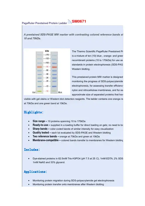
SM0671PageRuler Prestained Protein LadderA prestained SDS-PAGE MW marker with contrasting colored reference bands at10 and 70kDa.The Thermo Scientific PageRuler Prestained Proteis a mixture of ten (10) blue-, orange- and green-srecombinant proteins (10 to 170kDa) for use as sizstandards in protein electrophoresis (SDS-PAGE)Western blotting.This prestained protein MW marker is designed fomonitoring the progress of SDS-polyacrylamide geelectrophoresis, for assessing transfer efficiency onylon and nitrocellulose membranes, and for estimapproximate size of separated proteins that have b visible with gel stains or Western blot detection reagents. The ladder contains one orange refer at 70kDa and one green band at 10kDa.Highlights:•Size range– 10 proteins spanning 10 to 170kDa•Ready-to-use– supplied in a loading buffer for direct loading on gels; no need to boil •Sharp bands– color-coded bands of similar intensity for easy visualization•Quality tested – each lot evaluated by SDS-PAGE and Western blotting•Two reference bands– orange at 70kDa and green at 10kDa•Membrane-compatible– colored bands transfer to membranes for Western blottingIncludes:•Dye-stained proteins in 62.5mM Tris-H3PO4 (pH 7.5 at 25°C), 1mM EDTA, 2% SDS, 1 1mM NaN3 and 33% glycerol.Applications:•Monitoring protein migration during SDS-polyacrylamide gel electrophoresis•Monitoring protein transfer onto membranes after Western blotting•Sizing of proteins on SDS-polyacrylamide gels and Western blotsProduct Details:SDS-PAGE band profile of the Thermo ScientificPageRuler Prestained Protein Ladder. Images arefrom a 4-20% Tris-glycine gel (SDS-PAGE) andsubsequent transfer to membrane.Sm1841Spectra Multicolor Broad Range Protein LadderA prestained, 4-color, SDS-PAGE MW marker especially for proteins between 10 and 260kDa.The Thermo Scientific Spectra Multicolor Broad RangeProtein Ladder is a 4-color protein standard containing 10prestained recombinant prokaryotic proteins (10 to 260kDa)for use as size standards in gel electrophoresis and Westernblotting.This prestained protein MW marker is designed formonitoring the progress of SDS-polyacrylamide gelelectrophoresis, for assessing transfer efficiency onto PVDF,nylon and nitrocellulose membranes, and for estimating theapproximate size of separated proteins that have been made visible with gel stains or Western blot detection reagents. Four different chromophores (blue, orange, green, pink) are bound to the different component proteins, producing a brightly colored ladder with an easy-to-remember pattern.Highlights:•Size range– 10 proteins spanning 10 to 260kDa•Multicolor– four different colors for unambiguous band-size assignment•Ready-to-use– supplied in a loading buffer for direct loading on gels; no need to boil•Sharp bands– color-coded bands of similar intensity for easy visualization•Quality tested – each lot evaluated by SDS-PAGE and Western blotting•Membrane-compatible– colored bands transfer to membranes for Western blotting Includes:•Dye-stained proteins in 62.5mM Tris-H3PO4 (pH 7.5 at 25°C), 1mM EDTA, 2% SDS, 10mM DTT, 1mM NaN3 and 33% glycerol.Applications:•Monitoring protein migration during SDS-polyacrylamide gel electrophoresis•Monitoring protein transfer onto membranes after Western blotting•Sizing of proteins on SDS-polyacrylamide gels and Western blotsProduct Details:SDS-PAGE band profile of the Thermo ScientificSpectra Multicolor Broad Range Protein Ladder.Images are from a 4-20% Tris-glycine gel(SDS-PAGE) and subsequent transfer to membrane.S m1811PageRuler Plus Prestained Protein LadderA prestained SDS-PAGE MW marker with contrasting colored reference bands at 10, 25 and 70kDa.The Thermo Scientific PageRuler Plus Prestained ProteinLadder is a mixture of nine (9) blue-, orange- andgreen-stained recombinant proteins (10 to 250kDa) for useas size standards in protein electrophoresis (SDS-PAGE)and Western blotting.This prestained protein MW marker is designed formonitoring the progress of SDS-polyacrylamide gelelectrophoresis, for assessing transfer efficiency onto PVDF,nylon and nitrocellulose membranes, and for estimating theapproximate size of separated proteins that have been made visible with gel stains or Western blot detection reagents. A blue chromophore is bound to all proteins, except proteins of two reference bands of 70kDa and 25kDa that are colored with an orange dye and one green reference band of 10kDa. PageRuler Plus Prestained Protein Ladder is ready to use: no heating, further dilution or addition of a reducing agent is required before loading onto a gel.Highlights:•Size range– nine proteins spanning 10 to 250kDa•Ready-to-use– supplied in a loading buffer for direct loading on gels; no need to boil•Sharp bands– color-coded bands of similar intesity for easy visualization•Quality tested – each lot evaluated by SDS-PAGE and Western blotting•Bright reference bands– orange at 70 and 25kDa, and green at 10kDa•Membrane-compatible– colored bands transfer to membranes for Western blotting Includes:•Dye-stained proteins in 62.5mM Tris-H3PO4 (pH 7.5 at 25°C), 1mM EDTA, 2% SDS, 10mM DTT, 1mM NaN3 and 33% glycerol.Applications:•Monitoring protein migration during SDS-polyacrylamide gel electrophoresis•Monitoring protein transfer onto membranes after Western blotting•Sizing of proteins on SDS-polyacrylamide gels and Western blotsProduct Details:SDS-PAGE band profile of the Thermo Scientific PageRuler Plus Prestained Protein Ladder. Images are from a 4-20% Tris-glycine gel(SDS-PAGE) and subsequent transfer to membrane.。
CERTIFICATE OF ANALYSISPageRuler ™Prestained Protein Ladder#SM067210 x 250 µl(for 100 mini gel applications 5 µl per well or 50 large gel applications 10 µl per well)Lot: Expiry Date:Storage: stable at 4°C for up to 3 months. For long term storage, store at -20°C.SM067_57_9.docDescriptionPageRuler ™Prestained Protein Ladder is a mixture of 10 recombinant, highly purified colored proteins with apparent molecular weights of 10 kDa to 170 kDa. Ladder proteins are covalently coupled with a blue dye except for two reference bands prestained with different colors. The 72 kDa reference band is orange and 10 kDa reference band is green.The ladder is supplied in gel loading buffer and isready-to-use: no heating, further dilution or addition of a reducing agent is required.ContentsApproximately 0.1-0.2 mg/ml of each protein in the storage buffer (62.5 mM Tris-H 3PO 4 (pH 7.5 at 25°C), 1 mM EDTA, 2% (w/v) SDS, 10 mM DTT, 1 mM NaN 3 and 33% (v/v) glycerol).Applications∙ Monitoring of protein separation during SDS-PAGE (1). ∙ Verifying Western transfer efficiency (2, 3).∙ Approximate sizing of proteins on SDS-polyacrylamidegels and Western blots.Instruction for Use❶ Thaw the ladder at room temperature for a few minutes to dissolve precipitated solids. DO NOT BOIL!❷ Mix gently, but thoroughly, to ensure the solution is homogeneous.❸ Load the following volumes of the ladder on an SDS-polyacrylamide gel:– 5 µl per well for mini gel,– 10 µl per well for large gel.Use the same volumes for Western blotting.❹ After the run is complete, stain the gel or perform Western transfer procedure as desired.Note•Each lot of the PageRuler™ Prestained Protein Ladder is calibrated against a precisely sized, PageRuler™ Unstained Protein Ladder and calculated apparent molecular weights are reported in the picture.•For precise molecular weight determinations use PageRuler™ Unstained Protein Ladder, #SM0661, see.•In 8 or 10% gels low molecular weight proteins may migrate with the dye front.•Loading volumes are intended for use in gels with a thickness of 0.75 mm. For thicker gels, the recommended loading volume should be increased.•PageRuler™ Prestained Protein Ladder could be used in Western blotting with all common membranes: PVDF, nylon and nitrocellulose.•Longer transfer times or higher transfer voltages may be required for Western blotting of large (>100 kDa) proteins.Lot specific MW, kDa4-20% Tris-glycine SDS-PAGEcontinued on back pageQUALITY CONTROL5 µl of PageRuler™ Prestained Protein Ladder resolves 10 bands of equal intensities in 4-20% SDS-PAGE (Tris-glycine buffer) and after Western blotting onto PVDF membrane.Quality authorized by: Jurgita Zilinskiene References1. Laemmli, U.K., Cleavage of structural proteins during the assembly of the head of bacteriophage T4, Nature, 227, 680-685, 1970.2. Burnette, W.N., "Western blotting": electrophoretic transfer of proteins from sodium dodecyl sulfate – polyacrylamide gels to unmodified nitrocellulose and radiographic detection with antibody and radioiodinated protein A, Anal. Biochem., 112 (2), 195-203, 1981.3. Towbin, H., et al., E lectrophoretic transfer of proteins from polyacrylamide gels to nitrocellulose sheets: procedure and some applications, Proc. Natl. Acad. Sci. USA, 76, 4350-4354, 1979.This product is manufactured under the license forStrep-tag® technology covered by US patents Nos.5,506,121, 6,103,493 and foreign counterparts. Related Products∙DualColor™ Protein Loading Buffer Pack #R1011∙Loading Buffer Pack #R0891∙Spectra™ Multicolor Broad Range Protein Ladder #SM1841∙PageRuler™ Unstained #SM0661∙PageRuler™ Plus Prestained Protein Ladder #SM1811∙PageSilver™ Silver Staining Kit #K0681∙PageBlue™ Protein Staining Solution #R0571∙10X Tris-glycine-SDS Buffer #B46∙10X Tris-tricine-SDS Buffer #B48∙DTT #R0861 ∙ProteoJET™ Mammalian Cell Lysis Reagent #K0301∙ProteoJET™ Cytoplasmic and Nuclear ProteinExtraction Kit #K0311∙Bradford Reagent, ready-to-use #R1271∙Bovine Serum Albumin Standard Set, ready-to-use #R1281∙Bovine Gamma Globulin Standard Set, ready-to-use #R1291PRODUCT USE LIMITATION.This product is developed, designed and sold exclusively for research purposes andin vitro use only. The product was not tested for use in diagnostics or for drugdevelopment, nor is it suitable for administration to humans or animals.Please refer to for Material Safety Data Sheet of the product.。
蛋白marker标记颜色方法全文共四篇示例,供读者参考第一篇示例:蛋白marker标记颜色方法在生物科学研究中,标记蛋白是非常常见的实验技术之一。
蛋白marker的标记可以帮助研究人员在实验中追踪和定位蛋白,从而更好地理解蛋白的功能和相互作用。
而标记蛋白的颜色选择也是非常重要的,不同颜色的标记能够帮助实验者在实验过程中更清晰地识别和分析样品。
本文将介绍一些常用的蛋白marker标记颜色方法,希望能够帮助读者更好地进行蛋白标记实验。
一、荧光标记荧光标记是蛋白标记颜色中最常用的方法之一。
通过将蛋白与荧光物质结合,可以在光学显微镜下直接观察到蛋白标记的颜色。
常用的荧光标记包括FITC(荧光异硫氰酸酯),TRITC(罗丹明异硫氰酸酯)和Cy5(Cyanine5)。
这些荧光物质在实验中常用于免疫荧光染色、融合蛋白标记等实验中。
荧光标记的优势是信号强度高、灵敏度高且不受环境影响,适用于单细胞和组织的标记。
在实验中,荧光标记的颜色可以根据荧光物质的不同而有所区分。
比如,FITC标记的蛋白呈现绿色,TRITC标记呈现橙红色,Cy5标记呈现红色。
实验者可以根据实验需要选择不同的荧光标记物质,并通过显微镜观察到标记蛋白的颜色。
二、生物素标记生物素标记是一种基于生物素-亲和素相互作用的蛋白标记方法。
生物素是一种维生素H成分,具有与亲和素结合的特性。
在实验中,生物素可以与生物素素结合并形成稳定的结合物,从而实现蛋白的标记。
生物素标记的颜色在实验中常表现为紫色,实验者可以通过观察颜色变化来判断蛋白是否标记成功。
生物素标记的优势是其结合力强、稳定性高,适用于长时间的实验操作。
另外,生物素标记也可以用于有机溶剂、高温、酸碱环境等多种条件下的蛋白标记。
因此,在一些特殊实验条件下,生物素标记是一种非常理想的标记方法。
三、酶标记酶标记是另一种常用的蛋白标记方法。
通过将蛋白与酶结合,可以在实验中通过酶的催化作用来识别蛋白的存在。
常用的酶标记包括辣根过氧化物酶(HRP)、碱性磷酸酶(AP)等。

预染超低分子量蛋白质Marker(3.3kD-31.0kD)说明书货号:PR1900规格:20T(200μ1)保存:-20℃保存,有效期为一年。
避免反复冻融,建议分装冻存。
产品简介:本产品包含预染的3种多肽和2种低分子量蛋白质组成,分子量范围为3.3kD-31.0kD。
可以用于直接观察蛋白质电泳状况以及清晰地判断Western Blot的转膜效果。
经Tricine-甘油SDS-PAGE凝胶电泳时以及转移到PVDF或NC膜上可看到清晰的5条蓝色的蛋白条带。
本品为蛋白质和多肽混合物的冻干粉,每种预染蛋白含量约为40μg,配有一支1×上样缓冲液。
使用说明:1.开封后,溶于200μ11×上样缓冲液,可根据需要进行小量分装,每管10μl,-20℃贮存,每次取一管使用,避免反复冻融。
2.使用前取分装后的小管室温融化后即可上样电泳。
电泳示意图:凝胶的配置方法:分离胶夹层胶浓缩胶20%/4.5ml16.5%/4.5ml15.5%/4.5ml10%/2ml4%/2ml49.5%T3%C///0.407ml0.160ml49.5%T6%C1.82ml1.50ml 1.395ml//凝胶缓冲液 1.50ml 1.50ml 1.50ml0.667ml0.496ml 甘油0.48ml0.48ml0.48ml//ddH2O0.70ml1.02ml1.125ml0.926ml1.344ml 10%PAGE胶凝固剂40µl40µl40µl20µl20µlPAGE胶促凝剂5µl5µl5µl3µl3µl凝胶制备及染色注意事项:1.先配制分离胶,聚合后再配制夹层胶,最后配制浓缩胶,3种胶的制胶体积比为4:1.5:1。
电泳时,30V 跑1-2h后,待指示前沿到达分离胶上沿时,把电压调至100V,至电泳结束,整个电泳过程大约需要6-8h。


Western Blot Marker说明书货号:PR1800规格:10T(50μL)/20T(100μL)保存:-20℃可保存2年。
避免反复冻融,建议分装保存。
产品简介:传统的Western Blot试验需要使用预染蛋白Marker来跟踪检测阳性抗原信号和转膜效率,但预染蛋白Marker无法在胶片上曝光,曝光信号需要根据膜上的预染蛋白Marker来判断。
Western Blot Marker是专门为Western Blot试验设计的分子量标准,由9种发光蛋白和9种预染蛋白组成;9种发光蛋白分别为23、30、36、44、50、62、90、120和160KDa,这些蛋白均可与抗体结合,因此能与抗原在同一张膜上曝出信号,阳性信号可以直接通过Western Marker的位置来判断;9种预染蛋白分别为15、25、35、40、55、70、100、130和180KDa,其中70KDa为红色,其他为蓝色,转膜完毕后在膜上肉眼可见。
主要特征:1.可与抗原同时检测信号,使Western blot结果判断更加简便2.发光Marker没有偶联和标记其他分子,分子量更加准确3.综合发光Marker和预染Marker的优势,既能监测转膜又能与抗原同时检测使用说明:1.取出后于室温放置融化;2.温和地充分混匀,取5-10μL该Western Blot Marker至上样孔中,进行SDS-PAGE电泳。
3.电泳完毕后转至PVDF膜或NC膜,与一抗二抗孵育。
4.孵育完毕,将Western Blot Marker与抗原一起通过ECL曝光至胶片或荧光显色。
图示:1、HRP标记的Goat anti mouse IgG二抗(一抗:mouse anti p38)ECL化学发光检测第1页,共2页第2页,共2页2、多重荧光检测注意事项:1.本产品可直接使用,不需要加热。
2.可根据抗体的效价或曝光时间的长短调整Western Blot Marke的上样量。
PRODUCT INFORMATIONThermo ScientificPageRuler Prestained Protein Ladder #26616 2 x 250 µlLot: XXXXXXXX Expiry Date: __WARNING! HARMFUL IF INHALED OR ABSORBEDTHROUGH SKIN. CAUSES RESPIRATORY TRACT, EYEAND SKIN IRRITATION. MAY CAUSE ALLERGIC SKIN REACTION. MAY BE HARMFUL IF SWALLOWED.CONTAINS MATERIAL WHICH CAUSES DAMAGE TO THE FOLLOWING ORGANS: KIDNEYS, LUNGS, UPPER RESPIRATORY TRACT, SKIN, EYES. CONTAINS MATERIALWHICH MAY CAUSE DAMAGE TO THE FOLLOWINGORGANS: LIVER, GASTROINTESTINAL TRACT.Avoid exposure - obtain special instructions before use. Donot breathe vapor or mist. Do not ingest. Do not get in eyesor on skin or clothing. Use only with adequate ventilation. Keep container tightly closed and sealed until ready for use. Wash thoroughly after handling. Refer to MSDS.|2661®~26616|72559?~XXXXXXXXStore at -20°C/onebio /pierce Made in Lithuania IntroductionThe Thermo Scientific™ PageRuler™ Prestained Protein Ladder is a prestained mixture of ten recombinant proteins ranging from 10 kDa to 170 kDa. Three different chromophores are bound to the proteins, producing a brightly colored ladder. The protein ladder is conveniently packaged and ready to use with no heating, diluting or additional reducing agent necessary.Lot-to-lot variation of the apparent molecular weight of prestained proteins is ~5%.Storage Buffer: 62.5mM Tris•H3PO4 (pH 7.5 at 25°C),1mM EDTA, 2% (w/v) SDS, 10mM DTT, 1mM NaN3,33% (v/v) glycerol.Important Product Information•Do not boil the protein ladder.•The protein ladder can be stored at 4ºC for up to 3 month. •For precise protein MW determination uses the PageRuler Broad Range Unstained Protein Ladder (#26630). •Each lot of the PageRuler Prestained Protein Ladder is calibrated against PageRuler Unstained Protein Ladder and calculated apparent molecular weights are reported in the lot specific product information sheet provided with the product.Rev.5Migration Patterns of PageRuler Prestained Protein LadderRecommendations for Loading1. Thaw the ladder at room temperature for a few minutes to dissolve precipitated solids. Do not boil!2. Mix gently, but thoroughly, to ensure the solution is homogeneous.3. Load the following volumes of the ladder on anSDS-polyacrylamide gel:– 5 µl per well for mini gel,– 10 µl per well for large gel.Use the same volumes for Western blotting.The loading volumes listed above are recommended for gels with a thickness of 0.75-1.0 mm. The loading volume should be doubled for 1.5 mm thick gels.Important Notes•Prestained proteins can have different mobilities in various SDS-PAGE-buffer systems. However, they are suitable for approximate molecular weight determination when calibrated against unstained standards in the same system. See the table provided for migration patterns in different electrophoresis conditions.•In low-percentage gels (< 10%), the low-molecular weight proteins in the ladder may migrate with the dye front. •PageRuler Prestained Protein Ladder can be used in Western blotting with all common membranes: PVDF, nylon and nitrocellulose.•Longer transfer times or higher transfer voltages may be required for Western blotting of large (>100 kDa) proteins.Representative picture of PageRuler Prestained ProteinLadder4-20% Tris-glycine SDS-PAGEGeneral ReferencesBurnette, W.N. (1981). “Western blotting”: electrophoretic transfer of proteins from sodium dodecyl sulfate – polyacrylamide gels to unmodified nitrocellulose and radiographic detection with antibody and radioiodinated protein A. Anal Biochem 112(2):195-203.Kurien, B.T. and Scofield, R.H. (2003). Protein blotting: a review. J Imm Meth 274:1-15.Laemmli, U.K. (1970). Cleavage of structural proteins during the assembly of the head of bacteriophage T4. Nature 227:680-5. Towbin, H., et al. (1979). Electrophoretic transfer of proteins from polyacrylamide gels to nitrocellulose sheets: procedure and some applications. Proc Natl Acad Sci USA 76:4350-4.This product is manufactured under the license for Strep-tag® technology covered by US patents Nos. 5,506,121, 6,103,493 and foreign counterparts.PRODUCT USE LIMITATIONThis product is developed, designed and sold exclusively for research purposes and in vitro use only. The product was not tested for use in diagnostics or for drug development, nor is it suitable for administration to humans or animals.Please refer to /onebio for Material Safety Data Sheet of the product.© 2013 Thermo Fisher Scientific, Inc. All rights reserved. All trademarks are the property of Thermo Fisher Scientific, Inc. and its subsidiaries.。
Thermo Scientific PageRuler Pierce 26616 Fermentas SM0671预染蛋白 Marker/Ladder Westernblot 实验北京代理Thermo 26616(SM0671)Fermentas 预染蛋白Marker/Ladder26616 markerFermentas是一家经过ISO9002认证的来自立陶宛的生物技术公司,是世界第二大工具酶和分子生物学试剂盒生产商。
该公司现在以Fermentas替代原有的MBI Fermentas商标生产和出售分子生物学产品,通过其全球分销商网络推销其产品。
该公司产品包括限制性内切酶、DNA甲基化转移酶、DNA/RNA修饰酶、PCR 产品、核酸Markers等。
PageRuler 预染蛋白LadderThermo Scientific PageRuler 预染Ladder 是一种由10 个蓝染、橙染和绿染的重组蛋白 (10 - 170kDa) 组成的混合物,在SDS-PAGE( 蛋白电泳) 和Western blotting 中作为测定分子量大小的标准品使用。
该预染蛋白分子量marker 旨在监测SDS-PAGE 电泳进程,评估转移到PVDF、尼龙和硝酸纤维素膜上的转移率,并在凝胶染色或使用Western blot 检测试剂后估算可见的分离蛋白的大致分子量。
该蛋白Ladder 包含一个70kDa 的橙色参考条带和一个10kDa 的绿色参考条带。
产品特点:•分子量范围– 10 种蛋白分子量范围在10 到170kDa 之间•易于使用–与上样缓冲液预先混合,可直接凝胶上样,无需煮沸•条带清晰–亮度类似,但颜色不同的条带便于观察•质量检测–每批次均经过SDS-PAGE 和Western blotting 验证•两个参考条带– 70kDa 为橙色,10kDa 为绿色•与膜兼容– Western blotting 时彩色条带将转移到膜上应用:•监测SDS 聚丙烯酰胺凝胶电泳期间蛋白的迁移•监测Western blotting 蛋白转移到膜上的情况•测定SDS 聚丙烯酰胺凝胶和Western blots 上的蛋白分子量Thermo Scientific PageRuler 预染蛋白Ladder 的 SDS-PAGE 条带图谱。
凯基预染彩色蛋白分子量 MarkerCat Number:For Research Use OnlyStore at -20℃ for one yearExpire date:一、 试剂盒说明凯基预染彩色蛋白分子量 Marker是由共价偶联蓝色发光基团的9种纯化蛋白的混合物,其指示分子量范围约在11kDa~2500kDa,在变性聚丙烯酰胺SDS-PAGE凝胶电泳中显示出9条蓝色的蛋白条带,本Marker溶于上样Buffer中,可即开即用,适用于在无需染色的SDS-PAGE凝胶电泳上监测蛋白,亦可用于监测蛋白的电转膜。
其九条分离的蛋白条带分别为:共价偶联的发光基团影响蛋白的迁移率,本预染蛋白Marker只用于指示近似的蛋白质分子量,每条预染的蛋白条带都经与未预染的标准条带校准,其分子量如上表所示。
二、 试剂盒组份组份 50 tests蛋白分子量Marker 250μL上述Marker溶于Storage Buffer【62.5 mM Tris-H3PO4(pH 7.5 at 25°), 1 mM EDTA, 2% SDS, 10 mM DTT,1 mM NaN3 and 33% glycerol】建议将上述1000μL蛋白分子量Marker分装保存,每次取用一管,以避免污染,三、操作步骤a)取一管Marker于室温或加热37—40℃溶化,并温和混匀,勿煮沸;b)冷却后,SDS-PAGE胶上样,其上样体积为:四、 注意事项上样体积 胶 本品 tests5μL 0.75mm 厚 小迷你胶 50 tests10μL 1. 5mm 厚 小迷你胶0.75mm 厚 大胶25 tests20μL 1. 5mm 厚 大胶 12 tests由于本Marker的贮存Buffer中含有SDS,因此不能作为非变性聚丙烯酰胺电泳的分子量Marker。
五、 凯基相关产品(详见凯基网站)1、蛋白提取核蛋白和胞浆蛋白提取试剂盒全蛋白提取试剂盒膜蛋白和胞浆蛋白提取试剂盒线粒体蛋白提取试剂盒磷酸化蛋白提取试剂盒(简易型)活性蛋白提取试剂盒红细胞裂解液细菌蛋白提取试剂盒酵母蛋白提取试剂盒植物蛋白提取试剂盒蛋白裂解液系列2、蛋白定量BCA法蛋白定量检测试剂盒Bradford法蛋白定量检测试剂盒Lowry法蛋白定量检测试剂盒3、蛋白染色蛋白质银染检测试剂盒蛋白质考马斯亮蓝染色检测试剂盒考马斯亮蓝蛋白快速染液4、蛋白分子量Marker蛋白分子量Marker(14KD-116KD)蛋白分子量Marker(10KD-200KD)预染蛋白分子量Marker(20KD-118KD)预染彩色蛋白分子量Marker(11KD-250KD)5、Western bloting组装试剂盒凯基蛋白提取和SDS-PAGE凝胶电泳试剂盒凯基Western bloting检测试剂盒6、Western bloting化学发光试剂盒ECL检测试剂盒加强型ECL检测试剂盒。
蛋白Marker可分为:一、未预染的Marker即宽分子量蛋白标准、高分子量蛋白标准以及低分子量蛋白标准;二、预染的Marker即单色预染和多色预染。
在western blot 过程中,分子量Marker就像个螺丝钉一样没虽然是个小细节,然而就是这样一个小细节对实验结果有着不可忽视的作用。
这个Western Blot 参照家族的一员的作用主要是用来指示蛋白条带所对应的分子量大小,只有标准量精确无误了,实验结果才有说服力,除此之外,蛋白标准还有表示转移成功或者蛋白在凝胶上的电泳程度等等的作用,所以选择正确的蛋白Marker也是western blot实验成功的必要条件之一。
总体来说,蛋白分子量标准可以分成未染蛋白分子量标准、预染蛋白分子量标准二个级别。
以下是关于蛋白分子量标准的小叙:一. 未染色(pre mixed)蛋白分子量标准未染色的蛋白分子量标准是最简单,也是最准确的一种。
由于没有附带染料分子或者是标记分子,所示大小正好是蛋白原本的大小,是精确判断蛋白大小必须的。
现在的Marker多数都选用预混和的Marker,方便不同大小的蛋白比较。
预混的Marker通常有几条带加倍浓度作为指示,因为混合的条带越多,越不好记,谁知道哪条是那条!数到眼都花了。
所以当看到特别浓的那几条标志带就记得是哪里了。
不过要记得,小带通常都不那么容易看清楚的。
在选择上来说,当然是选择其中至少有一条条带和自己的目的蛋白大小相近的最好,越近越好。
如果你的蛋白不幸在两条跨度较大Marker条带之间,选别的Marker吧。
预混的Marker 使用上不如预染Marker(pre-stained)好用,因为电泳过程中完全看不到,要和目标蛋白一起等到最后染色才“开蛊”,无法对实验起预示参照作用。
完全属于“后知后觉”型的,当然还是比“不知不觉”不做对照的要好。
①宽分子量蛋白标准如果从实验室总体考虑的,就选范围尽量宽,条带分布比较均匀的,这样你的蛋白无论在哪个区间都容易判断,避免正好落在一段空白区的危险。
普利莱基因技术有限公司电话************,62027915 Email :************************Applygen Technologies Inc. DocRev: 201910 Page 1 of 1 预染色蛋白Marker (10KDa--180KDa ) P1103描述:预染色蛋白分子量标准Marker 在蛋白凝胶电泳和转膜时肉眼可见,因而可以动态指示蛋白条带的迁移位置。
预染色蛋白Marker 还可随目的蛋白一起转膜并在膜上清楚显现,因而可以用肉眼直接观察蛋白转膜是否成功。
同时还能为膜的切割提供肉眼可见的指示,方便而安全地把膜分割用于多重抗体检测。
在Western Blot 完成后,根据预染色蛋白Marker 的位置,可以方便地估计目的蛋白条带的分子量大小。
特征和应用:即开即用电泳时预染蛋白Marker 肉眼可见预染蛋白条带可转移到膜上免除凝胶染色,免除膜染色用于估计蛋白条带大小和位置。
适用:8-20% 非变性或变性聚丙烯酰胺凝胶电泳(还原或非还原条件)。
规格:100 µl 、250、500 µl,含62 mM Tris-Cl, pH6.8, 2% SDS,1% beta-巯基乙醇,10% Glycerol ,0.04% Bromphenolblue 。
即开即用,每次上样5µl 。
储存:常温运输,-20 o C 储存,12个月有效,开前离心。
无需过度分装以免造成丢失。
使用方法:1. 室温融化,开管前简短离心收集。
加样前勿热变性。
直接吸取5 l 上样 (5 mm 宽加样孔,1.5 mm 厚mini-gel)。
宽加样孔、长凝胶可酌情多加。
2.预染蛋白进入分离胶后将按条带大小分离,显示为肉眼可见的条带。
3. 转膜时预染蛋白Marker 将转移到膜上,肉眼可见。
注意事项:1.开管前简短离心以避免丢失。
即开即用,用前切勿加热变性。
2. 推荐加样量为:每5 mm 宽加样孔(1.5 mm 厚凝胶)加5-10µl 。
PageRuler™ Prestained Protein Ladder26616目录号# 规格、浓度分析报告MSDS2661650-100 applic. 26616产品信息特点▪锐利条带,颜色稳定。
▪两条参考条带– ~70 kDa的橙色条带和~10 kDa绿色条带。
▪即用型上样–无需煮沸。
▪在凝聚中易于示踪。
▪Western转移效果好。
应用▪监控蛋白的SDS-聚丙烯酰胺凝胶电泳迁移。
▪监控Western杂交的蛋白转膜效率。
▪SDS-聚丙烯酰胺凝胶电泳和Western杂交确定蛋白大小。
说明预染蛋白Ladders/marker设计用于监控蛋白的SDS-聚丙烯酰胺凝胶电泳分离、验证Western 时蛋白转移至PVDF、尼龙和硝酸纤维膜的效率、粗略鉴定蛋白大小等应用(1-3)。
这些预染的蛋白分子量标准由预染的原核生物重组蛋白组成,是预染的天然蛋白的混合物。
PageRuler™ Prestained Protein Ladder是一个三色分子量标准,含有10种蛋白,分子量范围为10 - 170 kDa。
该分子量标准含有两条参考条带,分别为70 kDa的橙色参考条带和10 kDa的绿色参考条带。
保存缓冲液62.5 mM Tris-H3PO4 (pH 7.5, 25°C), 1 mM EDTA, 2% SDS, 10 mM DTT, 1 mM NaN3和33%甘油。
质量控制测试:Western杂交检测和SDS-聚丙烯酰胺凝胶电泳。
保存4°C稳定保存3个月。
如需保存更长时间,请-20°C保存。
图1。
Migration patterns of PageRuler™ Pr estained Protein Ladder in different electrophoretic conditions.The apparent molecular weight of each protein (kDa) has been determined by calibration of each protein against an unstained protein ladder in specific electrophoresis conditions.* Migration patterns were determined using respective NuPAGE® precast gels.。
标准蛋白marker蛋白marker,又称蛋白质分子量标准,是一种广泛应用于蛋白质电泳和免疫印迹等实验中的生物学试剂。
它的主要作用是用于估算待测蛋白的分子量,以及判断蛋白电泳实验中蛋白分离情况的好坏。
标准蛋白marker是一种重要的实验试剂,在生物学研究中有着广泛的应用。
蛋白marker的种类繁多,不同的实验需要使用不同类型的蛋白marker。
一般来说,蛋白marker可以分为预染色和后染色两种类型。
预染色蛋白marker通常被染成蓝色或紫色,可以直接在凝胶中可见,方便实验操作。
后染色蛋白marker则需要在电泳后进行染色才能观察到。
根据实验需要,选择适合的蛋白marker对于实验结果的准确性和可靠性至关重要。
在选择蛋白marker时,需要考虑几个关键因素。
首先是蛋白marker的分子量范围,不同的实验需要使用不同范围的蛋白marker来进行分析。
其次是蛋白marker的稳定性和可重复性,优质的蛋白marker应该具有稳定的性能和可靠的重复性。
最后是蛋白marker的灵敏度,对于少量蛋白的检测,需要选择灵敏度较高的蛋白marker来进行实验。
除了以上几点考虑因素外,还需要注意蛋白marker的存储条件和使用方法。
一般来说,蛋白marker应该存储在干燥、阴凉的环境中,避免阳光直射和高温。
在使用时,需要根据实验要求稀释适量的蛋白marker,并严格按照说明书中的操作步骤进行实验操作,以确保实验结果的准确性。
总之,蛋白marker作为一种重要的生物学试剂,在蛋白质电泳和免疫印迹等实验中发挥着重要作用。
选择合适的蛋白marker对于实验结果的准确性和可靠性至关重要,因此在选择和使用蛋白marker时需要仔细考虑各种因素,并严格按照说明书中的操作步骤进行实验操作。
希望本文能够对您在蛋白marker的选择和使用上提供一些帮助,祝您实验顺利,取得理想的结果。
蛋白marker成分
蛋白marker指的是在蛋白质分离、纯化和凝胶电泳等二维电泳等实验中用来标记蛋白质分子的化学物质。
蛋白marker分为两种:分子量标记marker和等电点标记marker。
其中分子量标记marker(molecular weight marker)是一种根据分子量来标记蛋白质的化学物质,由于分子量与凝胶电泳迁移距离成正比,所以通常会将分子量marker输入到凝胶中,方便确定蛋白质样品的分子量。
等电点标记marker(isoelectric point marker)则是一种根据蛋白质等电点来标记蛋白质的化学物质,通常用于二维电泳中。
蛋白marker主要由蛋白质、胶体、染色剂和缓冲液组成。
其中蛋白质用于标记蛋白质分子,胶体用于产生凝胶进行电泳分离,染色剂用于染色标记分离的蛋白质,缓冲液则用
于维持凝胶电泳的稳定性。
总之,蛋白marker在蛋白质分离、纯化和凝胶电泳等实验中有着非常重要的作用,通过标记蛋白质分子的特定化学物质,可帮助研究者定位、鉴定和分析蛋白质样品。