邓思敏-颏下岛状瓣
- 格式:pptx
- 大小:7.56 MB
- 文档页数:16
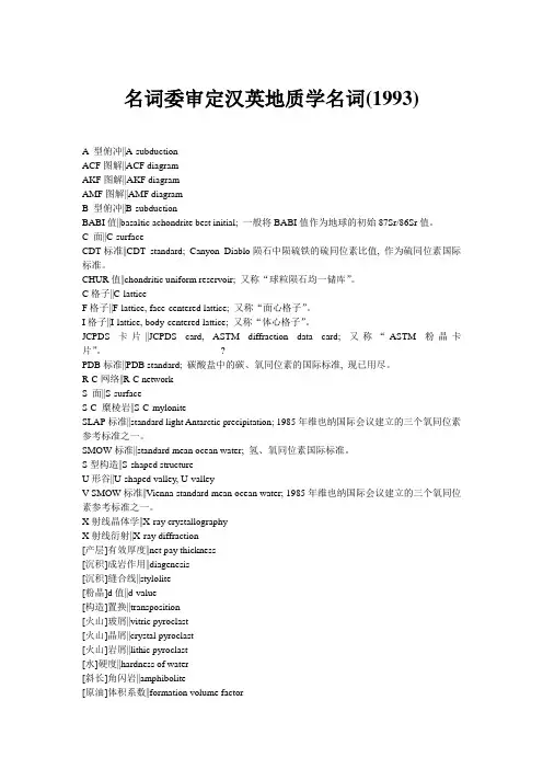
名词委审定汉英地质学名词(1993)A 型俯冲||A-subductionACF图解||ACF diagramAKF图解||AKF diagramAMF图解||AMF diagramB 型俯冲||B-subductionBABI值||basaltic achondrite best initial; 一般将BABI值作为地球的初始87Sr/86Sr值。
C 面||C-surfaceCDT标准||CDT standard; Canyon Diablo陨石中陨硫铁的硫同位素比值, 作为硫同位素国际标准。
CHUR值||chondritic uniform reservoir; 又称“球粒陨石均一储库”。
C格子||C-latticeF格子||F-lattice, face-centered lattice; 又称“面心格子”。
I格子||I-lattice, body-centered lattice; 又称“体心格子”。
JCPDS卡片||JCPDS card, ASTM diffraction data card; 又称“ASTM粉晶卡片”。
?PDB标准||PDB standard; 碳酸盐中的碳、氧同位素的国际标准, 现已用尽。
R-C网络||R-C networkS 面||S-surfaceS-C 糜棱岩||S-C-myloniteSLAP标准||standard light Antarctic precipitation; 1985年维也纳国际会议建立的三个氧同位素参考标准之一。
SMOW标准||standard mean ocean water; 氢、氧同位素国际标准。
S型构造||S-shaped structureU形谷||U-shaped valley, U-valleyV-SMOW标准||Vienna standard mean ocean water; 1985年维也纳国际会议建立的三个氧同位素参考标准之一。

延边大学医学学报 2022年3月 第45卷 第1期与疾病报告2020概要[J].中国循环杂志,2021,36(6):521-545.[2] MEDINA-LEYTE DJ,ZEPEDA-GARCíA O,DOMíNG-UEZPéREZ M,et al..Endothelial dysfunction,inflam-mation and coronary artery disease:potential biomark-ers and promising therapeutical approaches[J].Int JMol Sci,2021,22(8):3850.[3] HUANG C,HUANG WH,WANG R,et al..Ulinastatininhibits the proliferation,invasion and phenotypicswitching of PDGF-BB-induced VSMCs via Akt/eNOS/NO/cGMP signaling pathway[J].Drug Des Devel Ther,2020,14:5505-5514.[4] 赵高峰.延边地区朝鲜族SMARCA4基因SNPrs1122608与冠心病相关性研究[D].延吉:延边大学,2020.[5] IM PK,MILLWOOD IY,KARTSONAKI C,et al..Al-cohol drinking and risks of total and site-specific canc-ers in China:A 10-year prospective study of 0.5millionadults[J].Int J Cancer,2021,149(3):522-534.[6] ROSCH PJ.Could the benefits of drinking alcohol out-weigh all its risks[J].Stress Health,2013.[7] WALLERATH T,POLEO D,LI H,et al..Red wine in-creases the expression of human endothelial nitric oxidesynthase:a mechanism that may contribute to its bene-ficial cardiovascular effects[J].JACC,2003,41(3):471-478.[8] ZHAO F,JI Z,CHI J,et al..Effects of Chinese yellowwine on nitric oxide synthase and intercellular adhesionmolecule-1expressions in rat vascular endothelial cells[J].Acta Cardiologica,2016,71(1):27-34.[9] 金春花,刘文博,关宏锏,等.一氧化氮合酶各种亚型的信号通路对心力衰竭的作用机制与治疗的研究进展[J].中国循环杂志,2017,32(8):830-832.[10]WAGNER MC,YELIGAR SM,LOU AB,et al..PPARγligands regulate NADPH oxidase,eNOS,and barrierfunction in the lung following chronic alcohol ingestion[J].Alcohol Clin Exp Res,2012,36(2):197-206.[11]TIRAPELLI CR,LEONE AF,YOGI A,et al..Ethanolconsumption increases blood pressure and alters the re-sponsiveness of the mesenteric vasculature in rats[J].J Pharm Pharmacol,2008,60(3):331-341.[12]CHEN SS,WANG ZY,ZHOU H,et al..Icariin re-duces high glucose-induced endothelial progenitor celldysfunction via inhibiting the p38/CREB pathway andactivating the Akt/eNOS/NO pathway[J].Exp TherMed,2019,18(6):4774-4780.■■■■■■■■■■■■■■■■■■■■■■■■■■■■■■■■■■■■■■■■■■■■■■■■■■[基金项目] 国家自然科学基金项目(编号:81560179).[收稿日期] 2022-01-03[作者简介] 黄银珠(1995—),女,硕士,研究方向为口腔颌面外科.[通信作者] 金成日,Email:jcr1105@163.com.初级纤毛相关基因KIF3A通过Hedgehog信号通路影响口腔鳞状细胞癌增殖研究黄银珠,李京旭,金成日(延边大学附属医院口腔科,吉林延吉133000)[摘要] [目的]探讨初级纤毛相关基因KIF3A通过Hedgehog信号通路对口腔鳞状细胞癌增殖能力产生的影响.[方法]选择人舌鳞癌Tca8113细胞株作为研究对象,分为KIF3A-siRNA转染组和对照组.采用蛋白印迹实验和实时荧光定量实验检测各组KIF3A、Shh、Gli1蛋白及mRNA的相对表达水平;利用MTT细胞增殖实验观察Tca8113细胞增殖能力的变化.[结果]蛋白印迹实验结果显示,与对照组比较,KIF3A-siRNA转染组KIF3A、Shh、Gli1蛋白表达水平均明显下调(P<0.05);实时荧光定量实验结果显示,与对照组比较,KIF3A-siRNA转染组KIF3A、Shh、Gli1mRNA表达水平均明显下调(P<0.01);MTT细胞增殖实验结果显示,细胞培养24h时两组细胞增殖间差异无统计学意义(P>0.05),细胞培养48、72h时KIF3A-siRNA转染组增殖率较对照组明显降低(P<0.01).[结论]沉默初级纤毛相关基因KIF3A可能通过调控Hedgehog信号通路抑制口腔鳞癌的增殖能力.[关键词] 口腔鳞状细胞癌;初级纤毛;KIF3A;Hedgehog信号通路DOI:10.16068/j.1000-1824.2022.01.002[中图分类号] R 739.8 [文献标志码] A [文章编号] 1000-1824(2022)01-0007-04·7·■■■■■■■■■■■■■■■■■■■■■■■■■■■■■■■■■■■■■■■■■■■■■■■■■■Journal of Medical Science Yanbian University Mar.2022 Vol.45 No.1Effects of primary cilium-associated gene KIF3Aon oral squamous cellcarcinoma proliferation by Hedgehog signaling pathwayHUANG Yinzhu,LI Jingxu,JIN Chengri(Department of Stomatology,Affiliated Hospital of Yanbian University,Yanji 133000,Jilin,China)ABSTRACT:OBJECTIVETo explore the effect of primary cilium-associated gene KIF3Aon oral squamouscell carcinoma proliferation through Hedgehog signaling pathway.METHODS Human tongue squamous cellcarcinoma Tca8113cell line was selected and divided into KIF3A-siRNA transfection group and controlgroup.Western blot and real-time qPCR were used to detect the relative expression levels of KIF3A,Shhand Gli1proteins and mRNA in each group.MTT cell proliferation assay was used to observe theproliferation of Tca8113cells.RESULTS Western blot results showed that compared with the control group,the protein expression levels of KIF3A,Shh and Gli1in the KIF3A-SiRNA transfection group weresignificantly down-regulated(P<0.05).Real-time qPCR results showed that compared with the controlgroup,the mRNA expression levels of KIF3A,Shh and Gli1in the KIF3A-SiRNA transfection group weresignificantly down-regulated(P<0.01).MTT cell proliferation assay showed that there was no significantdifference in cell proliferation between the two groups after 24hcell culture(P>0.05).The proliferationrate of the KIF3A-SiRNA transfection group was significantly lower than that of the control group after cellculture for 48and 72h(P<0.01).CONCLUSIONSilencing primary cilium-associated gene KIF3Amayinhibit the proliferation of oral squamous cell carcinoma by regulating Hedgehog signaling pathway.Keywords:oral squamous cell carcinoma;primary cilia;KIF3A;Hedgehog signaling pathway 口腔癌人数约占全世界罹患恶性肿瘤人数的3%,其中鳞状细胞癌比例超过90%.2005年,WHO将口腔鳞状细胞癌(OSCC)定义为一种具有不同分化程度和侵袭性的肿瘤,早期可出现广泛的淋巴结转移,好发于40~70岁的烟酒爱好者[1].口腔癌患者5年生存率约为50%[2],患者术后预后差和5年生存率低的主要原因为患者未及时就诊、淋巴结转移及复发[3].控制OSCC的侵袭转移能力对预后至关重要,认为可偿试从分子水平出发找出更好的治疗方法[4].初生纤毛属于一种细胞表面的细胞器,在脊椎动物发育和人类遗传疾病中发挥着重要作用.纤毛对发育信号的响应是必需的,而越来越多的证据表明初级纤毛是专属于Hedgehog信号传导的[5],而Hedgehog信号传导对肿瘤的发生发展起着重要作用.研究[6]结果显示,多种肿瘤组织中均存在Hedgehog信号的异常激活,如肺小细胞癌、胰腺癌、前列腺癌、乳腺癌、恶性胶质瘤、髓母细胞瘤及多发性骨髓瘤等.KIF3A属于初级纤毛内运输系统,具有维持和发挥初级纤毛正常功能的作用[7].本研究探讨了初级纤毛相关基因KIF3A通过Hedgehog信号通路对OSCC增殖能力产生的影响.1 材料与方法1.1 细胞 人舌鳞癌细胞株Tca8113购自美国菌种保藏中心(ATCC),置于延边大学附属医院中心实验室封存.1.2细胞培养与转染 取Tca8113细胞置于温度37℃、二氧化碳体积分数5%、饱和湿度95%的恒温孵育箱中,用含10%(质量分数)FBS、青链霉素的DMEM高糖型培养基传代培养.在转染前1d,将Tca8113细胞分为对照组和KIF3A-siRNA转染组后接种至6孔板中,待生长至80%时进行细胞转染.KIF3A-siRNA转染组转染KIF3A-siRNA,对照组未进行转染.转染操作按照LipofectamineTM2000Re-agen转染说明书进行.KIF3A-siRNA序列(5′-3′):UAA GGA AUG CUG AAG AAG ACG AGU C.1.3 蛋白印迹实验 转染24h后分别收集两组细胞,并加入RIPA与PMSF混合(100:1)冰上裂解细胞,提取细胞总蛋白,采用BCA试剂盒测定总蛋白水平.取各组标本行8%(质量分数)十二烷基硫酸钠聚丙烯酰胺凝胶(SDS-PAGE)电泳,转膜,室温下用5%(质量分数)脱脂奶粉封闭2h.TBST洗涤3·8·■■■■■■■■■■■■■■■■■■■■■■■■■■■■■■■■■■■■■■■■■■■■■■■■■■延边大学医学学报 2022年3月 第45卷 第1期次,每次10min,加入兔抗人Kif3a一抗(1:1 000)、兔抗人Shh一抗(1:2 000)、兔抗人Gli1(1:1 000)及兔抗人GAPDH(1:1 000)后置于湿盒中,于4℃冰箱中孵育过夜;次日TBST洗涤3次,每次10min,分别加入山羊抗兔IgG二抗(1:10 000)后置于湿盒中室温孵育2h;TBST洗涤3次,每次10min,加入增强型化学发光剂ECL显影.以GAPDH为内参,采用Imagine J软件分析蛋白条带灰度值.1.4 实时荧光定量实验 转染24h后分别收集两组细胞,采用TRNzol法提取细胞中的总RNA,反转录为cDNA,按照RT-PCR试剂盒说明书以cDNA为模板进行扩增.测定循环阈值(Ct),采用2-△△Ct法进行统计.实验重复3次.引物序列,KIF3A-正向:5′-AGA GCG TCA ACG AGG TGT TT-3′;KIF3A-反向:5′-TAT TGA TCG GCA TCT TGG CCC-3′;Shh-正向:5′-CTC GCT GCT GGT ATG CTC G-3′;Shh-反向:5′-ATC GCT CGG AGT TTC TGGAGA-3′;Gli1-正向:5′-AGC GTG AGC CTG AATCTG TG-3′;Gli1-反向:5′-CAG CAT GTA CTGGGC TTT GAA-3′;GAPDH-正向:5′-GGA GCGAGA TCC CTC CAA AAT-3′;GAPDH-反向:5′-GGC TGT TGTCATACT TCTCATGG-3′.1.5 MTT细胞增殖实验 转染24h后分别收集两组细胞,以5 000个/孔接种于96孔板中,每组设置5个复孔.培养24、48、72h后弃去旧培养液,每孔加入100μL MTT(5 g/L),于37℃、5%(体积分数)二氧化碳、95%饱和湿度的恒温孵育箱中培养4 h,弃去上清液终止反应,每孔加入100μL DMSO,置于振荡器3 min溶解结晶后在酶标仪波长490 nm处测定各孔的光密度(OD)值,计算相对细胞增殖率.相对细胞增殖率(%)=(实验组OD值/对照组OD值)×100%.1.6 统计学分析 采用Graphpad Prism 7.0软件进行数据分析.计量数据以均数±标准偏差( x±s)表示,进行两个样本均数的t检验,以P<0.05为差异有统计学意义.2 结果2.1 沉默KIF3A基因后对Tca8113细胞Hedgehog信号通路中Shh、Gli1蛋白表达水平的影响 与对照组比较,KIF3A-siRNA转染组24h时KIF3A、Shh及Gli1蛋白表达水平均明显降低,差异均具有统计学意义(P<0.05),见Figure 1.compared with control,*P<0.05.Figure 1 The protein expression levels of KIF3A,Shhand Gli1decreased significantly aftertransfection of Tca8113cells2.2 沉默KIF3A基因后对Tca8113细胞中Hedgehog信号通路Shh、Gli1mRNA表达水平的影响 与对照组比较,KIF3A-siRNA转染24h组KIF3A、Shh及Gli1mRNA表达水平均显著降低,差异均具有统计学意义(P<0.01),见Figure 2.compared with control,**P<0.01.Figure 2 The mRNA expression levels of KIF3A,Shhand Gli1decreased significantly aftertransfection of Tca8113cells2.3 沉默KIF3A基因后对Tca8113细胞增殖能力的影响 MTT检测结果显示,细胞培养24h时KIF3A-siRNA转染组与对照组细胞增殖间差异无统计学意义(P>0.05);细胞培养48、72h时,KIF3A-siRNA转染组与对照组比较增殖显著受到抑制(P<0.01),见Figure 3.·9·■■■■■■■■■■■■■■■■■■■■■■■■■■■■■■■■■■■■■■■■■■■■■■■■■■Journal of Medical Science Yanbian University Mar.2022 Vol.45 No.1compared with control,**P<0.01.Figure 3 The proliferation rate of Tca8113cells aftertransfection3 讨论初级纤毛为多数分化型细胞表面上的感觉附属物,具有化学感觉和机械感觉功能,在细胞周期控制中具有重要的作用.以往的研究认为初级纤毛在快速增殖的细胞上是找不到的,如癌症细胞,但KOWAL等[8]通过一种初级纤毛标志物Arl13b在HeLa和MG63细胞中发现了初级纤毛.未在OSCC中查看是否存在初级纤毛是本研究的不足之处.KIF3A属于初级纤毛内运输系统,在维持和发挥初级纤毛功能中起重要作用.HOANG-MINH等[9]的研究结果显示,沉默KIF3A基因表达后可降低胶质母细胞瘤中初级纤毛的数量,最终促进肿瘤的发生.玄云泽等[10]首次在OSCC中发现有初级纤毛,后来有学者亦在口腔白斑与OSCC组织中发现了初级纤毛,EGFR-Aurora A异常信号被激活后,口腔黏膜癌变过程中原发性纤毛逐渐减少[11].本研究结果显示,利用siRNA技术沉默KIF3A基因后mRNA和蛋白表达水平明显降低,提示初级纤毛的检出率降低.Hedgehog信号通路对胚胎发育及肿瘤的发生发展具有重要意义.研究[12-13]结果显示,Hedgehog信号通路的异常激活与皮肤、乳腺、肺及消化道等多种恶性肿瘤的发生、增殖密切相关.程志芬等[12]利用Hedgehog信号通路特异性抑制剂环巴胺处理Tca8113细胞株后,Smoothened的mRNA和蛋白表达明显减少,且细胞增殖受到抑制.KURODA等[14]的研究结果显示,给予Hedgehog信号通路抑制剂可抑制肿瘤前沿血管内皮细胞的增殖、迁移,进而抑制肿瘤的增殖和迁移.在小细胞肺癌中,沉默KIF3A后初级纤毛明显减少,且Shh信号通路介导的Gli1mRNA表达水平明显降低[15],此结果与本研究结果相符.研究[16]结果显示,Hedgehog信号通路转录因子Gli1的激活是恶性肿瘤的一个不良预后因素,利用环巴胺阻断Hedgehog信号通路可抑制Gli1激活,并增强头颈部鳞癌对放疗的敏感性,提示Hedgehog信号通路影响头颈部鳞癌的放疗预后.总之,本研究揭示了初级纤毛相关基因KIF3A在人舌鳞癌Tca8113细胞Hedgehog信号通路中的作用,初步解释了沉默KIF3A后可能通过抑制Hedgehog信号通路影响OSCC细胞的增殖能力,为OSCC患者的靶向治疗提供了理论依据.[参 考 文 献][1] DI PARDO BJ,BRONSON NW,DIGGS BS,et al..Theglobal burden of esophageal cancer:a disability-adjustedlife-year approach[J].World J Surg,2016,40(2):395-401.[2] BROCKLEHURST PR,BAKER SR,SPEIGHT PM.Oralcancer screening:what have we learnt and what is therestill to achieve[J].Future Oncol,2010,6(2):299-304.[3] BAVLE RM,VENUGOPAL R,KONDA P,et al..Mo-lecular classification of oral squamous cell carcinoma[J].JCDR,2016,10(9):Z18-Z21.[4] 陈新,徐文华,周健,等.口腔鳞状细胞癌现状[J].口腔医学,2017,37(5):462-465.[5] GOETZ SC,ANDERSON KV.The primary cilium:a signalling centre during vertebrate development[J].Nat Rev Gene,2010,11(5):331-344.[6] 那玉岩,刘万林.纤毛相关疾病:细胞学机制及转化应用进展[J].中国组织工程研究,2016,20(24):3642-3648.[7] NIEWIADOMSKI P,NIEDZIółKA SM,MARKIEWICZŁ,et al..Gli proteins:regulation in development andcancer[J].Cells,2019,8(2):147.[8] KOWAL TJ,FALK MM.Primary cilia found on HeLaand other cancer cells[J].Cell Biol Int,2015,39(11):1341-1347.[9] HOANG-MINH LB,DELEYROLLE LP,SIEBZEH-NRUBL D,et al..Disruption of KIF3Ain patient-de-rived glioblastoma cells:effects on ciliogenesis,hedge-hog sensitivity,and tumorigenesis[J].Oncotarget,2016,7(6):7029-7043.[10]玄云泽,李书进,金成日,等.初级纤毛相关基因KIF3A通过调控EMT过程在口腔鳞状细胞癌中的作用机制研究[J].重庆医学,2020,49(1):7-12.[11]YIN F,CHEN Q,SHI Y,et al..Activation of EGFR-Au-rora A induces loss of primary cilia in oral squamous cellcarcinoma[J].Oral Dis,2022,28(3):621-630.[12]程志芬,崔演,玄延花.Ptch和Smo在舌鳞状细胞癌Tca8113细胞中的表达及其意义[J].口腔医学研究,2015,31(5):433-436.[13]KONSTANTINOU D,BERTAUX-SKEIRIK N,ZAVROSY.Hedgehog signaling in the stomach[J].Curr OpinPharmacol,2016,31:76-82.[14]KURODA H,KURIO N,SHIMO T,et al..Oral squamouscell carcinoma-derived sonic hedgehog promotes angiogene-sis[J].Anticancer Res,2017,37(12):6731-6737.[15]COCHRANE CR,VAGHJIANI V,SZCZEPNY A,et al..Trp53and Rb1regulate autophagy and ligand-dependentHedgehog signaling[J].J Clin Invest,2020,130(8):4006-4018.[16]GAN GN,EAGLES J,KEYSAR SB,et al..Hedgehogsignaling drives radioresistance and stroma-driventumor repopulation in head and neck squamous cancers[J].Cancer Res,2014,74(23):7024-7036.■·01·■■■■■■■■■■■■■■■■■■■■■■■■■■■■■■■■■■■■■■■■■■■■■■■■■■。

应用游离前锯肌瓣矫治半侧颜面萎缩的初步临床研究田卫东;王栋;刘磊;黄富国;谢倩;闫征斌;李逸松;李声伟【期刊名称】《中国修复重建外科杂志》【年(卷),期】2005(19)10【摘要】目的探讨应用游离前锯肌瓣矫治半侧颜面萎缩的可行性,并讨论前锯肌瓣的解剖、制作及手术中注意的问题。
方法2001年9月~2004年7月,对3例严重半侧颜面萎缩患者采用游离前锯肌瓣移植术,其前锯肌瓣以胸背动、静脉为蒂,与颞浅动、静脉,颌外动、静脉,舌动、静脉等吻合;术中同时解剖胸长神经,并与面神经上颊支吻合。
游离肌瓣大小12 cm×8 cm^16 cm×12 cm。
结果3例患者术后前锯肌瓣游离移植颜面部凹陷区后色泽血运均良好,肌瓣完全成活,面颊部丰满圆润,无明显突起或不平,颌面部切口愈合好,瘢痕不明显,肩部运动良好,无“翼状肩”。
随访1~3年,患者外形恢复满意。
结论应用游离前锯肌瓣矫治严重半侧颜面萎缩安全可靠,术后有利于改善患者外形。
远期疗效尚待进一步观察。
【总页数】4页(P799-802)【关键词】游离前锯肌瓣;半侧颜面萎缩;矫治【作者】田卫东;王栋;刘磊;黄富国;谢倩;闫征斌;李逸松;李声伟【作者单位】四川大学华西口腔医院口腔颌面外科;华西医院骨科【正文语种】中文【中图分类】R782.26;R616.2【相关文献】1.自体真皮脂肪瓣游离移植矫治半侧颜面萎缩 [J], 刘振中;张基勋;王魏;张敬;蔡景龙2.游离背阔肌肌瓣联合自体脂肪填充序列治疗修复半侧颜面萎缩 [J], 唐泓波;陈雪;周芳;冯幼平3.股前外侧脂肪筋膜瓣游离移植矫治半侧颜面萎缩 [J], 滕利;吴国平;张智勇;丁波;靳小雷;季滢4.游离背阔肌肌瓣在修复半侧颜面萎缩症中的应用 [J], 刘五一;隋良朋5.应用游离股前外侧脂肪筋膜瓣矫治半侧颜面萎缩的临床研究 [J], 任敏;滕利;归来;张智勇;冯国平;靳小雷;庄洪兴因版权原因,仅展示原文概要,查看原文内容请购买。
世界地质G lobal G eolog y,2003,22(4):366~372文章编号1004O5589(2003)04O0366O07马来西亚沙捞越邦达、什兰江控矿角砾岩筒构造对比研究及其找矿意义*赵财胜孙丰月李碧乐丁清峰吴尚昆吉林大学地球科学学院,长春130061摘要马来西亚沙捞越邦达、什兰江控矿角砾岩筒构造均为典型的浅成-半浅成角砾岩筒构造,角砾岩及控矿类型具有明显的垂向分带特点。
邦达控矿角砾岩筒构造发育角砾岩筒上段的塌陷角砾岩,角砾岩筒剥蚀深度较浅,控制典型的高硫化型浅成低温金铜矿床,且其深部具有较大的找矿潜力。
什兰江控矿角砾岩筒构造发育角砾岩筒下段的爆发角砾岩和中段的流化角砾岩,分别控制斑岩型金矿化和低硫化型浅成低温热液型金矿化,角砾岩筒剥蚀深度较大,对矿床的保存和找矿极为不利。
关键词浅成)半浅成角砾岩筒构造角砾岩沙捞越邦达什兰江马来西亚中图分类号P618.4文献标识码AC omparative Study of Punda and Selanjan Ore--control Breccia Pipe Structurein Sarawak,Malaysia and Its Ore--prospecting SignificanceCaisheng Zhao,Fengyue Sun,Bile Li,Qingfeng Ding,Shangkun WuCollege o f Earth S c iences,Jilin University,Cha ngchu n,130026ChinaAbstract Ore--control breccia pipe structure of Punda and Selanjan in Sarawak,Malaysia,is a typical hy-pabyssal breccia pipe structure.Breccias in hypabyssal breccia pipe and types of m ineralization controlled by hy-pabyssal breccia pipe possess distinct vertical zonation.In Punda ore--control breccia pipe,the breccias are char-acteristic collapse breccia in the superficial segment or topside of the hy pabyssal breccia pipe,indicating its de-nudation is superficial.Furthermore,ty pes of mineralization controlled by the hypabyssal breccia pipe belong to high sulphidation epithermal deposit and have an excellent exploration prospect in the deep.In Selanjan ore--con-trol breccia pipe,the breccias are characteristic ex plosive breccia at the bottom of the breccia pipe and rheonor-phic breccia in the middle segment of the breccia pipe.At the same time,the ty pes of mineralization controlled by ex plosive breccia and rheonorphic breccia are porphyry gold m ineralization and low sulphidation epithermal mineralization respectively.Denudation is deep and harmful to the preservation and exploration of the gold de-posit.Key words hypabyssal breccia pipe structure,collapse breccia,rheonorphic breccia,ex plosive breccia, Saraw ak Punda,Selanjan,M alaysia收稿日期2003O07O21;改回日期2003O10O13*吉林大学地球科学学院与加拿大T M C公司国际合作项目(3B6010014015)资助.作者简介赵财胜(1976-),男,内蒙古凉城人,博士研究生,从事热液矿床成矿理论及预测研究.通讯作者简介孙丰月(1963-),男,山东淄博人,教授,博士生导师,从事区域成矿作用、幔源流体及其成岩成矿研究.自1870年首次在金伯利岩角砾岩筒中发现金刚石以来,角砾岩筒构造作为一种重要的控矿构造,在国外已得到广泛的重视和详尽的研究[1-7]。
西南医科大学学报2021年第44卷第5期JournalofSouthwestMedicalUniversityVol.44No.52021
隐丹参酮对人下咽鳞癌细胞FaDu增殖及迁移的影响高琳1,费静2,刘毅1,李晓红1
1.西南医科大学附属医院健康管理中心(泸州646000);2.西南医科大学附属医院耳鼻咽喉头颈外科(泸州646000)
【摘要】目的本研究通过体外实验探讨隐丹参酮对人下咽鳞癌细胞的增殖及迁移的影响。方法用不同浓度(0μM、2.5μM、5μM、10μM)的隐丹参酮处理FaDu细胞。通过细胞克隆、细胞划痕与Transwell实验分析药物对细胞增殖、迁移能力
的影响。Westernblot法检测周期相关蛋白和EMT相关蛋白的表达水平。结果与对照组相比,隐丹参酮能有效抑制FaDu细胞增殖及克隆形成能力(P<0.01),且呈剂量依赖性。在用隐丹参酮处理24h后,处理组细胞的划痕愈合能力及细胞迁移能力较对照组明显降低,呈剂量依赖性。药物处理后,AKT蛋白、细胞周期相关蛋白(CDK4和CyclinD)及EMT相关蛋白Vimentin表达明显下调。结论隐丹参酮能有效抑制人咽鳞癌细胞FaDu的增殖和迁移,其作用机制可能与其下调AKT、CDK4、CyclinD及Vimentin蛋白的表达有关,进而抑制细胞,表现出对较强的细胞毒性。【关键词】隐丹参酮人下咽鳞癌细胞增殖细胞迁移【中图分类号】R453.9文献标志码ADOI:10.3969/j.issn.2096-3351.2021.05.021
EffectofcryptotanshinoneontheproliferationandmigrationofhumanhypopharyngealsquamouscellcarcinomaFaDucells
GAOLin1,FEIJing2,LIUYi1,LIXiao-hong1
1.DepartmentofHealthManagementCenter,theAffiliatedHospitalofSouthwestMedicalUniversity,Luzhou


Acta Protozool. (2006) 45: 27 - 39 Redescriptions of Three Marine Peritrichous Ciliates, Zoothamnium alternans Claparède et Lachmann, 1859, Z. sinense Song, 1991 and Z. commune Kahl, 1933 (Ciliophora, Peritrichia), from North ChinaDaode JI1, 2, Weibo SONG1 and Alan WARREN31Laboratory of Protozoology, KLM, Ocean University of China, Qingdao; 2Ocean School of Yantai University, Yantai, People’s Republic of China; 3Department of Zoology, Natural History Museum, London, UKSummary. Three marine peritrichous ciliates, Zoothamnium alternans Claparède et Lachmann, 1859, Z. sinense Song, 1991and Z. commune Kahl, 1933, were isolated from coastal waters near Qingdao, China. The living morphology, infraciliature and silverline system were studied in both living and silver-impregnated specimens. Based on the Qingdao populations, improved diagnoses and redescriptions are provided for each species: Z. alternans is distinguished from its congeners by the alternately branched stalk, the macrozooids on the main stalk trunk, the J-shaped macronucleus and in having 40-55 silverlines between the oral area and the trochal band and 20-30 between the trochal band and the scopula; Z. sinense can be recognized by the small zooid size, the alternately branched stalk, and in having 55-70 silverlines between the oral area and the trochal band and 20-35 between the trochal band and the scopula; Z. commune can be distinguished from other species of similar zooid shape and size by the form of peniculus 3 and the number of silverlines between the oral area and the trochal band (59-70) and between the trochal band and the scopula (38-43).Key words: marine ciliate, morphology, Peritrichida, Zoothamnium.INTRODUCTIONZoothamnium Bory de St. Vincent, 1826 is a well-known genus with more than 70 species, many of which are abundant in marine biotopes (Kent 1880-1882, Kahl 1935, Stiller 1971, Küsters 1974, Corliss, 1979, Jankowski 1985). In common with most other genera of sessile peritrichs, however, few Zoothamnium species have been studied using modern methods (e.g. silver impreg-nation) as recommended by Foissner and Schiffmann (1974). Furthermore, there has been no revision of this genus since that of Kahl (1935). Consequently species separation and identification among Zoothamnium spp. is often very difficult (Kahl 1933; Precht 1935; Sommer 1951; Stiller 1953a, b; Stiller and Stevèiæ 1967).In the present study we report on three marine Zoothamnium species collected from the littoral zone of coastal waters near Qingdao, China, following observa-tions on both living and silver- impregnated specimens.Address for correspondence: Weibo Song, Laboratory of Proto-zoology, KLM, Ocean University of China, Qingdao 266003, People’s Republic of China; Fax: +86 532 203 2283; E-mail: wsong@2Current address: Ocean School of Yantai University, Yantai 264005, People’s Republic of China28 D. Ji et al.All three species are known forms for which detailed information concerning the infraciliature and silverline system are lacking. Therefore, based on the Qingdao populations, an improved diagnosis and redescription of each species is presented here.MATERIALS AND METHODSSample collection. Ciliates were collected from coastal waters at three sites in the Qingdao region, P.R. China, (36°08’N; 120°43’E) from both natural (Zoothamnium alterans) and artificial (Zoothamnium sinense, Z. commune) substrates. The natural substrate was the shell of the scallop Chlamys sp. The artificial substrates were glass slides fixed to a frame that was immersed in water and left for 10 days to allow colonization to occur. After this time the slides were retrieved and transported to the laboratory for examination.Zoothamnium alternans was collected on 19 April 2004 from a scallop (Chlamys sp.) farming pond, water temperature 12°C, salin-ity about 30‰.Zoothamnium sinense was collected on 10 July 2002 from the littoral zone of coastal waters off Qingdao, water temperature 26°C, salinity 30‰.Zoothamnium commune was collected on 22 May 2002 from a shrimp-farming pond, water temperature 20°C, salinity 28‰.Observations: Ciliates were observed in vivo using bright field and differential interference contrast microscopy. The infraciliature was revealed with protargol impregnation according to Wilbert (1975). The “dry” silver nitrate method (Foissner 1976) was used to demon-strate the silverline system.Drawings and terminology: Drawings of impregnated speci-mens were made with the help of a camera lucida at ×1250 magnifica-tion. Terminology is mainly according to Foissner and Schiffmann (1974) and Warren (1986).Deposition of slides: Two slides of Zoothamnium alternans (No. 0404190101, 0404190102) with protargol-impregnated speci-mens are deposited at the Laboratory of Protozoology, OUC, China. Another slide (No. 2005:3:24:7) is deposited in the collections of the Natural History Museum, London, U.K.One slide of Zoothamnium sinense (No. 020*******) with protargol-impregnated specimens is deposited in the collection of the Laboratory of Protozoology, OUC, China.One slide of Zoothamnium commune (No. 020*******) with protargol-impregnated specimens is deposited at the Laboratory of Protozoology, OUC, China. Another slide (No. 2005:3:24:8) is de-posited in the collections of the Natural History Museum, London, U.K.RESULTSClass: Oligohymenophora de Puytorac et al., 1974 Subclass: Peritrichia Stein, 1859Order: Sessilida Kahl, 1933Family: V orticellidae Ehrenberg, 1838Genus: Zoothamnium Bory de St. Vincent, 1826Redescription of Zoothamnium alternans Claparède et Lachmann, 1859 (Figs 1, 2; Table 1) Synonym: Zoothamnium chlamydis Hu et Song, 2001 (p216)Although Zoothamnium alternans has been known for a long time (Claparède and Lachmann 1859; Kent 1880-1882; Kahl 1933, 1935), it has never been investi-gated using silver impregnation methods. Therefore its infraciliature and silverline system remain unknown. Based on the population found in Qingdao, a detailed redescription and an improved diagnosis are presented here.Amended diagnosis: Marine Zoothamnium with alternately branched stalk. Cells differentiated into mi-cro- and macrozooids. Macrozooids ovoid, ca 70-90 ×45-55 µm in vivo, located only on main stalk trunk; microzooids inverted bell-shaped, 40-56 × 26-32 µm in size. Both micro- and macrozooids have a J-shaped macronucleus and an apically located contractile vacu-ole. Number of silverlines from oral area to aboral trochal band ca 40-55, from aboral trochal band to scopula ca 20-30.Amended description: Colonies with micro- and macrozooids. Microzooid inverted bell-shaped, 40-56 ×26-32 µm in vivo, with thin, distinctly everted peristomial lip and moderately elevated peristomial disc (Figs 1A, G). Cytoplasm colourless to slightly grey, usually con-taining granules and several vacuoles (Fig. 1A). Macro-nucleus J-shaped in lateral view. Oral section of macro-nucleus elongate, cylindroid in cross-section, semicircu-lar, transversely oriented. End of oral section sometimes recurved aborally (Fig. 1G). Aboral continuation of macronucleus cylindroid, straight, oriented diagonally to longitudinal axis. Aboral extremity of macronucleus usu-ally recurved orally. Single contractile vacuole located slightly above peristomial lip and pulses slowly (3-5 min intervals). Pellicle appears smooth, usually covered with a layer of rod-shaped bacteria (Figs 1B, 2D). Fine pellicular striations can be observed occasionally under ×1000 magnification (Fig. 2H). Number of silverlines from aboral trochal band to scopula ca 20-30, from peristome to aboral trochal band ca 40-55.Macrozooid oval-shaped, 70-90 × 45-55 µm in vivo, widest at mid-body, with thick peristomial lip, J-shaped macronucleus and single, apically located contractile vacuole (Figs 1I, 2A). Macrozooids located only on main trunk of stalk, one to several per colony, but absent at newly formed colonies. Telotroch acetabuliform (sau-On three marine peritrichous ciliates29Figs 1A-I.Zoothamnium alternans from life (A, B, D, E, G-I) and after protargol impregnation (C, F). A - a micozooid; B - bacteria on pellicle;C - detail of infundibular polykineties;D - stalk structure;E - colony form, from Grell (1968);F - oral infraciliature, arrow marks the distal kinety fragment;G - lateral view of a microzooid; H- a telotroch; I - a macrozooid. EM - epistomial membrane; G - germinal kinety,H - haplokinety, P1-3 - infundibular polykinety 1-3, Po - polykinety. Scale bars: 30 µm (1A, G-I); 10 µm (1B); 20 µm (1D); 300 µm (1E).30 D. Ji et al.Figs 2A-H. Photomicrographs of Zoothamnium alternans from life (A-E) and after protargol impregnation (F-H). A- colony form; B - showing a telotroch, a macrozooid and several microzooids; C - a telotroch; D - to show bacteria covering the pellicle; E - to show structure of stalk;F - lateral view, arrowheads mark the aboral trochal band;G - to show the epistomial membrane (arrow);H - silverline system. Scale bars: 300 µm (2A); 200 µm (2B); 50 µm (2C).On three marine peritrichous ciliates31cer-shaped), 100-120 µm in diameter and 40-50 µm in height, with large C-shaped macronucleus (Figs 1H; 2B, C). Telotrochs only formed by macrozooids.Colonies up to 1.2 mm high, alternately branched, and containing up to 120 zooids (Figs 1E, 2A). Stalk with smooth surface, main stalk trunk 18 µm in diameter, 8 µm at distal end. Stalk spasmoneme 3-6 µm in width, com-prising a dark fibrous bundle within a transparent thecoplasmic sheath, sometimes with a few mitochon-dria (Figs 1D, 2E).Buccal apparatus of similar structure to most conge-ners. Haplokinety and polykinety describing one and one-third turns around peristomial disc before entering infundibulum, where they make another turn (Fig. 1F). One short kinety fragment is always visible at the peristomial end of the haplokinety (Fig. 1F, arrow).In infundibulum, polykinety (P1) is accompanied by two other polykineties, P2 and P3, each of which consists of three ciliary rows (Fig. 1C). P1 and P2 much longer than P3, and originate from mid-infundibulum. P1 extends posteriorly to end of infundibulum, where it converges with P3 and the two terminate at the same level. Three rows of P2 terminate at different levels and significantly above the posterior ends of P1 and P3 (Fig. 1C). Germinal kinety parallel to haplokinety within abstomal half of infundibulum (Fig. 1F). Epistomial mem-brane short, located at opening of oral cavity (Figs 1F; 2G, arrow).Aboral trochal band composed of a row of dikinetids which encircle the cell in aboral third of zooid (Fig. 2F).Remarks: Given its highly distinctive morphology, i.e. alternately branched stalk, zooid size and appear-ance, marine habitat and the presence of macrozooids, the identity of the Qingdao population as Z. alterans is beyond reasonable doubt (Claparède and Lachmann 1859, Grell 1968).Zoothamnium alternans is similar to Z. niveum Ehrenberg, 1838, which also has an alternately branched stalk, macrozooids on the main trunk of the stalk and a marine habitat, but the latter forms considerably larger colonies (> 1 cm high vs. 1.2 mm high in Z. alternans) and has a relatively shorter P2 (about equal to P3 in length vs. more than twice as long as P3 in Z. alternans), thus the two can be clearly separated (Bauer-Nebelsick et al. 1996).Another marine species, Zoothamnium plumula Kahl, 1933, also has enlarged zooids and an alternately branch-ing stalk, and so is somewhat similar as the current organism. However, these two species can be separated by the number of silverlines between the aboral trochal band and the peristomial border (ca 70 in Z. plumula vs. 40-55 in Z. alternans) and the location of the macrozooids which are located at the distal ends of the accessory branches in Z. plumula vs. on the main trunk in Z. alternans (Song et al. 2002).Additionally, two other forms of Z. alternans were reported by Greeff (1870) and Kent (1880-1882) re-spectively, both of which are considered to be misidentifications. The population described by Kent has many enlarged zooids on the accessory branches butTable 1. Morphometric characters of Zoothamnium alternans (first line, data for microzooids), Z. sinense (second line) and Z. commune (third line). Min - minimum, Max - maximum, Mean - arithmetic mean, SD - standard deviation, n - sample number.Min Max Mean SD n Body length in vivo in µm405648.4 4.9116364840.7 3.4696010484.412.910 Body width in vivo in µm263229.3 1.6516303833.3 2.659485650.0 2.839 No. of silverlines from oral area to aboral trochal band4055--455*70*--4597065.0 3.8010 No. of silverlines from aboral trochal band to scopula2030--320*35*--4384340.6 1.679* Detected from living cells.32 D. Ji et al.lacks typical macrozooids on the main stalk trunk while Greeff’s population possesses long cylindrical microzooids of a type that are not found in Z. alternans.Zoothamnium chlamydis Hu et Song, 2001 has a strong similarity to Z. alternans in all key characters (Table 2). Therefore Z. chlamydis is considered to be a junior synonym of Z. alternans (Hu and Song 2001). Redescription of Zoothamnium sinense Song, 1991 (Figs 3, 4; Table 1)Synonyms: Zoothamnium commune sensu Song, 1991 (p47); Z. truncatum Song, 1986 (primary homonym) (p229)Amended diagnosis: Marine Zoothamnium with alternately branched stalk; zooids inverted bell-shaped, measuring about 36-48 × 30-38 µm in vivo; peristomial disc flat, occasionally very slightly elevated above dis-tinctly everted peristomial lip; macronucleus C-shaped and transversely oriented; single contractile vacuole apically located; pellicle finely striated, with ca 20-35 striations from scopula to aboral trochal band and ca 55-70 striations from aboral trochal band to oral area.Amended description: Zooid inverted bell-shaped, usually measuring 36-48 × 30-38 µm in vivo, with maximum width just below everted, single-layered peris-tomial lip (Figs 3A, I; 4B). Peristomial disc flat, occa-sionally slightly elevated above peristomial lip (Figs 3I,4B). Pellicle appears smooth at low magnification, fine pellicle striations only visible at ×1000 magnification or higher.Cytoplasm colourless, usually containing granules and a few food vacuoles (Figs 3A, I). Single contractile vacuole apically positioned (Figs 3A, I; 4C, arrow). Macronucleus generally C-shaped, transversely oriented, located in oral 2/3 of body and surrounds the aboral portion of infundibulum (Figs 3A, I).Cells highly sensitive to stimuli and contract readily. Also zooids readily convert to telotrochs, which are acetabuliform, ca 25 µm high and 45 µm in diameter (Figs 3E; 4B, arrow).Colony ca 400 µm high, containing up to 40 zooids (Figs 3B, 4A); macrozooid formation was not observed. Stalk smooth, typically alternately branched, 6-9 µm in diameter. Spasmoneme 1.5-3 µm in diameter, mitochon-dria not observed.Oral infraciliature as shown in Figs 3J, K and 4D, F. Haplo- and polykinety both make one and one-third turns of peristome before entering infundibulum, where they make a further turn.At adoral end of polykinety, a short kinety fragment is always recognizable (Fig. 3J, double arrowhead). Each of three infundibular polykineties consisting of three ciliary rows. Rows of P1 about equal in length, terminating at end of infundibulum. P2 about half length of P1. Inner two rows of P2 lie close to P1 while outer one is separated from the other two; all threeTable 2. Morphological comparison between three Zoothamnium spp. and morphologically similar congeners. Measurements in µm. Species Body Body Number Number of Shape of Habitat Data source length width of silverlines silverlines macronucleusin vivo in vivo from peristo-from aboralme to aboral trochal bandtrochal band to scopulaZ. alternans40-5626-3240-5520-30J-shaped marine present studyZ. chlamydis50-60-27-4719-29vertically marine Hu and Song (2001)C-shapedZ. commune60-10448-5659-7038-43C-shaped marine present studyZ. hentscheli63-8435-42--C-shaped freshwater Kahl (1935)Z. niveum54-6616-22--C-shaped marine Bauer-Nebelsicket al. (1996)Z. plumula50-10030-507024-28C-shaped marine Song et al. (2002) Z. sinense36-4830-3855-7020-35C-shaped marine present studyZ. sinense*33-6129-53--C-shaped marine Song (1991)Z. thiophilum90-95---C-shaped freshwater Stiller (1946)*Called Z. commune sensu Song, 1991On three marine peritrichous ciliates33Figs 3A-K.Zoothamnium sinense Song, 1991 from life (A-I) and after protargol impregnation (J, K). A - a typical zooid; B - colony form; C, D - zooid (C) and colony (D), after Song (1991) (called Z. commune); E -telotroch; F-H - colony (F) and zooid (H), after Song (1986) (called Z. truncatum); I - zooid, lateral view; J - general oral infraciliature, arrow depicts the epistomial membrane, double arrowhead marks the distal kinety fragment; K - detail of infundibular polykineties. G - germinal kinety, H - haplokinety, P1-3 - infundibular polykinety 1-3, Po - polykinety. Scale bars: 20 µm (3A, C, E, G-I); 150 µm (3B, D, F).34 D. Ji et al.Figs 4A-I. Photomicrographs of Zoothamnium sinense from life (A-C, G, H) and after protargol (D-F) and silver nitrate impregnation (I).A - a small colony;B - to show zooid shape and a developing telotroch (arrow);C - zooid at high magnification, to show the contractile vacuole (arrow);D - oral infraciliature, arrow marks the epistomial membrane;E - to show aboral trochal band;F - to show infundibular polykineties.G - photomicrograph of holotype of Z. sinense as described in Song (1991); H - photomicrograph of specimen from population described by Song (1991) (called Z. commune); I - silverline system of specimen from population described by Song (1991), arrows mark the aboral trochal band. P1-3 - infundibular polykinety 1-3. Scale bars: 150 µm (4A, G); 50 µm (4B, C, H).On three marine peritrichous ciliates35rows of P2 end conspicuously above the posterior end of P1(Figs 3J, K). P3 about 2/3 length of P2, anterior end considerably below those of P1 and P2 and terminates posteriorly above P1 but below P2. Abstomal half of inner row of P3 separated from the other two ciliary rows but converges with them adstomally (Figs 3J, K;4F). Haplokinety passes around infundibulum on wall opposite to peniculi; germinal kinety runs parallel to haplokinety in abstomal half of infundibulum before terminating in mid-region of infundibulum (Fig. 3J). Epistomial membrane located near opening of infundibu-lum as commonly seen in other peritrichs (Figs 3J arrow; 4D, arrow).Aboral trochal band appears as a row of loosely arranged dikinetids that encircle the body about ¼ of the body length from the scopula (Fig. 4E).Silverline system typical of genus, consisting of many parallel, transverse rows about 0.5 µm apart, ca 55-70 between peristomial area and aboral trochal band, ca 20-35 between aboral trochal band and scopula, and with sparsely distributed pelliclular pores (Fig. 4I). Aboral trochal band stains heavily with silver, probably com-posed of two rows of closely arranged silverlines (Fig. 4I, arrows).Remarks:Zoothamnium sinense was originally re-ported by Song (1986) under the name Z. truncatum (Figs 3F-H, 4G), and later renamed Z. sinense (Song 1991). According to the original description, Z. sinense has small body (33-59 × 35-64 µm), an alternately branched stalk, a single-layered peristomial lip, a C-shaped macronucleus and a marine habitat, all of which correspond well with the present population (Song 1986).Considering general living appearance, the following congeners should be compared with Z. sinense: Z. alternans Claparède et Lachmann, 1859, Z. com-mune Kahl, 1933 and Z. plumula Kahl, 1935 (Table 2). According to our present study, Zoothamnium alternans is similar to Z. sinense in zooid size, stalk branching style and habitat, but can be easily recognized from the latter by the presence of several large macrozooids (vs. absent in Z. sinense), its J-shaped macronucleus (vs. C-shaped in Z. sinense) and in having 40-55 silverlines between the peristome and the aboral trochal band (vs. 55-70 in Z. sinense).Zoothamnium commune and Z. plumula, like Z. sinense, also possess alternately branching stalks, inverted bell-shaped zooids and marine habitats. How-ever, both can be separated from Z. sinense by their distinctly larger body size (>50 µm vs. 36-48 µm long in vivo in Z. sinense) and differences in the arrangement of the kineties of P3 (all three parallel in Z. commune and Z. plumula vs. innermost kinety separated and with a different orientation in Z. sinense)(Song et al. 2002). Furthermore, Z. commune has 38-43 silverlines between the aboral trochal band and the scopula (vs. 20-35 in Z. sinense).Another marine Zoothamnium, which also has a small body size (33-61 µm long) and an alternately branched stalk, was reported by Song (1991) under the name Z. commune (Figs 3C, D; 4H). Considering the apparent difference from the original description of Z. commune by Kahl (1933) and the similarity with the current form, we consider that the population described by Song (1991) was misidentified and should be Z. sinense.Redescription of Zoothamnium commune Kahl, 1933 (Figs 5, 6; Table 1)Amended diagnosis: Marine Zoothamnium with alternately branched stalk. Zooids elongate, measuring about 60-104 × 48-56 µm in vivo. Single contractile vacuole apically located; macronucleus C-shaped and transversely to obliquely oriented. About 59-70 silverlines between aboral trochal band and peristomial lip, and 38-43 between aboral trochal band and scopula. Three ciliary rows in the third infundibular polykinety (P3) lie parallel to each other; P3 about half-length of P2 and terminates posteriorly conspicuously above P1.Amended description: Zooids elongate, inverted bell-shaped to conical; 60-104 × 48-56 µm in vivo (Figs 5A-C, E, F; 6A-C). Body widest just below peristomial lip. Peristomial disc slightly elevated above peristomial lip and contains the single contractile vacuole. Macro-nucleus C-shaped, more or less transversely oriented in third of body length to oral area and surrounding adstomal portion of infundibulum. Cytoplasm colourless and trans-parent, usually with granules and several food vacuoles (Figs 5A; 6B, C).Pellicle appears smooth at low magnification; fine pellicular striations observed only above ×400 magnifica-tion (Fig. 6D). After staining with silver nitrate, many tiny pellicular pores visible along pellicular striations (Fig. 6J). Aboral trochal band not detectable in living zooids and only faintly stained with silver nitrate. About 59-70 pellicular striations between aboral trochal band and peristomial lip, and 38-43 between aboral trochal band and scopula.Colony alternately branched, up to 1mm or more in height and contains up to 50 zooids (Figs 5D, I; 6A).36 D. Ji et al.Figs 5A-I.Zoothamnium commune from life (A-F, I) and after protargol impregnation (G, H). A - a typical zooid at high magnification; B, C, E - shape variability; D - colony; F - zooid (after Kahl, 1933); G - oral infraciliature, arrow marks the epistomial membrane; H - detail of infundibular polykineties, arrow marks the separated anterior end of outer ciliary row in P2. I - colony (after Kahl, 1933). F - filamentous reticulum; G - germinal kinety, H - haplokinety, P1-3 - infundibular polykinety 1-3, Po - polykinety. Scale bars: 50 µm (5A); 300 µm (5D, I);30 µm (5F).On three marine peritrichous ciliates37Figs 6A-J. Photomicrographs of Zoothamnium commune from life (A-D, F), after protargol (E, G-I) and silver nitrate impregnation (J). A- colony form; B, C - zooids at low magnification; D - zooid at high magnification, to show pellicular striations; E - to show epistomial membrane (arrow) and germinal kinety (arrowhead); F - detail of stalk, to show mitochondria in spasmoneme; G - to show the separated anterior end of outer kinety in peniculus 2 (arrow); H - lateral view, to show the aboral trochal band; I - to show infundibular polykineties; J - silverline system. P1-3 - infundibular polykinety 1-3. Scale bars: 300 µm (A); 100 µm (B); 50 µm (C).38 D. Ji et al.Stalk with smooth surface, 16 µm in diameter in main trunk and 10 µm in diameter at distal ends of branches. Spasmoneme 7 µm in diameter in main trunk and 4 µm in diameter at distal ends of branches, and with numer-ous dark mitochondria (Fig. 6F).Oral infraciliature of Z. commune formed of peristo-mial and infundibular parts separated by the epistomial membrane (Figs 5G, H; 6E arrow, G., I). Peristomial part consisting of haplo- and polykinety, which are parallel to one another throughout their length and make one and one-quarter turns of the peristome before entering the infundibulum. Within infundibulum haplokinety and polykinety spiral on opposite walls and end at border of cytostome. Infundibular part of haplokinety accompa-nied by germinal kinety in adoral half (Figs 5G; 6E, arrowhead) and a filamentous reticulum in aboral half (Fig. 5G). Infundibular polykinety transforms into three polykineties at adstomal half, designated P1-3 (Figs 5G, H; 6I). P1 consists of three parallel rows of kinetosomes that extend to the edge of the cytostome. The three rows of P2 originate in mid-infundibulum, adjacent to point where the polykinety differentiates into P1, the outer-most row being slightly separated from the other two at this point (Figs 5H arrow; 6G, arrow); P2 terminates between and above adstomal ends of P1 and P3 (Fig. 5H). P3 is about half the length of P2. It originates in aboral half of infundibulum and comprises three parallel ciliary rows that terminate slightly above those of P1 (Fig. 5H).Aboral trochal band composed of a row of loosely arranged double kinetosomes that encircle the aboral quarter of the cell (Fig. 6H).Remarks: The current population is identified as Zoothamnium commune based on the following: (1) the stalk is alternately branched; (2) accessory branches are unequal in length giving the colony an asymmetrical shape; (3) zooids are similar in shape; (4) marine habitat (Kahl 1933).In terms of the stalk branching pattern, body shape and size, Zoothamnium hentscheli Kahl, 1935 and Z. thiophilum Stiller, 1946 are similar to Z. commune (Table 2), although their infraciliature and silverline system remain unknown. However, both of these taxa are freshwater forms unlike the Qingdao population which is marine (Kahl 1935, Stiller 1946).Zoothamnium sinense also has an alternately branched stalk and marine habitat, but differs from Z. commune in having distinctly smaller size zooids (ca 36-48 × 30-38 µm vs. 60-104 × 48-56 µm).Zoothamnium plumula Kahl, 1933 also possesses an alternately branched stalk and is marine, but differs from the Qingdao population in having enlarged zooids at the distal ends of the stalk branches (vs. absent) in Z. commune and fewer silverlines between scopula and aboral trochal band (24-28 vs. 38-43 in Z. commune) (Song et al. 2002).In general, the present study contains three alterna-tively branched species, which have similar living appearance and can be easily confused with each other as well as related congeners. Their previous misidentifications indicate that the criterion for identifi-cation on Zoothamnium species should be adequately improved.Based on our study, we suggest that the following characters should be considered for species identifica-tion/separation: The branching pattern, the length of the main trunk below the first ramification, zooid differentia-tion (micro-/macrozooid), zooid size, appearance of oral border (single/double-layered), shape of macronucleus, location of contractile vacuole, arrangement of the third oral polykinety, number of silverline and habitat. Acknowledgements. This work was supported by the “Natural Science Foundation of China” (Project number: 30430090) and by theDarwin Initiative Programme (Project number: 14-015). The authors would like to thank Ms. Xiaofeng Lin, OUC, for her help with sampling.REFERENCESBauer-Nebelsick M., Bardele C. F., Ott J. A. (1996) Redescription of Zoothamnium niveum (Hemprich & Ehrenberg, 1831) Ehrenberg, 1838 (Oligohymenophora, Peritrichida), a ciliate with ectosymbiotic, chemoautotrophic bacteria. Eur. J. Protistol.32: 18-30Bory de St. Vincent J. B. (1826) Essai d’une Classification des Animaux Microscopiques. de l’imprimerie de Mme. veuve Agasse, ParisClaparède É., Lachmann J. (1859) Etudes sur les infusoires et les rhizopodes. Mém. Inst. Nat. Genev. 6 (year 1858): 261-482 Corliss J. O. (1979) The Ciliated Protozoa. Characterization, Clas-sification and Guide to the Literature. 2nd ed. Pergamon Press, OxfordEhrenberg C. G. (1838) Die Infusionsthierchen als V olkommene Organismen. LeipzigFoissner W. (1976) Erfahrungen mit einer trockenen Silberimprägnationsmethode zur Darstellung argyrophiler Strukturen bei Protisten. Verh. Zool.-bot. Ges. Wien115: 68-79 Foissner W., Schiffmann H. (1974) Vergleichende studien an argyrophilen strukturen von vierzehn peritrichen ciliaten.Protistologica 10: 489-508Greeff R. (1870) Untersuchungen über den Bau und die Naturgeschlichte der V orticellen. Arch. Naturgesch.37: 353-384 Grell K. G. (1968) Protozoologie. 2nd ed. Springer, Berlin, Heidel-berg, New York. 9-11Hu X., Song W. (2001) Description of Zoothamnium chlamydis sp.n. (Protozoa: Ciliophora: Peritrichida), an ectocommensal peritri-chous ciliate from cultured scallop in North China. Acta Protozool.40: 215-220。
DOI: 10.1126/science.281.5384.1860, 1860 (1998);281 Science , et al.Howard Y. Chang Adapter Protein Daxx Activation of Apoptosis Signal-Regulating Kinase 1 (ASK1) by theThis copy is for your personal, non-commercial use only.clicking here.colleagues, clients, or customers by , you can order high-quality copies for your If you wish to distribute this article to othershere.following the guidelines can be obtained by Permission to republish or repurpose articles or portions of articles): January 15, 2012 (this infomation is current as of The following resources related to this article are available online at/content/281/5384/1860.full.html version of this article at:including high-resolution figures, can be found in the online Updated information and services, /content/281/5384/1860.full.html#ref-list-1, 4 of which can be accessed free:cites 20 articles This article 386 article(s) on the ISI Web of Science cited by This article has been /content/281/5384/1860.full.html#related-urls 100 articles hosted by HighWire Press; see:cited by This article has been/cgi/collection/cell_biol Cell Biologysubject collections:This article appears in the following registered trademark of AAAS.is a Science 1998 by the American Association for the Advancement of Science; all rights reserved. The title Copyright American Association for the Advancement of Science, 1200 New York Avenue NW, Washington, DC 20005. (print ISSN 0036-8075; online ISSN 1095-9203) is published weekly, except the last week in December, by the Science o n J a n u a r y 15, 2012w w w .s c i e n c e m a g .o r g D o w n l o a d e d f r o mActivation of Apoptosis Signal–Regulating Kinase 1(ASK1)by the Adapter Protein DaxxHoward Y.Chang,*Hideki Nishitoh,*Xiaolu Yang,†Hidenori Ichijo,‡David Baltimore ‡The Fas death receptor can activate the Jun NH 2-terminal kinase (JNK)pathway through the receptor-associated protein Daxx.Daxx was found to activate the JNK kinase kinase ASK1,and overexpression of a kinase-deficient ASK1mutant inhibited Fas-and Daxx-induced apoptosis and JNK activation.Fas activation induced Daxx to interact with ASK1,which consequently relieved an inhibitory intramolecular interaction between the amino-and carboxyl-termini of ASK1,activating its kinase activity.The Daxx-ASK1connection completes a signaling pathway from a cell surface death receptor to kinase cascades that modulate nuclear transcription factors.Fas is a cell surface receptor that induces apoptosis upon oligomerization (1).Fas be-longs to a family of related death receptors,including the receptors for tumor necrosis factor–␣(TNF-␣)and the cytotoxic ligand TRAIL (1,2).Fas-induced apoptosis has a critical role in maintaining peripheral im-mune tolerance (1).Fas can activate two in-dependent signaling pathways.One well-characterized pathway involves the adapter protein FADD,which recruits procaspase-8and activates a protease cascade leading to apoptosis (1,3).The second pathway is me-diated by Daxx,which can enhance Fas-in-duced apoptosis by activating the JNK kinase cascade,culminating in the phosphorylation and activation of transcription factors such as c-Jun (4,5).Because Daxx might activate JNK through a mitogen-activated protein (MAP)kinase kinase kinase (MAP3K)(6–8),we focused on ASK1.It is a MAP3K that can activate apoptosis,is activated by TNF-␣,and the dominant negative form of which can block TNF-␣Ϫinduced death (8).Us-ing an immunoprecipitation (IP)-kinase as-say after expression in human embryonic kidney 293cells (8,9),we found that ASK1activity was potentiated by coex-pression with Daxx (Fig.1A).Another MAP3K that can activate the same kinase cascade,TAK1(6),was not activated by Daxx.The Daxx domain that encodes its JNK activation and apoptotic activities (amino acids 501to 625)and fragments incorporating it (4),but not other parts of Daxx,also increased ASK1activity (Fig.1B).These data implicate ASK1as a down-stream target of Daxx.Consistent with this notion,endogenous ASK1activity was ac-tivated rapidly by Fas cross-linking in a dose-dependent manner in Jurkat cells;lowbut detectable ASK1activation was evident 5min after Fas cross-linking (Fig.1C).To determine the functional role of ASK1in Daxx and Fas signaling,we tested the effect of altering ASK1activity on the apo-ptotic activities of Daxx and Fas (Fig.2).An activated deletion mutant of Daxx,DaxxC501,can induce cell death in a Fas-independent manner in 293cells but not in HeLa cells (4).However,coexpression of ASK1and DaxxC501in HeLa cells synergistically induced apoptosis (Fig.2A).A conser-vative point mutation in the ATP binding loop of ASK1(K709R)completely abro-gated cell killing (Fig.2A).ASK1(K709M),which has less residual kinase activity than ASK1(K709R),inhibited apoptosis by Fas and DaxxC501in a dose-dependent manner (Fig.2,B and C).ASK1(K709M)also in-hibited the ability of DaxxC501and Fas to activate JNK,whereas the caspase inhibitor crmA did not (Fig.2D).Collectively,these results imply a critical role for the ASK1kinase in JNK activation and apoptosis in-duced by Fas binding of Daxx.Because MAP3Ks such as Raf directly interact with upstream signaling proteins (5),we assayed physical interaction between Daxx and ASK1by coimmunoprecipitation from transfected 293T cells.Full-length hu-man Daxx specifically coimmunoprecipitated with ASK1(Fig.3A),indicating that these two proteins physically interact in mammali-H.Y.Chang and X.Yang,Department of Biology,Massachusetts Institute of Technology,Cambridge,MA 02138,USA.H.Nishitoh and H.Ichijo,Depart-ment of Biomaterials Science,Faculty of Dentistry,Tokyo Medical and Dental University,1-5-45Yushi-ma,Bunkyo-ku,Tokyo 113-8549,and Department of Biochemistry,Cancer Institute,Tokyo,Japanese Foun-dation for Cancer Research,1-37-1Kami-Ikebukuro,Toshima-ku,Tokyo 170,Japan.D.Baltimore,Depart-ment of Biology,Massachusetts Institute of Technol-ogy,Cambridge,MA 02138,and California Institute of Technology,Pasadena,CA 91125,USA.*These authors contributed equally to this work.†Present address:Department of Molecular and Cel-lular Engineering and Institute for Human Gene Ther-apy,University of Pennsylvania,Philadelphia,PA 19104,USA.‡To whom correspondence should beaddressed.tion of ASK1.(A )Daxx activates ASK1.pcDNA3-Myc-ASK1(0.5g)or pCS3-Myc-TAK1(0.5g)was cotransfected with pEBB-Daxx (1.5g)into 293cells (23).ASK1and TAK1were im-munoprecipitated by anti-Myc.The immune complex was incubated with GST-MKK6and GST-SAPK/p38␥,and the kinase activity was measured with the substrate ATF2(1–109)peptide.(Top)Phosphorylation of ATF2after in vitro kinase (IVK)assay.(Bottom)Immunoblotting (WB)of immunopre-cipitated Myc-ASK1and Myc-TAK1.Fold activation of ASK1and TAK1kinase activities is indicated below.Kinase activities relative to the amount of ASK1or TAK1proteins were calculated,and the activities are shown as fold activation relative to the activities of ASK1or TAK1from Daxx-negative cells.(B )ASK1activation by Daxx deletion mutants.pcDNA3-FLAG-ASK1(0.5g)and each Daxx mutant (1.5g)were cotransfected into 293cells (left)or HeLa cells (right),and ASK1was immuno-precipitated with anti-FLAG.The immune complex was incubated with GST-MKK6,and then the kinase activity was measured with the substrate GST-SAPK/p38␥(KN).The sequences incorporated in each Daxx construct are as follows:Daxx [amino acids (aa)1to 739],Daxx ⌬C (aa 1to 625),Daxx1–501(aa 1to 501),DaxxC501(aa 501to 739),Daxx501–625(aa 501to 625),DaxxC (aa 626to 739).(Top)Phosphorylation of GST-SAPK3/p38␥(KN).(Bottom)Expression of FLAG-ASK1.Fold activation of ASK1kinase activities is indicated below.(C )Fas-induced activation of ASK1.Jurkat cells (5ϫ106)were treated with CH-11anti-human Fas (MBL,Nagoya,Japan)(100ng/ml)for the indicated times (left)or with the indicated concentrations for 30min (right).The endogenous ASK1was immunoprecipitated with anti-ASK1(DAV)(24),and the ASK1kinase activity was measured as described in (B).18SEPTEMBER 1998VOL 281SCIENCE 1860 o n J a n u a r y 15, 2012w w w .s c i e n c e m a g .o r g D o w n l o a d e d f r o man cells.FLAG-tagged FADD was not copre-cipitated by ASK1under the same condition.To evaluate the observed Daxx-ASK1inter-action under more physiological conditions,we examined the association of endogenous Daxx and ASK1by coimmunoprecipitationin L/Fas cells,a mouse fibroblast cell line expressing murine Fas (4).Daxx became as-sociated with ASK1after Fas ligation byanFig.2.Role of ASK1in Daxx-and Fas-induced apoptosis and signaling.(A )Synthetic lethality of ASK1with DaxxC501.HeLa cells were transfected with 0.5g of pcDNA3-ASK1or pcDNA3-ASK1(K709R)(23)and 1.0g of pEBB-DaxxC501along with 0.5g of pCMV-lacZ reporter by calcium phosphate precipitation.Total amount of transfected DNA was made con-stant by adding vector DNA.Twenty-four hours after transfection,the cells were stained with X-Gal and scored for apoptotic morphology (4).Specific apoptosis was calculated as the percentage of apoptotic blue cells in each experimental condition minus the percentage of apoptotic blue cells (ϳ5%)in parallel vector-transfected cells.The data shown are the mean ϮSD of two to four independent experiments.(B )Inhibition of Fas-induced apopto-sis by ASK1(K709M).HeLa cells were transfected with 0.5g of pEBB-Fas and pCMV-lacZ and the indicated amount (in micrograms)of ASK1(K709M).Jo2antibody (12.5ng/ml)was added 16hours later.X-Gal staining was done at 24hours after transfection.Specific apoptosis was calculated as in (A).(C )Inhibition of DaxxC501-induced apoptosis by ASK1(K709M).pEBB-DaxxC501(2.0g)and the indicated amount (in micro-grams)of pcDNA3-ASK1(K709M)were cotransfected with 0.5g of pCMV-lacZ in 293cells.Twenty hours after transfection the cells were stained with X-Gal and specific apoptosis scored as in (A).(D )Inhibition of DaxxC501-and Fas-induced JNK activation by ASK1(K709M).Expression constructs for each indicated protein (1.0g)were cotransfected with 1.0g of pCMV-FLAG-JNK1in 293cells.Cells in lanes 7to 10were treated with Fas mAb (Jo2,0.5g/ml)for 30min before assay.JNK1was immunoprecipitated with anti-FLAG,and in vitro kinase assay with 1g of GST-cJun(1–79)was performed as described (4).(Top)Phospho-rylation of GST-cJun(1–79).(Bottom)Immunoblotting of immunoprecipitated FLAG-JNK1.Fig.3.Daxx interacts with ASK1.(A )As-sociation of Daxx and ASK1in 293T cells.Four micrograms of pRK5-FLAG-hDaxx,pcDNA3,or pcDNA3-Myc-ASK1(23)were cotransfected with 2.0g of pRK5-crmA in 293T cells by calcium phosphate precipitation.(CrmA prevents the induction of apoptosis and allows the accumulation of tranfected proteins.)After 24hours,cells were extracted in IP-lysis buffer (25),immunoprecipitated with anti-Myc coupled to agarose beads (Santa Cruz)for 3hours at 4°C,and washed three times with 500l of IP-lysis buffer.The IP samples as well as portions of the extracts (10%of IP input)were resolved by SDS-PAGE and immunoblotted with M2anti-FLAG (Kodak)as described (4).(B )Fas-induced interaction of Daxx and ASK1.(Left)Identification of endogenous Daxx protein in L/Fas cells.Lysate from 3ϫ107L/Fas cells was immunoprecipitated with poly-clonal anti-Daxx (DSS)(24)in the absence or presence of blocking peptide (5g/ml)and immunoblotted with DSS.(Right)L/Fas cells (3ϫ107)were treated with mAb Jo2(immunoglobulin G,100ng/ml)(26)for the indicated times (lanes 4to 7)or left untreated (lane 3).Cell lysates were immunoprecipitated with anti-ASK1(lanes 3to 7)and immunoblotted with DSS (top)(25).Equivalent IP of ASK1was confirmed by immunoblotting of the same membrane with anti-ASK1(bottom).(C )Recruitment of endogenous ASK1to Fas.L/Fas cells (1.5ϫ107)were incubated in the presence or absence of Jo2(2g/ml)for 30min at 37°C.Cells were washed once with ice-cold PBS and lysed in IP-lysis buffer.The postnuclear supernatant was immunoprecipitated with 40l of protein A/G-agarose (Santa Cruz)for 3hours at 4°C.In samples that were not first incubated with Jo2,isotype-matched control antibody (2g/ml,lane 1)or Jo2(lane 2)were added after cell lysis.Immunopre-cipitates were washed five times with lysis buffer,resolved by 7.5%SDS-PAGE,and immunoblotted for ASK1with the DAV antiserum.Positions of molecular size standards (in kilodaltons)are shown on the left.(D )Requirement of Daxx for Fas-ASK1interaction.Two micrograms of pcDNA3-ASK1(K709R),1.0g of pCI-AU1-hFas,and 4.0g of pRK5-hDaxxC (23)in the indicated combinations were transfected into 293T cells along with 2.0g of pRK5-crmA and vector DNA as needed to equalize total DNA.Transfected cells were extracted,immunoprecipitated with anti-AU1(Babco)and protein A/G-agarose (Santa Cruz),and immunoblotted for HA-ASK1as in (A).(E )Schematic diagram of ASK1mutants.Amino acid number of domain boundaries is indicated.pcDNA3-⌬N,⌬C,and kinase each contain a COOH-terminal hemagglutinin (HA)epitope tag.pcDNA3-ASKN contains an NH 2-terminal Myc epitope tag (23).(F )Daxx interacts with the NH 2-terminus of ASK1.Four micrograms of each ASK1mutant was cotransfected with 4.0g of pRK5-FLAG-hDaxx and 2.0g of pRK5-crmA in 293T cells.Samples:ASK1(lanes 1and 5);⌬N (lanes 2and 6);⌬C (lanes 3and 7);kinase (lanes 4and 8).Twenty-four hours after transfection cells were extracted in IP-lysis buffer and immunoprecipitated with M2anti-FLAG coupled to agarose beads (Kodak).IP samples and extract aliquots were immunoblotted by anti-HA as in (A).Positions of molecular size standards (in kilodaltons)are shown on theright. SCIENCE VOL 28118SEPTEMBER 19981861o n J a n u a r y 15, 2012w w w .s c i e n c e m a g .o r g D o w n l o a d e d f r o magonistic monoclonal antibody (mAb);this interaction peaked after 15min and decreased thereafter (Fig.3B).The Daxx-ASK1inter-action raised the possibility that ASK1may interact indirectly with Fas through Daxx.In L/Fas cells,the endogenous ASK1was spe-cifically coimmunoprecipitated with Fas after mAb cross-linking (Fig.3C,lane 3),indicat-ing that ASK1does interact with Fas and therefore may be a component of the Fas receptor signaling complex.In contrast,ad-dition of mAb to Fas after cell lysis,which immunoprecipitates monomeric Fas (10),did not coprecipitate ASK1(Fig.3C,lane 2).The Fas-ASK1interaction is apparently mediated by Daxx because coexpression of DaxxC,the COOH-terminal 112amino acid Fas-binding domain of Daxx,blocked the Fas-ASK1in-teraction,presumably by competing out en-dogenous Daxx (Fig.3D,lane 3).The ability of DaxxC to block ASK1recruitment to Fas can explain the documented dominant nega-tive effects of DaxxC on both Fas-induced apoptosis and JNK activation (4).In the yeast two-hybrid system,ASK1interacted with Daxx but not with Fas (11),suggesting that Daxx interacts directly with ASK1and bridg-es ASK1and Fas.Deletion mutagenesis showed that the NH 2-terminal 648amino ac-ids of ASK1,termed ASKN,could interactwith Daxx (Fig.3E and F,lane 7),whereas other parts of ASK1could not interact.Deletion of the NH 2-terminal 648amino acids of ASK1,forming ASK1⌬N,caused the constitutive activation of kinase activity (12)as it does in other MAP3Ks (6).Purified recombinant glutathione S-transferase (GST)–ASKN inhibited the in vitro kinase activity of ASK1but not ASK1⌬N immunoprecipitated from cells (Fig.4A),suggesting that one or more interacting cellular factors regulate ASKN autoinhibition.ASK1⌬N exhibited constitutive cell death activity in HeLa cells in the absence of added Daxx (Fig.4B).Apoptosis induced by ASK1⌬N was quanti-tatively similar to that induced by ASK1plus DaxxC501and was not enhanced by coex-pression with DaxxC501(Fig.4B).These results indicate that an activated allele of ASK1functions as a genetic bypass of Daxx and suggests that with regard to ASK1acti-vation,the function of Daxx is to relieve the inhibition caused by the NH 2-terminal regu-latory domain.We tested this model directly by in vivo interaction assays.ASKN interact-ed with Daxx (Fig.4C,lane 2).It also spe-cifically coimmunoprecipitated ASK1⌬N (Fig.4C,lane 4),implying an intramolecular interaction in full-length ASK1.Importantly,when an excess of Daxx was coexpressedwith ASKN and ASK1⌬N,ASKN associated with Daxx but not ASK1⌬N (Fig.4C,lane 6).This supports a model whereby Daxx activates ASK1activity by displacing an in-hibitory intramolecular interaction between the NH 2-and COOH-termini of the kinase and “opening up”the kinase into an active conformation.In support of this model,ASKN can inhibit the constitutive apoptotic activity of ASK1⌬N in trans,and this inhibi-tion is fully reversed by the coexpression of Daxx (Fig.4D).The present results suggest a Fas-Daxx-ASK1axis in activating JNK and p38MAP kinase cascades.The mechanism by which ASK1is activated by Daxx is similar to that described for the activation of Byr2,a MAP3K in the Schizosaccharomyces pombe mating pheromone pathway,by its activators Ste4and Shk1(13).Fas activation has been reported to activate JNK by caspase-depen-dent (14)and -independent pathways (4,15).During apoptosis,caspases can cleave and activate PAK2and MEKK (16,17),two kinases that can activate the JNK pathway;JNK activation in this context is believed to effect morphologic changes associated with apoptosis (16).The Daxx-ASK1connection provides a mechanism for caspase-indepen-dent activation of JNK by Fas and perhaps other stimuli.In mice deficient for JNK3,hippocampal neurons are protected from ap-optosis after excitotoxic injury,illustrating that in certain circumstances JNK is essential for the apoptotic program (18).In this study,we have used several tumor-derived cell lines where JNK activation by the Fas-Daxx-ASK1axis led to apoptosis.Because FADD -deficient embryonic fibroblasts and T cells are blocked for Fas-induced apoptosis (19),at least in these cells Daxx does not provide an independent death pathway.The physiologic role of the Daxx-ASK1axis and its cell spec-ificity in vivo remain to be addressed.References and Notes1.S.Nagata,Cell 88,355(1997);A.K.Abbas,ibid.84,655(1996).2.P.Golstein,Curr.Biol.7,R750(1997).3.M.P.Boldin,T.M.Goncharov,Y.V.Goltsev,D.Wallach,Cell 85,803(1996);M.Muzio,et al.,ibid.,p.817.4.X.Yang,R.Khosravi-Far,H.Y.Chang,D.Baltimore,ibid.89,1067(1997).5.J.M.Kyriakis and J.Avruch,BioEssay 18,567(1996);C.J.Marshall,Cell 80,279(1995).6.K.Yamaguchi et al.,Science 270,2008(1995).7.L.A.Tibbles et al.,EMBO J.15,7026(1996).8.H.Ichijo et al.,Science 275,90(1997).9.293and HeLa cells were maintained in Dulbecco’s minimum essential medium (DMEM)supplemented with 10%fetal bovine serum (FBS),glucose (4.5g/ml),and penicillin (100U/ml)and transfected with Tfx-50(Promega).Jurkat cells were cultured in RPMI 1640medium containing 10%FBS and antibi-otics in an atmosphere of 5%CO 2at 37°C.SAPK3/p38␥and ATF2peptide (1–109)were provided by M.Goedert and Z.Yao,respectively.Cells were extracted and immunoprecipitated with Myc mAb (Ab-1,Calbiochem),FLAG mAb (M2,Kodak),or antiserum to ASK1(DAV)(19)with protein G(forFig.4.Mechanism of Daxx activation of ASK1.(A )ASKN inhibition of ASK1activity in vitro.pcDNA3-ASK1-HA orpcDNA3-ASK1⌬N-HA were transfected into 293cells and immunoprecipitated with anti-HA (12CA5)and protein A–Sepharose.Equalized input kinase activities were incubated with the indicated amount of GST or GST-ASKN for 60min at 4°C,and subjected to the immune complex kinase assay as described in Fig.1B.G-ASKN,GST-ASKN.(B )Constitutive apoptotic activity of ASK1⌬N.HeLa cells were transfected with 0.5g of each indicated ASK1mutant,1.0g of pEBB or pEBB-DaxxC501,and 0.5g of pCMV-lacZ reporter.Twenty-four after transfection,cells were stained with X-Gal and scored for specific apoptosis as in Fig.2A.(C )Daxx releases the COOH-terminus of ASK1from the NH 2-terminus of ASK1.293T cells were transfected with pcDNA3-Myc-ASKN,pcDNA3-ASK1⌬N,and pRK5-FLAG-hDaxx as indicated along with 2.0g of pRK5-crmA and vector DNA as needed.Two micrograms of each indicated DNA was transfected in lanes 1to 4;in lanes 5and 6,0.5g of ASKN,2.0g of ASK1⌬N,and 4.0g of Daxx were transfected.Transfected cells were extracted,immunoprecipitated with anti-Myc coupled to agarose beads,and immunoblotted with anti-HA and anti-FLAG as in Fig.3A.(D )One microgram each of pcDNA3-ASK1⌬N,pcDNA3-ASKN,and pRK5-FLAG-hDaxx was cotransfected as indicated with 0.5g of pCMV-lacZ and vector DNA as needed in HeLa cells.Twenty-four hours after transfection cells were stained with X-Gal and scored for specific apoptosis as in Fig.2A.18SEPTEMBER 1998VOL 281SCIENCE 1862 o n J a n u a r y 15, 2012w w w .s c i e n c e m a g .o r g D o w n l o a d e d f r o mAb-1or M2)–or protein A (for DAV)–Sepharose.Immune complex assays were performed essential-ly as described (12).Phosphorylation of ATF2pep-tide or GST-SAPK3/p38␥was analyzed by a Fuji BAS2000image analyzer.ASK1or TAK1protein was detected by immunoblotting and enhanced chemiluminescence (ECL),which in exposures less than 10min did not detect 32P radioactivity from kinase autophosphorylation.Protein levels from immunoblot were quantified by densitometry (Quantity One program,pdi).10.F.C.Kischkel et al .,EMBO J.14,5579(1995).11.EGY48yeast strain was tranformed with EG202-ASK1(K709R),pJG4-5vector,pJG4-5-mFas(192–295),or pJG4-5-mDaxx,and JK101reporter plasmids,and quantitative liquid -galactosidase (-Gal)assay was performed (4).Relative -Gal units ϮSD for ASK1(KR)alone,2.4Ϯ0.2;ASK1(KR)plus Daxx,119Ϯ8.8;ASK1(KR)plus Fas,1.8Ϯ0.4.12.M.Saitoh et al .,EMBO J.17,2596(1998).13.H.Tu,M.Barr,D.L.Dong,M.Wigler,Mol.Cell.Biol.17,5876(1997).14.M.A.Cahill et al.,Oncogene 13,2087(1996);F.Toyoshima,T.Moriguchi,E.Nishida.J.Cell Biol.139,1005(1997);S.Huang et al.,Immunity 6,739(1997).15.E.Goillot et al .,Proc.Natl.Acad.Sci.U.S.A.94,3302(1997);H.Wajant et al .,Curr.Biol.8,113(1998).16.T.Rudel and G.M.Bokoch,Science 276,1571(1997);N.Lee et al .,Proc.Natl.Acad.Sci.U.S.A.94,13642(1997).17.M.H.Cardone,G.S.Salvesen,C.Widmann,G.John-son,S.M.Frisch,Cell 90,315(1997).18.D.D.Yang et al .,Nature 389,865(1997).19.W.-C.Yeh,et al .Science 279,1954(1998);J.Zhang,D.Cado,A.Chen,N.H.Kabra,A.Winoto,Nature 392,296(1998).20.H.Hsu,J.Xiong,D.V.Goeddel,Cell 81,495(1995).21.K.Tobiume et al .,mun.239,905(1997).22.Abbreviations for the amino acid residues are as follows:A,Ala;C,Cys;D,Asp;E,Glu;F,Phe;G,Gly;H,His;I,Ile;K,Lys;L,Leu;M,Met;N,Asn;P,Pro;Q,Gln;R,Arg;S,Ser;T,Thr;V,Val;W,Trp;and Y,Tyr.23.pEBB-Daxx (4),Daxx mutants (4),pEBB-Fas (4),pcDNA3-ASK1(8),pcDNA3-ASK1(K709R)(8),Myc-TAK1(6),pCMV-FLAG-JNK1(4),pRK5-crmA (20),EG202-ASK1(K709R)(12),and pJG4-5-mDaxx (4)were as described.ASK1⌬N,⌬C,kinase,FLAG-ASK1,Myc-ASK1,ASK1(K709M)-HA,and Myc-ASKN were constructed in pcDNA3(Invitrogen)by polymerase chain reaction (PCR).FLAG-tagged human Daxx and hDaxxC were derived from EST clone AA085057and constructed in pRK5(20)by PCR.pCI-AU1-hFas was constructed by J.Wang and M.J.Lenardo.The plas-mids of GST–human MKK6,GST SAPK3/p38␥(KN),and GST-ASKN for bacterial fusion protein were con-structed in pGEX-4T-1(Pharmacia Biotech)by PCR.24.Antiserum to ASK1(DAV)was raised to the peptide sequence DAVATSGVSTLSSTVSHDSQ,amino acids 1217to 1236in human ASK1,as described (21).Rabbit polyclonal antibody to mouse Daxx (DSS)was raised against the peptide sequence DSSTRVDSP-SHELVTSSLC (amino acids 680to 698)(22).25.293T cells (2ϫ106)[grown in DMEM supplemented with 10%FBS,penicillin-streptomycin (100U/ml),and glutamine (1mM)]were plated onto a 60-mm dish the day before transfection.Twenty-four hours after transfection,cells were washed once in ice-cold phosphate-buffered saline (PBS)and lysed in 300l of IP-lysis buffer [50mM Hepes (pH 7.4),1%NP-40,150mM NaCl,10%glycerol,1mM EDTA,2mM dithiothreitol]supplemented with 1mM phenyl-methylsulfonyl fluoride and 1%aprotinin.Extract (50l)was diluted in IP-lysis buffer (500l)and immu-noprecipitated with antibody reagents as described in the figure legends.In Fig.3B,L/Fas cells were lysed in 1ml of lysis buffer.Cell lysates were immunoprecipi-tated with antiserum to ASK1through use of protein A–Sepharose.The beads were washed twice with the washing buffer,separated by SDS—polyacrylamide gel electrophoresis (PAGE),and immunoblotted with anti-Daxx (DSS).26.J.Ogasawara et al .,Nature 364,806(1993).27.We are grateful to P.Svec for technical support.We thank S.Nagata,K.Matsumoto,J.Wang,M.J.Le-nardo,D.V.Goeddel,M.Goeddert,and Z.Yao for reagents,and A.Hoffmann for valuable advice and critical review of the manuscript.H.I.thanks K.Miya-zono for valuable discussion.H.Y.C.is supported by the Medical Scientist Training Program at Harvard Medical School.H.I.is supported by Grants-in-Aid forscientific research from the Ministry of Education,Science,and Culture of Japan.X.Y.is a fellow of the Leukemia Society of America.Supported by NIH grant CA51462.16March 1998;accepted 13August 1998Promotion of Dendritic Growth by CPG15,an Activity-InducedSignaling MoleculeElly Nedivi,*†Gang-Yi Wu,Hollis T.ClineActivity-independent and activity-dependent mechanisms work in concert to regulate neuronal growth,ensuring the formation of accurate synaptic con-nections.CPG15,a protein regulated by synaptic activity,functions as a cell-surface growth-promoting molecule in vivo.In Xenopus laevis ,CPG15enhanced dendritic arbor growth in projection neurons,with no effect on interneurons.CPG15controlled growth of neighboring neurons through an intercellular sig-naling mechanism that requires its glycosylphosphatidylinositol link.CPG15may represent a new class of activity-regulated,membrane-bound,growth-promoting proteins that permit exquisite spatial and temporal control of neu-ronal structure.The cpg15gene was identified in a forward genetic approach designed to isolate activity-regulated genes that mediate synaptic plasticity (1).In the adult rat,cpg15is induced in the brain by kainate (KA)and in visual cortex by light (2).During development,cpg15expres-sion is correlated with times of afferent in-growth,dendritic elaboration,and synaptogen-esis (2).Sequence analysis predicts a small,secreted protein (2)that is membrane-bound by a glycosylphosphatidylinositol (GPI)linkage (3).Antiserum generated against bacterially ex-pressed rat CPG15recognizes a protein from rat brain dentate gyrus extracts (Fig.1A)(4)of the size predicted by sequence analysis.A pro-tein of similar size is induced in Xenopus laevis after KA injections into the brain ventricle (Fig.1A)(5).In situ hybridizations using a partial clone of Xenopus cpg15indicate that the CPG15mRNA is expressed in retinal ganglion cells and in differentiated neurons throughout the central nervous system (CNS)of stage-47tadpoles (6).Xenopus CPG15protein is present in neurons and axons throughout the CNS (7,8).In the optic tectum,differentiated neurons label in a honeycomb pattern similar to N-CAM (neural cell adhesion molecule)and other cell-surface antigens,while cells in the proliferativezone have undetectable levels of CPG15(Fig.1C).To investigate the cellular function of CPG15,we used a recombinant vaccinia virus (VV)to express CPG15in optic tectal cells of albino Xenopus tadpoles (9,10).Tadpoles were infected by ventricular injection with VV car-rying rat cpg15and -galactosidase (-gal)cDNAs in a dual promoter vector,or with a control virus containing only the -gal cDNA (11).Two days after viral infection and approx-imately 24hours after the beginning of expres-sion of foreign protein (9),single tectal cells were labeled with DiI (10,12).Confocal imag-es through the entire structure of each neuron were collected at 24-hour intervals over a peri-od of 3days,and three-dimensional (3D)im-ages were reconstructed from this (13).The most prominent effect of CPG15on the morphology of tectal projection neurons was that the dendritic arbors of neurons from CPG15VV-infected animals increased their total dendritic branch length (TDBL)and be-came more complex than arbors of neurons from -gal–infected or uninfected animals (Fig.2)(14).This effect was quantified as an increase in averaged TDBL (Fig.3A)and by Sholl analysis (Fig.3B).We measured the distribution of dendritic arbor sizes,expressed as TDBL,within the population of neurons from CPG15VV-infect-ed animals and from control animals (Fig.3C).All three populations of neurons showed a gradual shift toward larger TDBLs as their dendritic arbors grow.The shift toward larger TDBLs was greatest in neurons from CPG15VV-infected animals.This analysis also demonstrates the presence of a subpopulationCold Spring Harbor Laboratory,1Bungtown Road,Cold Spring Harbor,NY 11724,USA.*To whom correspondence should be addressed.E-mail:nedivi@†Present address:Department of Brain and Cognitive Sciences and Center for Learning and Memory,Mas-sachusetts Institute of Technology,Cambridge,MA 02139,USA. SCIENCE VOL 28118SEPTEMBER 19981863o n J a n u a r y 15, 2012w w w .s c i e n c e m a g .o r g D o w n l o a d e d f r o m。
庙岛群岛海域网采浮游植物种类组成及分布喻龙;王磊;王文君;李峰;郝彦菊【摘要】于2015-05(春季)、2015-08(夏季)对北至北隍城岛、南至蓬莱近岸的庙岛群岛海域进行网采浮游植物调查.样品共鉴定出67种浮游植物物种,其中硅藻60种,甲藻5种,甲藻孢囊1种,金藻1种.该海区春季浮游植物群落优势种为具槽帕拉藻(Paralia sulcata,优势度0.384)、刚毛根管藻(Rhizosolenia setigera,优势度0.319)、冰河拟星杆藻(Asterionellopsis glacialis,优势度0.054)、布氏双尾藻(Ditylum brightwellii,优势度0.021);夏季优势种为高齿状藻(Odontella regia,优势度0.787),庙岛海峡南部海区出现高齿状藻的密集分布区域.夏季浮游植物总丰度是春季的2.17倍.春、夏季浮游植物多样性指数和均匀度指数都较低,且其数量的时间、空间分布变化明显.春季,浮游植物细胞丰度、主要优势种数量、多样性指数、种类丰富度指数的空间分布特征为海区北部高,南部低;夏季与春季相反,为海区南部高,北部低.%The net-phytoplankton of the Miaodao Archipelago waters was investigated in May (spring) and August (summer),2015.67 phytoplankton species were identified,including 60 diatom species,5 dinoflagellate species,1 dinoflagellates cysts,and 1 chrysophyta species.In spring,Paralia sulcata (dominance 0.384),Rhizosolenia setigera (dominance0.319),Asterionellopsis glacialis (dominance 0.054),and Ditylum brightwellii (dominance 0.021) were the dominant species.In summer,Odontella regia (dominance 0.787) was the only dominant species,and there was mass Odontella regia distributing in the south of Miaodao strait.The total phytoplankton cell abundance in summer was 2.17 times of that in spring.Both phytoplankton diversity index and uniformity index were lowin spring and summer,the temporal and spatial distributions of phytoplankton quantities were obviously variational.In spring,the spatial distributions of phytoplankton abundance,the quantities of main dominant species,diversity index,and species richness index were lower in the south and higher in the north of the sea area.However,the characteristic of spatial distribution of these parameters were opposite insummer,demonstrating higher in the south and lower in the north of the sea area.【期刊名称】《海洋科学进展》【年(卷),期】2017(035)003【总页数】10页(P404-413)【关键词】庙岛群岛海域;网采浮游植物;种类组成;优势种;空间分布【作者】喻龙;王磊;王文君;李峰;郝彦菊【作者单位】烟台市海洋环境监测预报中心,山东烟台264003;烟台市海洋环境监测预报中心,山东烟台264003;烟台市海洋环境监测预报中心,山东烟台264003;烟台市海洋环境监测预报中心,山东烟台264003;中国农业大学烟台研究院,山东烟台264670【正文语种】中文【中图分类】Q178.53海洋浮游植物作为初级生产者是海洋食物链的第一环节,是大多数浮游动物和滤食性贝类的饵料,在海洋生态系统的物质循环和能量转换过程中起到重要的作用。
云南大学学位评定委员会学位授予公示
经云南大学学位评定委员会2016年6月21日审定,同意授予曹务坤等119名研究生博士学位,同意授予阿杜等1279名研究生硕士学位,同意授予施京等93名研究生法律硕士硕士学位,同意授予龚琴等42名研究生翻译硕士学位,同意授予乐人铭等697名研究生工程硕士学位,同意授予汪涉源等13名研究生工程管理硕士硕士学位,同意授予马骏等72名研究生工商管理硕士学位,同意授予王永康等31名研究生工商管理硕士硕士学位,同意授予唐平等88名研究生公共管理硕士学位,同意授予王婧等20名研究生国际商务硕士学位,同意授予宋圆圆等113名研究生汉语国际教育硕士学位,同意授予杨晶晶等37名研究生会计硕士学位,同意授予李欣窈等37名研究生教育硕士学位,同意授予李梓等7名研究生旅游管理硕士学位,同意授予何亚玲等30名研究生社会工作硕士学位,同意授予陈虹燚等43名研究生审计硕士学位,同意授予王缘等40名研究生图书情报硕士学位,同意授予李炜等18名研究生文物与博物馆硕士学位,同意授予方鑫等47名研究生新闻与传播硕士学位,同意授予刘禹含等28名研究生艺术硕士学位,同意授予乔丹娜等25名研究生应用统计硕士学位,同意授予孙宁等38名研究生资产评估硕士学位。
现将学位授予名单公示如下:
博士学位
硕士学位
专业硕士学位
注:姓名后划“※”者为同等学力申请硕士学位人员。
若对授予以上人员学位有异议,博士在十五个工作日内、硕士在三个工作日内以书面的形式向云南大学学位评定委员办公室提出。
特此公示。
云南大学学位评定委员会
二Ο一六年六月二十一日。