形貌可控钒酸铜纳米晶的水热合成及其电化学传感性能
- 格式:pdf
- 大小:5.69 MB
- 文档页数:12
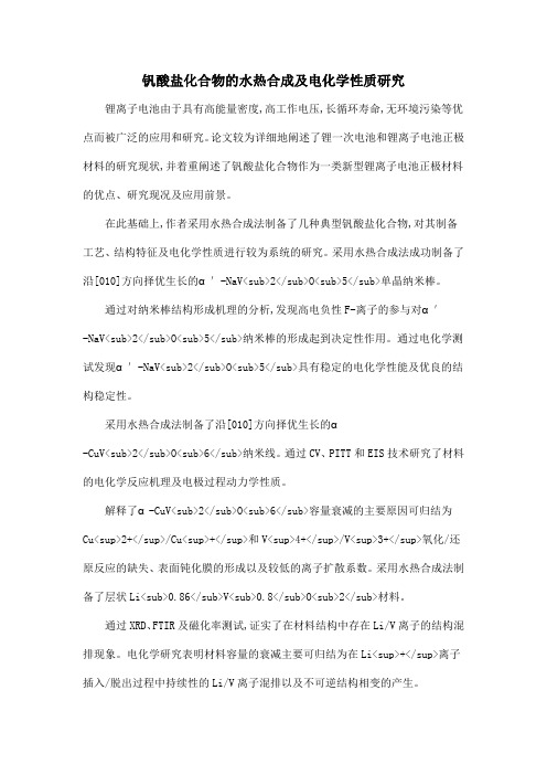
钒酸盐化合物的水热合成及电化学性质研究锂离子电池由于具有高能量密度,高工作电压,长循环寿命,无环境污染等优点而被广泛的应用和研究。
论文较为详细地阐述了锂一次电池和锂离子电池正极材料的研究现状,并着重阐述了钒酸盐化合物作为一类新型锂离子电池正极材料的优点、研究现况及应用前景。
在此基础上,作者采用水热合成法制备了几种典型钒酸盐化合物,对其制备工艺、结构特征及电化学性质进行较为系统的研究。
采用水热合成法成功制备了沿[010]方向择优生长的α′-NaV<sub>2</sub>O<sub>5</sub>单晶纳米棒。
通过对纳米棒结构形成机理的分析,发现高电负性F-离子的参与对α′-NaV<sub>2</sub>O<sub>5</sub>纳米棒的形成起到决定性作用。
通过电化学测试发现α′-NaV<sub>2</sub>O<sub>5</sub>具有稳定的电化学性能及优良的结构稳定性。
采用水热合成法制备了沿[010]方向择优生长的α-CuV<sub>2</sub>O<sub>6</sub>纳米线。
通过CV、PITT和EIS技术研究了材料的电化学反应机理及电极过程动力学性质。
解释了α-CuV<sub>2</sub>O<sub>6</sub>容量衰减的主要原因可归结为Cu<sup>2+</sup>/Cu<sup>+</sup>和V<sup>4+</sup>/V<sup>3+</sup>氧化/还原反应的缺失、表面钝化膜的形成以及较低的离子扩散系数。
采用水热合成法制备了层状Li<sub>0.86</sub>V<sub>0.8</sub>O<sub>2</sub>材料。

微波水热法制备钒酸铁粉体及光催化性能的研究微波水热法制备钒酸铁粉体及光催化性能的研究钒酸铁(FeVO4)是一种具有优异光催化活性和电化学性能的功能性材料,广泛应用于环境污染物的去除和能源转换领域。
然而,传统的合成方法往往需要高温、长时间,且难以控制晶体形貌和尺寸。
近年来,微波水热合成方法因其快速、能耗低、晶型可控等优势逐渐被应用于制备钒酸铁粉体。
本研究旨在利用微波水热法制备钒酸铁粉体,并研究其光催化性能。
首先,准备了钒酸铁前驱体溶液,该溶液中含有适量的钒酸铵和铁盐。
然后,将前驱体溶液加入到适量的水中,并在微波辐射下进行水热反应。
通过调节反应温度、时间和前驱体比例等参数,可以控制钒酸铁粉体的形貌和尺寸。
实验结果表明,通过微波水热法制备的钒酸铁粉体呈现出纳米级的颗粒大小,并具有较高的结晶度。
在不同反应条件下,钒酸铁粉体的形貌和尺寸均有所变化。
通过SEM观察,发现在较低的反应温度下,钒酸铁晶体的形貌呈现出近似纳米棒状结构;而在较高的反应温度下,钒酸铁晶体的形貌则更趋向于球状结构。
进一步研究发现,制备的钒酸铁粉体对甲基橙等有机染料具有较高的光催化降解性能。
实验中,通过调节反应条件和染料浓度,研究了钒酸铁粉体的光催化降解效果。
结果显示,钒酸铁粉体对有机染料的光催化效果受到反应温度和时间的影响较大。
在较低的温度和时间条件下,钒酸铁粉体呈现出优异的光催化活性,可在短时间内高效降解有机染料。
此外,研究还对钒酸铁粉体进行了光电化学性能的测试。
结果显示,制备的钒酸铁粉体具有良好的光电化学性能,可用作光电催化剂。
在光电解水实验中,钒酸铁粉体表现出良好的光电转化效率,可有效地将光能转化为化学能。
综上所述,本研究成功地通过微波水热法制备了纳米级的钒酸铁粉体,并研究了其光催化性能和光电化学性能。
所得结果表明,通过微波水热法合成的钒酸铁粉体具有较高的晶型控制能力和优异的光催化活性。
这一研究为利用钒酸铁材料进行环境水处理和光电催化转化提供了有力的支持和指导,具有重要的应用价值综上所述,通过微波水热法成功合成了纳米级的钒酸铁粉体,并研究了其形貌、尺寸及其对有机染料的光催化降解性能和光电化学性能。


低维钒氧化合物纳米材料的水热制备及性能研究的开题报告一、研究背景和意义低维纳米材料具有体材料所没有的独特性质和应用前景,因此在材料科学和纳米技术领域受到广泛关注。
钒氧化物作为一种重要的功能材料,具有丰富的物理、化学与电化学性质,被广泛应用于催化剂、电化学器件、能源存储与转换等领域。
近年来,纳米钒氧化物在这些领域中的应用也逐渐受到了关注。
另一方面,水热法作为钒氧化物纳米材料制备的一种有效方法,因其简便、环保、高效等优势,在制备低维纳米材料方面具有广泛应用前景。
因此,针对水热法制备的低维纳米钒氧化物材料,对其制备、结构和性能进行深入研究和探究,对于进一步开发和应用这些材料具有重要的科学和应用价值。
本研究旨在通过水热法制备钒氧化物纳米材料,探究不同反应条件下(如反应时间、温度、酸度等)对制备材料的结构、形貌和性能的影响,为该类材料的研究提供新的方法和认识。
二、主要研究内容1. 选择合适的前驱体和反应条件,成功制备高质量的低维钒氧化物纳米材料。
2. 利用XRD、TEM、SEM等手段对制备的材料进行表征和分析,研究不同反应条件对制备材料的结构、形貌以及晶体结构的影响。
3. 通过电化学和光学测试,对制备材料的电化学和光学性能进行研究和评价,探究不同反应条件下对材料性能的影响,并分析其物理和化学机制。
三、预期研究成果1. 成功制备高质量的低维钒氧化物纳米材料,并确定其结构和形貌;2. 发现不同反应条件对制备材料的结构、形貌和性能的影响,并了解其物理和化学机制;3. 对该类纳米材料的电化学和光学性能进行深入研究和评价,为其在催化、电化学和光催化等领域中的应用提供理论和实验基础。
四、研究方法和实验方案1. 制备方法:采用水热法制备低维纳米钒氧化物材料。
2. 实验步骤:(1)选择合适的前驱体、反应条件(如反应时间、温度、酸度等),制备低维钒氧化物纳米材料;(2)采用XRD、TEM、SEM等多种表征手段对制备的材料进行表征和分析,探究不同反应条件对其性质的影响;(3)通过电化学和光学测试,对材料的电化学和光学特性进行评价,并分析其物理和化学机制;(4)对实验数据进行分析和处理,总结结论和研究成果。

纳米结构MnO2 的水热合成、晶型及形貌演化李英品;周晓荃;周慧静;沈铸睿;陈铁红【期刊名称】《高等学校化学学报》【年(卷),期】2007(28)7【摘要】以水热合成方法制备了具有不同微观形貌的纳米结构MnO2, 并以X射线衍射(XRD), 扫描电镜(SEM)和X射线光电子能谱(XPS)等方法对其进行了表征. 跟踪考察了二氧化锰的晶型及微观形貌随水热反应时间的演变过程, 在Ostwald ripening机理作用下, MnO2晶型转化过程为γ-MnO2, α-MnO2和β-MnO2, 同时形貌由微米球转变为海胆结构、空心海胆结构和纳米线.【总页数】4页(P1223-1226)【作者】李英品;周晓荃;周慧静;沈铸睿;陈铁红【作者单位】南开大学化学学院,材料化学系,天津,300071;南开大学化学学院,材料化学系,天津,300071;南开大学化学学院,材料化学系,天津,300071;南开大学化学学院,材料化学系,天津,300071;南开大学化学学院,材料化学系,天津,300071【正文语种】中文【中图分类】O614【相关文献】1.形貌可控钒酸铜纳米晶的r水热合成及其电化学传感性能 [J], 韩桂洪;杨淑珍;黄艳芳;杨晶;柴文翠;张锐;陈德良2.纳米MnO2:水热法制备及盐对晶型和形貌的影响 [J], 桂义才;钱立武;钱雪峰3.形貌可控钒酸铜纳米晶的水热合成及其电化学传感性能 [J], 韩桂洪;杨淑珍;黄艳芳;杨晶;柴文翠;张锐;陈德良;;;;;;;4.水热合成温度及Na^+对二氧化锰晶型和形貌的影响 [J], 刘院英;徐亚周;魏秀格;常照荣5.形貌可控Co_3O_4纳米晶的水热合成及表征 [J], 朱振峰;吕景;刘辉;段聪越;邓璐因版权原因,仅展示原文概要,查看原文内容请购买。
《微波水热制备二氧化钒结构及形貌调控》篇一一、引言随着材料科学的发展,二氧化钒(VO2)因其独特的相变特性和在光电器件、热致变色器件等领域的广泛应用,逐渐成为研究热点。
本文旨在探讨微波水热法制备二氧化钒的工艺,以及通过调控实验参数实现其结构及形貌的优化。
二、文献综述传统的二氧化钒制备方法主要包括溶胶-凝胶法、化学气相沉积法等。
然而,这些方法往往存在能耗高、周期长、产物形貌难以控制等问题。
近年来,微波水热法因其快速、高效、可控性强的特点,被广泛应用于纳米材料的制备。
因此,采用微波水热法制备二氧化钒具有很高的研究价值。
三、实验部分1. 材料与设备实验所需材料包括钒源(如偏钒酸铵)、溶剂(如去离子水)等。
设备包括微波反应器、离心机、扫描电子显微镜(SEM)、X射线衍射仪(XRD)等。
2. 实验方法(1)制备过程:将钒源溶解在去离子水中,加入适量的表面活性剂或添加剂,然后置于微波反应器中进行水热反应。
通过调整微波功率、反应时间、溶液浓度等参数,得到不同形貌的二氧化钒产物。
(2)表征方法:利用SEM和XRD对产物的形貌和结构进行表征。
SEM可以观察产物的微观形貌和尺寸,XRD则可以分析产物的晶体结构和相纯度。
四、结果与讨论1. 结构分析通过XRD分析,我们可以得到产物的晶体结构信息。
当微波功率适中、反应时间适宜时,可以得到纯度较高的二氧化钒产物,其XRD图谱与标准VO2图谱吻合较好,表明成功制备了VO2。
此外,通过调整实验参数,还可以得到不同晶型的VO2,如单斜相和四方相等。
2. 形貌调控SEM结果表明,通过调整微波功率、反应时间、溶液浓度等参数,可以实现对二氧化钒形貌的调控。
例如,降低微波功率和延长反应时间有助于得到粒径较小的二氧化钒纳米颗粒;而提高溶液浓度和微波功率则可以得到片状或棒状的二氧化钒产物。
这些不同形貌的二氧化钒在光电器件、催化剂等领域具有不同的应用潜力。
五、结论本文采用微波水热法制备了二氧化钒,并通过对实验参数的调控实现了对其结构和形貌的优化。
《微波水热制备二氧化钒结构及形貌调控》篇一一、引言随着科技的发展,二氧化钒(VO2)因其独特的相变性质和在光电器件、热致变色器件等领域的广泛应用,受到了广泛的关注。
制备高质量的二氧化钒材料并对其进行形貌调控,是当前研究的热点之一。
微波水热法作为一种新兴的制备方法,具有快速、高效、绿色环保等优点,因此被广泛应用于材料制备领域。
本文采用微波水热法制备二氧化钒,对其结构及形貌进行调控,旨在为相关研究提供一定的理论和实践参考。
二、实验部分1. 材料与试剂实验所需材料包括钒源(如偏钒酸铵)、溶剂(如去离子水)等。
所有试剂均为分析纯,使用前未进行进一步处理。
2. 实验方法采用微波水热法制备二氧化钒。
首先,将钒源溶解在溶剂中,然后加入适量的去离子水,在微波反应器中进行反应。
通过调整反应条件(如反应时间、温度、功率等),实现对二氧化钒结构及形貌的调控。
3. 样品表征利用X射线衍射(XRD)、扫描电子显微镜(SEM)、透射电子显微镜(TEM)等手段对制备的二氧化钒进行结构及形貌分析。
三、结果与讨论1. 结构分析通过XRD分析,可以确定制备的二氧化钒的晶体结构。
调整反应条件,可以获得不同晶体结构的二氧化钒。
例如,在较低的反应温度下,可以得到单斜相二氧化钒;在较高的反应温度下,则可能得到四方相或其他相态的二氧化钒。
2. 形貌调控通过调整微波水热法的反应条件,可以实现二氧化钒的形貌调控。
例如,在较低的功率下反应较长时间,可以得到片状或棒状的二氧化钒;而在较高的功率下反应较短时间,则可能得到颗粒状或球状的二氧化钒。
此外,通过添加表面活性剂等手段,还可以进一步调控二氧化钒的形貌。
利用SEM和TEM等手段对制备的二氧化钒进行形貌分析。
从SEM图像中可以看到不同形貌的二氧化钒;而TEM则可以更清晰地展示其内部结构。
通过对形貌的分析,可以进一步理解反应条件对二氧化钒形貌的影响。
3. 性能分析对不同结构及形貌的二氧化钒进行性能测试,如光学性能、电性能等。
Trans.Nonferrous Met.Soc.China27(2017)1105−1116Hydrothermal synthesis and electrochemical sensing properties ofcopper vanadate nanocrystals with controlled morphologiesGui-hong HAN1,Shu-zhen YANG1,Yan-fang HUANG1,Jing YANG2,Wen-cui CHAI1,Rui ZHANG2,De-liang CHEN21.School of Chemical Engineering and Energy,Zhengzhou University,Zhengzhou450001,China;2.School of Materials Science and Engineering,Zhengzhou University,Zhengzhou450001,ChinaReceived12March2016;accepted1September2016Abstract:Morphology-controlled synthesis of copper vanadate nanocrystals is of great significance in electrochemical sensing applications.A facile hydrothermal process for synthesizing copper vanadate nanocrystals with various morphologies(e.g., nanoparticles,nanobelts and nanoflowers)was reported.Phase,morphology and electrochemical performance of the as-synthesized copper vanadate nanocrystals were characterized by X-ray diffraction(XRD),scanning electron microscope(SEM)and cyclic-voltammogram(CV)techniques.The results revealed that the morphologies of the Cu3V2O7(OH)2·2H2O(CVOH) nanocrystals could be controlled by changing copper salts,surfactants and pH values.The CVOH samples showed enhanced electrochemical response to ascorbic paratively,the CVOH nanobelts had the higher electrochemical sensing performance than those of CVOH nanoparticles and nanoflowers.The CVOH-nanobelts-modified GCEs had a linear relationship between the peak currents in their CVs and ascorbic acid concentration.The CVOH nanocrystals can be used as potential electrochemical active materials for the determination of ascorbic acid.Key words:copper vanadate nanocrystals;hydrothermal synthesis;electrochemical sensors;ascorbic acid1IntroductionAscorbic acid(AA)is one of the most important vitamins due to its antioxidant and other benefits for human bodies[1].AA is often added to various foods and pharmaceutical products for prevention of some diseases.The detection and quantitative determination of AA for the quality control in producing pharmaceuticals is essentially important.Therefore,there is an urgent need in developing easy-to-use and inexpensive methods to detect AA[2].Electrochemical methods for accurate determination of analytes have attracted increasing attention because of their intrinsic advantages of rapid response,high sensitivity,easy operation and low cost[3].Glassy carbon electrodes(GCE),carbon paste (CP)electrodes and gold arrays microelectrodes,which were functionalized by various active materials,have been used to detect various analytes[4−6].Nanocrystals of metal oxides,carbon and metals were widely used to modify the electrodes’surfaces to facilitate the electron transfer between target analytes and electrode surfaces and then to improve their performance[7−12].The morphologies and sizes of the nanocrystals highly affect the modification[13]and electrochemical property of electrodes[7].Metal vanadate nanocrystals have been reported in electrochemical applications[14−18].Among those metal vanadates,copper vanadate nanocrystals showed a great potential in the surface modification of electrodes because of their unique electrochemical performance[11,16].Controlled synthesis and electrochemical applications of copper vanadate nanocrystals with unique morphologies and sizes are the most interesting research topics.Their electrochemical properties are highly influenced by the morphologies and sizes that are largely determined by synthetic processes and starting reactants. As a typical phase,CuV2O6nanocrystals have been(51404213,51404214,51574205,51172211)supported by the National Natural Science Foundation of China;Projects (14HASTIT011,154100510003)supported by the Program for University Science and Technology Innovation Talents of HenanProvince,China;Projects(2013M531682,2014T70682)supported by the China Postdoctoral Science Fund;Project(1421324065)supported by the Development Fund for Outstanding Young Teachers of Zhengzhou University,ChinaCorresponding author:De-liang CHEN;Tel:+86-371+67881046;Fax:+86-371+67881593;E-mail:dlchen@DOI:10.1016/S1003-6326(17)60129-8Gui-hong HAN,et al/Trans.Nonferrous Met.Soc.China27(2017)1105−1116 1106synthesized via various processes(i.e.,solid-state reaction,sol-gel process,hydrothermal method)using different starting materials(i.e.,V2O5+Cu(NO3)2[19], NH4VO3+Cu(NO3)2[20],V2O5hydrogel+Cu2O[21]). Hollow Cu0.95V2O5microspheres and CuV2O6 nanoparticles have been synthesized using VO2and Cu(NO3)2as precursors in the polyvinyl pyrrollidone solutions[22].Also,Cu2V2O7nanoparticles have been synthesized using a thermal-decomposition method[11]. Recently,Cu3(OH)2V2O7·n H2O nanoparticles have been synthesized by hydrothermal methods using V2O5+ Cu(NO3)2[23]or V2O5+CuSO4·7H2O[24]as the starting materials.Cu3(OH)2V2O7·2H2O samples with different morphologies and sizes by a sol-gel process or chemical precipitation method were also reported[25,26]. In addition,Cu3V2O8nanoparticles have been gotten with CuSO4·5H2O and NH4VO3as materials under the assistance of different Schiff base ligands via a simple precipitation approach[27,28].However,the synthesis of copper vanadate nanocrystals with controlled morphologies using simple and cost-effective methods is still full of challenges.In this work,a new and facile hydrothermal process was developed to synthesize copper vanadate nanomaterials with various morphologies,and their applications in electrochemical detection of AA were systematically investigated.The optimal hydrothermal conditions were explored by adjusting acid radicals, additives and pH values,which are the key factors affecting the morphologies[29−32].The copper vanadate nanocrystals with typically unique morphologies(i.e.,nanoparticles,nanobelts,and nanoflowers)have been used as the active materials to modify GCEs,which were used as the working electrodes to detect ascorbic acid on the basis of the electrochemically sensing mechanism.2Experimental2.1Materials and methodsNH4VO3(AR grade)and Cu(CH3COO)2·H2O(AR grade)were purchased from Tianjin Guangfu Fine Chemical Research,China.CuSO4·5H2O(AR grade, Tianjin Kemiou Chemical Reagent Co.,Ltd,China), CuCl2·H2O(AR grade,Tianjin Fengchuan Chemical Reagent Co.,Ltd.,China)were also used as copper sources.Cu(NO3)2·3H2O(AR grade)and poly-vinylpyrrolidone(PVP)were obtained from Sinopharm Chemical Reagent Co.,Ltd.,sodium dodecyl benzene sulfonate(SDBS)and hexadecyltrimethy ammonium bromide(CTAB)were purchased from Tianjin Fu Chen Chemical Reagents Factory and China National Pharmaceutical Group Corporation,respectively.All chemicals were used as received.Copper vanadate samples were synthesized under different conditions,by varying copper sources(CuSO4, Cu(NO3)2,Cu(CH3COO)2,or CuCl2),Cu2+concentration (0.03−0.075mol/L),surfactants(PVP,SDBS or CTAB), pH values(3−11)and hydrothermal reaction time (12−24h).The details for hydrothermal synthesis of copper vanadate nanocrystals are listed in Table1.In a typical synthesis(S6in Table1),0.1404g (~1.2mmol)of NH4VO3was dissolved in30mL of distilled water at80°C under magnetic stirring,and4S7CuSO40.03PVP/0.3247Non-uniform nanoparticles S8CuSO40.03PVP/0.32411Irregular particlesS9CuSO40.03SDBS/0.3205Pieces with particlesS10CuSO40.03CTAB/0.3205Pieces with particlesS11CuSO40.03−205Strings with particlesS12CuSO40.075−125Irregular particles with microrobs S13Cu(NO3)20.075−125Irregular particlesS14Cu(CH3COO)20.075−125NanoparticlesS15CuCl20.075−125Irregular particlesGui-hong HAN,et al/Trans.Nonferrous Met.Soc.China27(2017)1105−111611070.4494g(~1.8mmol)of CuSO4·5H2O was simulta-neously dissolved in another30mL of distilled water at room temperature.The above NH4VO3solution was then added slowly to the CuSO4solution under a strong magnetic stirring,and then0.18g of PVP was finally added into the above mixture,in which the concentrations of Cu2+([Cu2+]),3VO-([3VO-]),and content of PVP(w(PVP))were0.03mol/L,0.02mol/L, and0.3%,respectively.The pH value of the solution was adjusted to~3using sulfuric acid.The resulting suspension was stirred for another10min,and then transferred into a Teflon-lined stainless steel autoclave with a volume of100mL.After carefully sealed,the autoclave was heated to180°C and kept at180°C for 24h,followed by natural cooling in air.The dark green precipitate was collected by centrifugation,washed with water and then freeze-dried in air.The obtained sample with a dark green color was marked as S6in Table1, consisting of copper vanadate nanobelts,and then used for further characterization.2.2Characterization2.2.1Phase composition and morphologyThe phase composition was recognized using X-ray diffraction(XRD)patterns,recorded on a D3X-ray diffractometer equipped with a graphite mono-chromatized Cu Kαradiation source(λ=1.5406Å)at a scan rate of0.05(︒)/s in a2θrange of11°−50︒.The morphology was observed by scanning electron microscopy(SEM)using a JSM-7001F microscope.The sample was coated with a thin Au film prior to SEM observation.2.2.2Electrochemical measurementTo prepare electrodes,the suspensions containing copper vanadate nanocrystals were firstly prepared:1mg of the as-obtained copper vanadate nanocrystals and5μL of nafion solution(5%)were mixed with1mL of N,N-dimethylformamide(DMF)with a ultra-sonication treatment for15min.A glassy carbon electrode(GCE) with a diameter of3mm was polished to a mirror finish using polishing papers containing alumina powders with different sizes of firstly50−70μm and then30−50nm, respectively.The polished GCE was sequentially washed using alcohol and distilled water with an ultrasonic cleaner.Approximately10μL of copper vanadate suspensions was finally carefully coated on the surface of the polished GCE.After the solvents evaporated at room temperature,the modified GCEs were used to evaluate their electrochemical properties.The electrochemical(EC)measurement was performed on an electrochemical working station(model CS310,Wuhan Correst Instruments Co.,Ltd.,China). The GCEs modified with copper vanadate nanocrystals were served as the working electrodes,and the platinum plate and saturated calomel electrode(SCE)were served as the counter electrode and reference one,respectively. All potentials were with respect to the SCE.Cyclic voltammograms(CVs)were recorded in a potential range of−1.0V to+1.0V at a potential scan rate of 50mV/s in an aqueous solution consisting of0.1mol/L KCl and ascorbic acid with a given concentration (0.005−2.0mmol/L).All electrochemical measurements were carried out at room temperature.3Results and discussion3.1Effects of copper sources on morphology ofcopper vanadate nanocrystalsCopper vanadate(Cu3V2O7(OH)2·2H2O)nano-crystals were synthesized via the reaction between acidic radical copper sources and ammonium met-vanadate under a hydrothermal condition.We firstly checked the effects of acidic radical copper sources on the formation of copper vanadate nanocrystals.Figure1shows the XRD patterns of the samples (S12,S13,S14and S15in Table1)obtained by hydrothermal treatment at180°C for12h using ammonium met-vanadate and different copper sources (Cu(CH3COO)2·H2O,Cu(NO3)2·3H2O,CuCl2·2H2O or CuSO4·5H2O)as the reactants(no surfactants and pH=5). As Fig.1shows,the characteristic peaks of the four samples are similar,and all the diffraction peaks can be readily indexed to the pure phase of Cu3V2O7(OH)2·2H2O with a monoclinic structure (JCPDS card No.80−1170,a=10.607Å,b=5.864Å, c=7.214Å).No other impurities are detected,suggesting that the samples obtained are highly pure.The crystallite diameter of Cu3V2O7(OH)2·2H2O nanostructures obtained using the Scherrer equation[33]:D=Kλ/βcosθ;Fig.1XRD patterns of Cu3V2O7(OH)2·2H2O nanocrystals synthesized using various copper salts as precursors: (a)CuSO4·5H2O;(b)Cu(NO3)2·3H2O;(c)CuCl2·H2O;(d)Cu(CH3COO)2·H2OGui-hong HAN,et al/Trans.Nonferrous Met.Soc.China27(2017)1105−1116 1108whereβis the breadth of the observed diffraction line at its half intensity maximum,K is the so-called shape factor,which usually takes a value of about0.9,andλis the wavelength of X-ray source used in XRD.Calculated crystalline domain sizes have been found to be51nm for sample S14.The peak intensities of sample S14derived from Cu(CH3COO)2·H2O are stronger than those of the other samples,whereas peak intensities of sample S15 from CuCl2·H2O are the weakest ones.The intensities in the XRD peaks indicate that the sample derived from Cu(CH3COO)2·H2O is of higher crystalline degree than that from CuCl2·H2O.The morphologies of the Cu3V2O7(OH)2·2H2O samples obtained with various copper precursors at 180°C for12h were observed by SEM microscopy. Figure2shows the typical SEM images.One can see that the surfaces of Cu3V2O7(OH)2·2H2O nanoparticles are greatly affected by the sources of copper salts.Figures 2(a)and(b)show the SEM images of sample S14 derived from Cu(CH3COO)2·H2O,and this product is mainly composed of nanoparticles with an average size of~75nm.Its corresponding particle-size distribution determined by the Nano Measurer software according the SEM result in Fig.2(b)is shown in Fig. 3.The Gaussian-fitting curve of the particle-size distribution indicates that the diameters of the nanoparticles range from55to80nm.NI et al[23,24]reported a similar result,but they used different starting materials and reaction conditions(i.e.,V2O5,hexamethylenetetramine, CuSO4·7H2O or Cu(NO3)2and sodium sulfate,at140°C for24h).The SEM images of the Cu3V2O7(OH)2·2H2O samples derived from CuSO4,Cu(NO3)2,and CuCl2are shown in Figs.2(c),(d),and(e),respectively.One can see that these samples mainly consist of nanoparticles with some of one-dimensional species(Fig.2(c)). According to the SEM images,one can find that copper salts highly influence the morphology of copper vanadate samples.ZHANG et al[34]reported similar influence of copper salts,and thought that the acidity played a key role in the formation of the copper vanadate nanocrystals. The acidity may cause the products to become more agglomerated under special conditions but the purities of them were little affected[29].3.2Effects of surfactants and pH valuesVarious surfactants,including anionic surfactant, cationic surfactant and nonionic surfactant,were used for the growth of copper vanadate nanocrystals.In this work,sodium dodecyl benzene sulfonate(SDBS), hexadecyltrimethy ammonium bromide(CTAB)and Fig.2Typical FE-SEM images of Cu3V2O7(OH)2·2H2O nanocrystals using various copper salts as precursors(pH=5,at180°C for 12h):(a,b)Cu(CH3COO)2·H2O;(c)CuSO4·5H2O;(d)Cu(NO3)2·3H2O;(e)CuCl2·H2OGui-hong HAN,et al/Trans.Nonferrous Met.Soc.China 27(2017)1105−11161109Fig.3Particle-size distribution of Cu 3V 2O 7(OH)2·2H 2O nanocrystals obtained using Cu(CH 3COO)2·H 2O as precursor (pH=5,at 180°C for 12h)polyvinylpyrrolidone (PVP)were selected as the anionic,cationic and nonionic surfactants,respectively [35−38].Figure 4shows the typical SEM images of the Cu 3V 2O 7(OH)2·2H 2O samples.Figure 4(a)shows theSEM image of the Cu 3V 2O 7(OH)2·2H 2O sample obtained with PVP,and the sample consists of copper vanadate nanobelts,30−50nm in width and more than several micrometers in length.Figures 4(b)−(d)show the SEM images of Cu 3V 2O 7(OH)2·2H 2O samples obtained with SDBS and CTAB.One can see that the Cu 3V 2O 7(OH)2·2H 2O nanocrystals obtained with SDBS and CTAB consist of small nanoparticles and some large particles of irregular morphologies.The effects of the contents of PVP w (PVP)and pH values on the morphology of Cu 3V 2O 7(OH)2·2H 2O nanocrystals were further investigated.Figure 5shows the typical SEM images of copper vanadate samples obtained using CuSO 4as the Cu source at 180°C for 24h with different pH values and PVP concentrations.As Figs.5(a)−(b)show,the Cu 3V 2O 7(OH)2·2H 2O samples obtained with conditions of pH=3and w (PVP)=0.3%−3%take on a belt-like morphology,several-micrometers long and less than 100nm in the apparent width.The change of the PVP concentration shows a slight impact on their morphology at pH=3,and a higher PVP concentration results in shorter nanobelts [35].AsFig.4Typical FE-SEM images of copper vanadate nanocrystals obtained with various surfactants (CuSO 4,at 180°C for 20h,pH=5,w (surfactant)=0.3%):(a)Without surfactant;(b)PVP;(c)CTAB;(d)SDBSGui-hong HAN,et al/Trans.Nonferrous Met.Soc.China27(2017)1105−11161110Fig.5Typical FE-SEM images of copper vanadate samples obtained using CuSO4at180°C for24h with different pH values and PVP contents:(a)pH=3,w(PVP)=0.3%;(b)pH=3,w(PVP)=3%;(c)pH=7,w(PVP)=0.3%;(d)pH=11,w(PVP)=0.3%Figs.5(a)−(d)show,the pH value is a key factor that influences the morphology of the copper vanadate nanocrystals.When the pH value is3,uniform nanobelts are obtained(Fig.5(a)),whereas when the pH value is7, small nanoparticles with a size range of50−100nm are the major products(Fig.5(c));when the pH value increases to11,irregular particles with a sheet-like morphology are formed(Fig.5(d)).Figures6(a)−(b)show the SEM images of Cu3V2O7(OH)2·2H2O samples obtained when the pH value is4and the PVP content is2.7%.As Figs.6(a)and (b)show,the product takes on a flower-like morphology, formed by assembling Cu3V2O7(OH)2·2H2O nanosheets. The diameters of the copper vanadate flowers are 3−5μm.The formation of flower-like morphology is influenced by the w(PVP)and pH values[35].Figures 6(c)and(d)show the SEM images of the Cu3V2O7(OH)2·2H2O samples obtained when the pH value was changed to5and when the PVP content was changed to 1.5%,respectively.It can be seen that as-obtained samples consist of broken nanorods and particles.The formation of the Cu3V2O7(OH)2·2H2O nanocrystals in the present hydrothermal process may be controlled by the synergistic effects of Ostwald ripening and PVP adsorption growth[16,34,35,39,40].The tiny crystalline nuclei are formed at the initial stage in a supersaturated medium,followed by a crystal growth. With the increase of reaction time,the irregular particles disappear and the long rods grow.Figures2(c)and4(a) indicate that the long rods grow at the expense of smaller particles due to the energy difference between them based on the Gibbs−Thompson law[41].In addition, PVP can act as the rod-like micelle template which plays a critical role in the formation of nanobelts and flowers. The PVP content can influence the length of nanobelts. The pH value is also a vital factor influencing the formation of Cu3V2O7(OH)2·2H2O nanocrystals.Firstly, the reaction for the formation of Cu3V2O7(OH)2·2H2O nanocrystals involves H+ions;thus when the pH value isGui-hong HAN,et al/Trans.Nonferrous Met.Soc.China27(2017)1105−11161111 Fig.6Typical FE-SEM images of copper vanadate nanocrystals obtained at180°C for24h(using CuSO4as raw material): (a,b)pH=4,w(PVP)=2.7%;(c)pH=5,w(PVP)=2.7%;(d)pH=4,w(PVP)=1.5%high,i.e.,there is a large amount of OH−ions in the reaction medium,the reaction rate is accelerated greatly and the morphology is hard to control.Secondly,the behavior of the PVP species may greatly change with the change of the pH value,and then the growth of Cu3V2O7(OH)2·2H2O nanocrystals is influenced by the pH value.3.3Electrochemical performance of copper vanadatenanocrystalsCopper vanadate nanocrystals can act as an electrode-modifying material to accelerate electron transport because of theirs low internal resistance and high discharge capacity[11,16,42].Ascorbic acid(AA) is sensitive to the physical and chemical properties of the glass carbon electrodes[43,44].We therefore use copper vanadate nanocrystals to modify the glassy carbon electrodes(GCEs)to modulate the electrochemical response to AA molecules.The as-synthesized Cu3V2O7(OH)2·2H2O nanocrystals with various morphologies(i.e.,nanoparticles,nanobelts and nanoflowers)were used to modify the GCEs.Figure7shows the electrochemical redox behavior (CV profiles)of ascorbic acid at the interfaces of the Cu3V2O7(OH)2·2H2O/GCEs in the presence of the ascorbic acid aqueous solution(2mmol/L).As Fig.7(a) shows,the bare GCE has no redox peaks.The GCE modified with Cu3V2O7(OH)2·2H2O nanobelts(Fig.7(d)) shows double well-defined redox peaks(i.e.,peaks1and 1′,peaks2-3and2-3′)with enhanced peak-current intensities,and their anodic and cathodic peak potentials are0.39V and−0.17V,respectively.As Figs.7(b)and(c) show,the GCEs modified with Cu3V2O7(OH)2·2H2O nanoparticles and nanoflowers are lowly sensitive to ascorbic acid,and only one pair of redox peaks are observed in their CVs.This suggests that the Cu3V2O7(OH)2·2H2O nanobelts show the best electrochemical activity and the fastest electron transfer under the present neutral conditions.Some similar results with only one pair of relatively weak redox peaks have been reported in the literature[1,2,12].The electro-chemical behavior of the electrodes modified by CuGeO3Gui-hong HAN,et al/Trans.Nonferrous Met.Soc.China27(2017)1105−1116 1112nanowires and CuGeO3/polyaniline nanowires to tartaricacid has also shown similar characteristic[45,46].PEI et al[16]reported that the Cu2.33V4O11nanobelts/GCEs show two pairs of redox peaks located at different voltages.The difference in the electrochemical redox behavior may result from the difference in the morphology and size of the active materials used to modify theirGCEs.Fig.7Cyclic voltammetric profiles of various electrodes at scan rate of50mV/s in0.1mol/L KCl solution containing 2mmol/L ascorbic acidFigure8shows the oxidation process of ascorbic acid(AA).AA molecules firstly diffuse to the nearest active sites,and then they are adsorbed onto the surfaces of the copper vanadate nanobelts.The AA molecules adsorbed are finally oxidized to be dehydroascorbic acid with the catalytic effect of the copper vanadate nanobelts. The enhanced electrocatalytical performance of GCEs modified with Cu3V2O7(OH)2·2H2O nanocrystals can be attributed to the enlarged surface areas of the Cu3V2O7(OH)2·2H2Onanobelts.Fig.8Schematic of oxydehydrogenation of ascorbic acid oxide Figure9shows the effect of scan rates on the electrochemical behavior of the GCEs modified with Cu3V2O7(OH)2·2H2O nanobelts in the presence of AA and KCl aqueous solutions([AA]=2mol/L and[KCl]= 0.1mol/L).As Fig.9(a)shows,when the scan rate increases from25mV/s to125mV/s,the anodicpeak Fig.9Cyclic voltammetric profiles of Cu3V2O7(OH)2·2H2O nanobelts/GCE in0.1mol/L KCl solution containing2mmol/L ascorbic acid at different scan rates(a)and plot of peak cathodic current(1)versus root square mean of scan rate(b) potentials shift positively slightly,whereas the cathodicpeak potentials move negatively.Figure9(b)shows the plot of the cathodic peak current versus the square root of the potential scan rate in the range of25to125mV/s. The obtained linear fitting equation is described as Eq.(1),and its linear correlation coefficient is as high as 0.993.The good linear relationship between the cathodic peak current and the mean square root of the scan rate indicates that the overall oxidation of ascorbic acid at the GCEs modified with Cu3V2O7(OH)2·2H2O nanobelts is controlled by the diffusion of AA molecules under the present conditions.The peak potentials are not changed obviously,suggesting that the electrode system is reversible to some extent.Thus the diffusion coefficient at room temperature can be calculated using the Randlese−Sevcik equation(Eq.(2))[7,47,48].I=−0.001ν1/2−4.63572×10−5A(1)53/21/2*1/2p00(2.6910)i n AD C v=⨯A(2)Gui-hong HAN,et al/Trans.Nonferrous Met.Soc.China 27(2017)1105−11161113where i p is the peak current,n is the number of electrons,A (cm 2)is the electrode area,*0c (mol/cm 3)is the concentration of ascorbic acid,D 0(cm 2/s)is the diffusion coefficient,and ν(V/s)is the potential scan rate.When n ,A ,*0c ,D 0and νare constants,the 1/2p 0i D ∝.It can be concluded that the diffusion coefficient of AA molecules at the Cu 3V 2O 7(OH)2·2H 2O nanobelts/GCE interface is much larger than those of the other two electrodes modified with Cu 3V 2O 7(OH)2·2H 2O nanoparticles and nanoflowers,judged by the larger cathodic peak current (i p in Fig.9).Figure 10(a)shows the CV curves of the GCEs modified with the copper vanadate nanobelts in the presence of 0.1mol/L KCl aqueous solutions containing ascorbic acid with various concentrations (0.005−2mmol/L)with a potential scan rate of 50mV/s.Figure 10(b)shows the plot of the cathodic peak current versus the AA concentration.One sees that the absolute values of the peak currents increase linearly with the increase in AA concentrations.The linear fitting equationobtainedFig.10Cyclic voltammetric profiles of copper vanadatenanobelts modified GCE in 0.1mol/L KCl solution containing ascorbic acid with different concentrations with scan rate of 50mV/s (a),plot of the peak cathodic currents (1)versus ascorbic acid concentrations (b)according to the parameters of peak 1is shown as Eq.(3),where c (mmol/L)is the concentration of ascorbic acid,with a linear correlation coefficient (R 2)as high as 0.994in the [AA]range of 0.005−2mmol/L.The comparison of the liner range of the electrochemical determination of ascorbic acid with other electrodes and modifiers is shown in Table 2,indicating that every research done in recent years had their own characteristics.The copper vanadate nanobelts obtained in the present work can be used as a potential sensing platform for electro-chemically quantitative detection of ascorbic acid.I =−1.02(4)×10−4c −6.9(3)×10−5A(3)Table 2Comparison of liner range of electrochemical determination of ascorbic acid other researchesElectrodeModifier Linearrange/(mmol·L −1)Time/Ref.Au PAH/polysaccharide films confined36Fe(CN)-1−502007/[49]Carbon nanotube paste electrode Silver nanoparticles0.03−22009/[50]Glassy carbon electrode Pd nanowire 0.025−0.92010/[51]Glassy carbon electrode Palladium nanoparticles supported on graphene oxide 0.02−2.282012/[52]3D graphene foam CuO nanoflowers0.00043−0.22014/[2]Glassy carbon electrode Hierarchical nanoporous (HNP)PtTi alloy0.2−12016/[53]Glassy carbon electrodeCu 3V 2O 7(OH)2·2H 2O nanobelts 0.005−2ThisworkThe electrochemical behavior of the GCEs modified with the copper vanadate nanobelts in the presence of ascorbic acid aqueous solution was also investigated under various pH conditions,e.g.,in the electrolytes including NaOH (pH=12)and H 2SO 4(pH=2).Figure 11shows the typical results.The ascorbic acid molecules in the acidic or alkaline conditions have no redox peaks,indicating that the [H +]concentration in the solution influences the oxidation of ascorbic acid.Ascorbic acid is an acid relatively stable under the acidic condition,less prone to oxidation−reduction reactions.AA is not stable in the alkaline environment,leading to the irreversibility of oxidation−reduction reactions.Therefore,the determination of AA using the GCEs modified with the copper vanadate nanobelts should be limited to a neutral condition.Gui-hong HAN,et al/Trans.Nonferrous Met.Soc.China27(2017)1105−11161114Fig.11Cyclic voltammetric profiles of copper vanadate nanobelts modified GCE electrodes in H2SO4(a)and NaOH(b) solutions containing2mmol/L ascorbic acid with scan rate of 50mV/s4Conclusions1)The Cu3V2O7(OH)2·2H2O nanocrystals with various morphologies(e.g.,nanoparticles,nanobelts and nanoflowers)have been synthesized via a hydrothermal process by adjusting the experimental conditions. Cu(CH3COO)2·H2O is the suitable copper source for the synthesis of nanoparticles.Cu3V2O7(OH)2·2H2O nanobelts are synthesized using CuSO4·5H2O as the copper source with PVP(0.3%−3%)and pH=3.The Cu3V2O7(OH)2·2H2O nanoflowers are formed under the condition of pH=4and w(PVP)=2.7%.2)The GCEs modified with the Cu3V2O7(OH)2·2H2O nanobelts show higher electrochemical activity to ascorbic acid than those modified with Cu3V2O7(OH)2·2H2O nanoparticles and nanoflowers.The absolute values of the peak currents in the CVs increase linearly with the ascorbic acid concentrations in the range of0.005−2mmol/L.3)The Cu3V2O7(OH)2·2H2O nanobelts can be a promising candidate of electrochemically active material to modify glassy carbon electrodes that are applied in electrochemical determination of ascorbic acid or other organic molecules qualitatively or quantitatively. References[1]KARIMI-MALEH H,MOAZAMPOUR M,YOOSEFIAN M,SANATI A L,TAHERNEJAD-JAV AZMI F,MAHANI M.An electrochemical nanosensor for simultaneous voltammetric determination of ascorbic acid and sudan I in food samples[J].Food Analytical Methods,2014,7(10):2169−2176.[2]MA Y,ZHAO M,CAI B,WANG W,YE Z,HUANG J.3D graphenefoams decorated by CuO nanoflowers for ultrasensitive ascorbic acid detection[J].Biosensors and Bioelectronics,2014,59:384−388. [3]LEONARDI S G,ALOISIO D,DONATO N,RATHI S,GHOSH K,NERI G.Electrochemical sensing of ascorbic acid by a novel manganese(III)complex[J].Materials Letters,2014,133:232−235.[4]SAKIRA A K,SOMC I T,ZIEMONS E,DEJAEGHER B,MERTENS D,HUBERT P,KAUFFMANN J.Determination of arsenic(III)at a nanogold modified solid carbon paste electrode[J].Electroanalysis,2015,27(2):309−316.[5]HUANG X,LI Y,CHEN Y,WANG L.Electrochemicaldetermination of nitrite and iodate by use of gold nanoparticles/ poly(3-methylthiophene)composites coated glassy carbon electrode [J].Sensors and Actuators B:Chemical,2008,134(2):780−786. [6]IDRIS A O,MABUBA N,AROTIBA O A.Electroanalysis ofselenium in water on an electrodeposited gold-nanoparticle modified glassy carbon electrode[J].Journal of Electroanalytical Chemistry, 2015,758:7−11.[7]SONG M J,HWANG S W,WHANG D.Non-enzymaticelectrochemical CuO nanoflowers sensor for hydrogen peroxide detection[J].Talanta,2010,80(5):1648−1652.[8]PEI L Z,LIU H D,LIN N,XIE Y K,CAI Z Y.Electrochemicalanalysis of cyanuric acid using polyaniline/CuGeO3nanowires as electrode modified materials[J].Current Pharmaceutical Analysis, 2015,11(1):16−24.[9]LI H,WANG Y,YE D,LUO J,SU B,ZHANG S,KONG J.Anelectrochemical sensor for simultaneous determination of ascorbic acid,dopamine,uric acid and tryptophan based on MWNTs bridged mesocellular graphene foam nanocomposite[J].Talanta,2014,127: 255−261.[10]QIAN J,YANG X,YANG Z,ZHU G,MAO H,WANG K.Multiwalled carbon nanotube@reduced graphene oxide nanoribbon heterostructure:Aynthesis,intrinsic peroxidase-like catalytic activity, and its application in colorimetric biosensing[J].Journal of Materials Chemistry B,2015,3(8):1624−1632.[11]SIVAKUMAR V,SURESH R,GIRIBABU K,MANIGANDAN R,MUNUSAMY S,KUMAR S P,MUTHAMIZH S,NARAYANAN V.Copper vanadate nanoparticles:Synthesis,characterization and its electrochemical sensing property[J].Materials in Electronics,2014, 25(3):1485−1491.[12]CHANG K C,CHU C M,CHANG C H,CHENG H T,HSU S C,LAN C C,CHEN H H,PENG Y Y,YEH J M.Photoisomerization of electroactive polyimide/multiwalled carbon nanotube composites on the effect of electrochemical sensing for ascorbic acid[J].Polymer International,2015,64(3):373−382.[13]MANDAL D,MONDAL S,SENAPATI D,SATPATI B,SANGARANARAYANAN M V.Charge density modulated shape-dependent electrocatalytic activity of gold nanoparticles for the oxidation of ascorbic acid[J].The Journal of Physical Chemistry C, 2015,119(40):23103−23112.[14]AFONSO R,EISELE A P P,SERAFIM J A,LUCILHA A C,DUARTE E H,TARLEY C R T,SARTORI E R,DALL'ANTONIA LH.BiVO4-Bi2O3/ITO electrodes prepared by layer-by-layer:。