肾嫌色细胞癌影像
- 格式:pptx
- 大小:57.40 KB
- 文档页数:11

动态增强CT扫描对肾嫌色细胞癌的诊断价值分析作者:汪裕聪陈锋施承达李盛来源:《中国现代医生》2013年第35期[摘要] 目的探讨动态增强CT扫描对肾嫌色细胞癌的诊断价值及其临床应用。
方法回顾性分析12例经手术和病理证实为肾嫌色细胞癌的动态增强CT影像资料,对肿瘤的大小、形态、密度及动态增强特点进行分析。
结果 12例病例动态增强CT表现,原发病灶均为单发,呈软组织密度,3例见假包膜,5例有分叶,肿瘤密度均匀5例,不均匀7例,内部可见线状、小片状坏死或星芒状纤维灶,坏死区一般较小,边缘光整,1例见钙化,动态增强扫描皮髓期肿瘤强化明显低于肾皮质,而高于肾髓质,实质期强化明显低于肾实质,瘤体≤7 cm×6.5 cm的均未见转移。
结论肾嫌色细胞癌属膨胀性生长、少血供、低度恶性肿瘤,其恶性程度与肿瘤大小和坏死程度有关,其动态增强CT表现具有一定特征性,动态增强CT扫描对肾嫌色细胞癌的发现、术前临床评估和指导治疗有着重要的价值。
[关键词] 肾脏;嫌色细胞癌;体层摄影术;X线计算机[中图分类号] R816.7 [文献标识码] B [文章编号] 1673-9701(2013)35-0055-03肾嫌色细胞癌(chromophobe renal cell carcinoma,CRCC)是肾癌中相对少见的恶性肿瘤,恶性程度较其他肾细胞癌亚型都低。
近年来随着影像学的发展,人群体检数量的增多,很多病例在出现临床症状前被B超检查发现,肿瘤尚处在的早期阶段,对于瘤体较小,预后较好的早期CRCC临床可采用肾脏部分切除术、肿瘤挖除术或射频消融术等相对保守治疗,因此术前准确的诊断尤为重要,本文收集2004年5月~2012年11月经手术病理证实的12例CRCC 病例,对其动态增强CT表现进行回顾性分析,并复习文献,进一步探讨动态增强CT对CRCC的诊断价值。
1 资料与方法1.1 临床资料12例肾嫌色细胞癌患者,其中男7例,女5例,发现瘤体时年龄35~82岁,平均约52岁。
肾透明细胞癌与嫌色细胞癌、嗜酸细胞腺瘤CT 表现陈文新;朱庆强;孙骏;李洁;吴晶涛【期刊名称】《医学影像学杂志》【年(卷),期】2014(000)011【摘要】目的:探讨C T扫描对肾癌亚型的鉴别诊断价值。
方法收集29例经手术病理证实的肾癌患者资料,其中肾透明细胞癌(CCRCC)14例,肾嫌色细胞癌(CRCC)8例,嗜酸细胞腺瘤(OA )7例,对比分析各亚型肿瘤患者CT 平扫及三期增强扫描表现并与病理对照。
结果 CCRCC和OA多数密度不均(分别为12/14和5/7)、常见坏死(11/14和5/7);CRCC密度多均匀(6/8)。
CCRCC淋巴结及远处器官转移最常见(11/14),CRCC节段增强反转最常见(5/8)。
CT 平扫期,CCRCC密度稍高于CRCC、OA及正常肾皮质(45.8±3.6与41.4±2.4与43.7±3.3与41.5±5.1,F值=2.458,P >0.05)。
增强CT三期扫描CCRCC强化程度最高,强化模式呈“快进快退”,OA呈中度‐明显强化,CRCC强化程度最轻,两者均呈“渐进性”延迟强化。
CCRCC强化程度接近于各期肾皮质强化,OA强化稍低于各期肾皮质强化,CRCC强化程度明显低于肾皮质强化( P <0.01)。
OA强化程度高于CRCC ( P <0.01)。
皮质期及皮髓质期,OA强化高于肾髓质( P <0.01),但肾盂期低于肾髓质( P <0.01)。
皮质期,CRCC强化值高于肾髓质,但皮髓质期及肾盂期低于肾髓质强化。
结论 CCRCC、CRCC和OA的CT扫描有一定的特征性表现,与其病理特点密切相关,在肾癌亚型的鉴别诊断中有着较高的临床应用价值。
【总页数】5页(P2037-2040,2041)【作者】陈文新;朱庆强;孙骏;李洁;吴晶涛【作者单位】扬州大学临床医学院江苏省苏北人民医院医学影像科江苏扬州225001;扬州大学临床医学院江苏省苏北人民医院医学影像科江苏扬州 225001;扬州大学临床医学院江苏省苏北人民医院医学影像科江苏扬州 225001;扬州大学临床医学院江苏省苏北人民医院医学影像科江苏扬州 225001;扬州大学临床医学院江苏省苏北人民医院医学影像科江苏扬州 225001【正文语种】中文【中图分类】R737.1;R814.42【相关文献】1.肾透明细胞癌、嫌色细胞癌及嗜酸细胞腺瘤的MSCT鉴别诊断 [J], 吴娟;顾红梅;王欣全;李敏达;沈海霞;葛敏2.小肾嗜酸细胞腺瘤的CT增强表现及与小肾透明细胞癌的鉴别 [J], 马丽娅;胡道予;李佳丽;沈亚琪;李震;罗彦3.对比分析肾脏嫌色细胞癌和嗜酸细胞腺瘤的CT表现 [J], 熊文娟;谭永明;龚洪翰;李志;章晨蕾;钟军;何来昌4.增强CT在肾嫌色细胞癌和肾嗜酸细胞腺瘤中的鉴别诊断价值 [J], 黎亚娟; 黄夏玲; 龙莉玲5.基于增强CT影像组学在鉴别肾嫌色细胞癌与肾嗜酸细胞腺瘤中的应用价值 [J], 鲍远照;葛亚琼;程琦;刘欣;严立;韦炜因版权原因,仅展示原文概要,查看原文内容请购买。
肾嫌色细胞癌的CT和MRI诊断朱庆强;朱文荣;王中秋;史玉振;童明敏;林艳飞【摘要】Objective:To investigate the CT and MRI features of chromophobe cell renal carcinoma (CCRC), and learn more about of the disease. Methods: 16 cases of chromophobe cell renal carcinoma were retrospectively analyzed. All of them were confirmed by pathology and performed CT scan. Six of them involved in MR. CT and MR features of the tumor were observed and analyzed. Results:CT values of the normal renal cortex,medulla and CCRC were (30. 7±3. 2) ,(28. 5± 2. 8) and (32. 7±5.6)HU respectively on plain scan;CT values of them were (132. 3±17. 7),(58. 4±9. 4) and (68. 6± 12. 4)HU respectively in cortical phase;CT values of them were (166. 6±26. 8),(80. 6±5. 8) and (87. 8±25. 0)HU respectively in medullary phase;CT v alues of them were (132. 3±20. 7),(69. 6±4. 8) and (79. 4±18. 4)HU respectively in renipelvic phase. The enhancement of CCRC was lower than the normal renal cortex (cortical, medullary, renipelvic phase P<0. 01) ,but higher than the normal renal medulla(cortical,medullary,renipelvic phase P<0. 05). The tumor presented isointensity or hypointesity on Ti-weighted image and isointensity or hyperintensity on T2-weighted image. Conclusion:The CT and MR characters of CCRC have special features so that they do some help to CCRC diagnosis.%目的:探讨肾嫌色细胞癌的CT和MRI表现特点,提高对本病的认识。
36《上海医学影像》2013年第22卷第1期Sh an gh ai Me dical Imag in g,2013,Vo l.22,No.1肾嫌色细胞癌的MRI 特征沈丽娟 周良平 彭卫军 刘晓航 岳磊 甘华磊复旦大学附属肿瘤医院放射科,复旦大学上海医学院肿瘤学系,上海200032【摘要】目的 探讨肾嫌色细胞癌(ChRCC )的M RI 表现特征,以提高术前影像诊断的准确率。
方法 复习15例ChRCC 患者的术前M RI 资料,并与手术病理结果进行对照。
分析C hRCC 的M RI 表现,尤其从T2W I 信号特征,肿瘤的强化程度、强化均匀性及强化方式。
结果 15例患者中,单病灶14例、多病灶1例。
肿瘤呈球形7例、分叶状5例、不规则形3例。
肿瘤最大径为13~105m m ,平均44m m 。
T1W I 为等或稍低信号,T2W I 为稍低信号11例、等或稍高信号4例;9例信号较均匀、6例信号欠均匀。
10例病灶周边见完整低信号包膜、3例包膜不完整、2例包膜不明显。
增强扫描8例为中度强化、6例为轻度强化、1例为高度强化;7例病灶为较均匀强化、3例为强化欠均匀、5例为强化不均匀;12例病灶为平台型强化方式、2例为减退型、1例为持续强化型。
结论 ChRCC 多为T2W I 稍低信号,信号较均匀,轻至中度平台型强化,结合多期动态增强扫描的特点可获得正确的术前诊断。
【关键词】肾嫌色细胞癌;肾细胞癌;磁共振成像;影像诊断学【中图分类号】R445.2 【文献标识码】A 【文章编号】1008-617X (2013)01-0036-06MRI features o f chromophob e renal cell carcinomaSHE N Li-juan,ZHOU Liang-pin g,PENG Wei-jun,LIU Xiao-hang,YUE-Lei,GAN Hua-lei(Department of Diagnostic Radiology,Fudan University Shang hai Cancer Center;Department of Oncology,Shanghai MedicalCollege,Fudan University,Shanghai 200032,China)Corresponding author:ZHOU L iang-ping E-mail:zhoulp2003@【Abstract 】Objective To analyze the MRI features of chromophobe renal cell carcinoma (ChRCC),and to improvethe diagnostic accuracy.Methods MR images of 15ChRCC patients confirmed by surgical pathology were retrospectively reviewed.MR I features ,es pecially T2WI signal intensity and homogeneousness,and enhancement degrees and patterns,were analyzed and compared to surgical pathological findings.Results Among 15cases ,there were 14cases with unilateral and solitary lesions,only 1case with multifocality.Seven lesions appeared to be ball-like,5lesions lobulated,and 3lesions irregu-lar in shape.T he mean maximum diameter of the lesions was 44mm (range 13-105mm).All cas es pres ented hypointensity or isointensity signal on T1WI.O n T2WI,11cases showed hypointensity signal,and 4cases showed isointensity or hyperinten-sity s ignal.Nine cases showed homogeneous signal,and 6cas es s howed partially heterogeneous signal.Ten cases had complete pseudocapsule with hypointensity s ignal,3cases had incomplete pseudocapsule,and 2cases had no obvious pseudocaps ule.O n contrast-enhanced images,8cases showed moderate enhancement,6casees s howed slight enhancement,and only 1case showed obvious enhancement.Seven cases showed homogenous enhancement,3cases showed partially heterogeneous enhancement (<10%of the lesion),and 5cases showed heterogeneous enhancement.T welve cases had plat enhancement pattern,2cases had decreas ed enhancement pattern,and 1case had increased enhancement pattern.Conclusion ChRCC usually manifests with hy-pointensity and homogeneous s ignal on T 2WI with moderate or slight plat enhancement pattern.The MRI features may play an important role in the diagnosis of ChRCC before surgery.【Key words 】Chromophobe renal cell carcinoma;Renal cell carcinoma;Magnetic res onance imaging;Image diagnostics论著通信作者:周良平 z 3@y 肾嫌色细胞癌(chrom ophobe renal cell carci -noma ,ChRCC )是肾细胞癌中较为少见的亚型,占4%~10%。
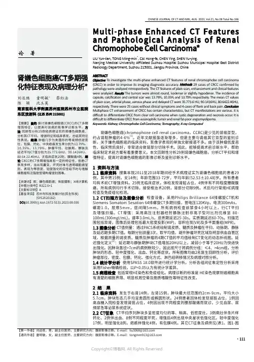
·111CHINESE JOURNAL OF CT AND MRI, AUG. 2023, Vol.21, No.08 Total No.166【通讯作者】童明敏,女,副主任医师,主要研究方向:腹部影像诊断。
E-mail:*****************Enhanced CT Features112·中国CT和MRI杂志 2023年08月 第21卷 第08期 总第166期2)。
增强后肾皮质、髓质及肾肿瘤的多期CT平均值及肿瘤CT值强化趋势如图3。
肿瘤平扫、动脉期、静脉及延迟平均CT值分别为35.773.6HU、90.1416HU、80.64 22.46HU。
3组间在密度均匀性、形态、出血、强化程度方面无明显统计学差异性(如表2)。
2.3 手术与病理 肿瘤肉眼观察呈灰黄褐色、金黄色,有包膜3例。
免疫组化:CK7(+)、CD117(+/-)、E-cad(+)、EMA(+)、CD117(+)。
肾脏部分切除9例 ,肾癌根治术20例。
无远处脏器转移、无后腹膜明显淋巴结肿大。
图1A-图1E 患者,男,49岁,右肾CRCC。
a平扫、b-d增强后三期横断位图,e皮质期冠状位重建。
体检发现有肾包块。
CT示右肾上极类圆形软组 织肿块,最大层面大小约59*67mm,平扫内见稍低密度区,增强扫描呈囊实性,实性部分肿块轻-中强化,CT平扫及三期增强CT值分 别为29-58-56-47HU,其内低密度区未见明显强化。
病理:嫌色性肾细胞癌,免疫组化结果癌细胞示EMA+++,CD10浆+++,CK7+++, CD117+++,E-cad+++,Vim++。
图2A-图2F 患者,女,72岁,CRCC,因“间歇性右腰区酸胀不适半年”入院。
平扫右肾中极见结节样等密度灶(a,CT值34HU),直径约2cm,密度较 均匀,增强后动脉期强化明显(b,CT值102HU),较肾皮质略低,中央可见疤痕,静脉其强化少减弱,持续性强化(c,83HU),冠状位重 建,肿块主要位于肾皮质,突出肾轮廓外(d)。
病史:患者,女,44岁,因“左侧腰痛1周,检查发现左肾肿物2天”入院。
如图1-4(图1轴位平扫,图2增强皮质期,图3增强实质期,图4增强排泄期)图1图2图3图4 基础解剖影像:图5图6图7图8图5-8所示分别为正常成人肾脏平扫及三期增强。
图5所示为双肾平扫,双肾实质密度均匀(黄色箭头),肾周脂肪间隙清晰,肾周筋膜未见增厚。
同层面可见肝脏(绿色箭头)、胆囊(蓝色箭头)、胰腺头部(红色箭头)。
图6所示为肾脏增强皮质期,注意肾皮质明显强化(黄色箭头),与轻度强化肾髓质界限清晰。
同层面可见胰头左侧肠系膜上动脉强化血管影。
图7所示为肾脏增强实质期,肾脏皮髓质强化程度近似,分辨困难,注意左肾门见囊性未强化影。
同层面可见胰头左侧肠系膜上静脉、肠系膜上动脉强化血管影。
图8所示为肾脏增强排泄期,双肾肾盂内见高密度对比剂影,以右肾明显(白色箭头),左肾门囊性低密度影未见对比剂,并非肾盂,考虑肾盂囊肿。
图1-4所示双肾平扫、皮质期、实质期、排泄期:左肾下极可见类圆形稍低密度影,局部突出于包膜外,边缘可见蛋壳样钙化影,密度不均,中心呈稍低密度影(绿色箭头),增强皮质期病灶呈轻度强化(黄色箭头),实质期强化达到峰值,排泄期病灶强化程度减低,始终低于肾实质密度,中心稍低密度影呈延迟强化,呈不典型星芒状。
影像诊断:左肾下极占位病变,考虑肾嫌色细胞癌或嗜酸细胞瘤。
知识点:肾脏增强分级及不同血供病变鉴别肾脏增强可以分析肿瘤的血供程度,判断肾细胞癌亚型,对治疗提供进一步指导意见。
1.增强分级:肾脏增强分为皮质期、实质期、排泄期,三者影像表现明显不同。
皮质期可见明显强化的肾皮质,与轻度强化的肾髓质分界清晰;实质期见皮髓质分界不明显,强化程度基本一致;排泄期可见皮髓质强化程度明显减低,肾盂及输尿管内可见高密度对比剂影,注意不要误以为是病变。
有文献对强化程度进行分级,主要分为轻度、中度、明显强化,其CT值范围为。
一般认为增强绝对值大于10Hu认为有强化,10-30Hu为轻度强化,30-50Hu为中度强化,50Hu以上为明显强化。