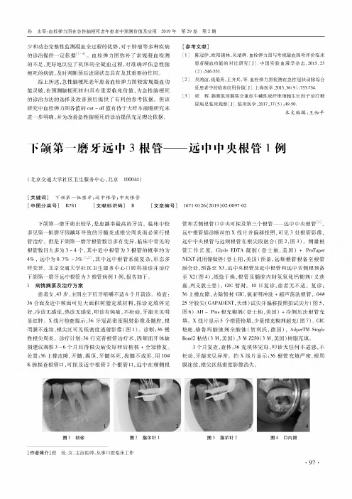
下颌第一磨牙5个根管1例
- 格式:pdf
- 大小:319.80 KB
- 文档页数:2


■论著・老年人群下颌第一磨牙根管结构和近中根根面形态研究吴宓勳吕娇张雪梅【摘要】目的:使用CBCT观察老年人下颌第一磨牙根管和牙根牙面形态。
方法:纳入98名老年患者的98颗下颌第一磨牙CBCT影像,观察根管结构及近中根根面凹陷情况。
结果:老年人下颌第一磨牙的近中根根管结构以IV型为主,占比78.6%;远中根以I、II型为主,占比分别为44.9%和12.2%;所有的近中根远中面都存在凹陷,56%近远中面均有凹陷,且根面凹陷程度与年龄呈负相关。
结论:老年人下颌第一磨牙根管常有钙化,根管结构变异较多;近中根的远中面都存在不同程度的凹陷,半数以上近中根的近远中面均有凹陷。
关键词:老年人;下颌第一磨牙;牙根形态;根管系统[中国图书分类号]R781.05[文献标识码]A D01:10.19749/.cjgd.l672-2973.2021.02.004A survey of root canal configuration and root morphology of the mandibular first molars in elderly populationWU Mi—hsun*LV Jiao,ZHANG X(e—Mei.(Beijing Tsinghua Changgung Hospital,School of Clinical Medicine*Tsinghua University,Beijing102218*China)[Abstract]Objective:This study was to investigate the distribution of root canal configuration and root surface morphology of the mandibular first molars in elderly Chinese population using CBCT.Methods:CBCT images of98mandibular first molars from98elderly patients were included to observe root canal configuration and the root surface morphology.Results: The most common root canal configuration found in the mesial roots was type IV(78.6%),while the most common configuration found in the distal roots was type I(44.9%)and type II(12.2%).On the mesial roots,root concavities were observed on all thedistal surfaces and in56%of both mesial and distal surfaces.Conclusion:The root canal of the first mandibular molar in the elderly is often calcified and has many root canal variations.The distal and mesial surfaces of the proximal and mesial roots had concavities,and more than half of the proximal and mesial roots had concavities.Key words:elderly population;mandibular first molar;root canal;root concavity下颌第一磨牙由于萌出早且牙面的窝沟点隙复杂,容易发生:坏。

下颌第一磨牙形态特征
下颌第一磨牙是人体口腔中最后萌出的恒牙之一,其形态特征包括以下几个方面:
1. 牙冠:下颌第一磨牙的牙冠相对较大,呈扁平状,前后宽度相近,呈椭圆形。
2. 牙合面:下颌第一磨牙的牙合面形态复杂,为牙合面尖、嵴、窝、沟、斜面最多的牙。
外形轮廓略似长方形。
3. 颊面:略似梯形,牙合缘长于颈缘,近远中径大于牙合颈径,近中缘直,远中缘突。
近、远中颊尖的颊轴嵴与颊沟平行,颊沟末端形成点隙。
4. 舌面:似梯形,比颊面小且稍圆突。
牙合缘可见近中舌尖、远中舌尖,舌沟从两舌尖捡通过,舌轴嵴不明显。
5. 邻面:约似四边形,牙冠向舌侧倾斜,颊尖较舌尖低。
远中面小于近中面。
近中颊颈角和近中舌牙合角较锐。
近、远中接触区均靠近牙合1/3偏颊侧。
6. 根:下颌第一磨牙的颈部较粗,根部长而弯曲,有时存在两个根管。
以上信息仅供参考,如需了解更多信息,建议咨询口腔科医生或查阅口腔医学相关书籍。
下颌第一磨牙近中三根管的临床观察徐海;张光东【摘要】Objective To investigate the types of mesial middle root canals ( MM) and correlate the clinical incidence of mesial mid⁃dle root canals of mandibular first molars with variables of gender, age, tooth type. Methods 12 mature permanent mandibular first molars with mesial middle root canals from August 2014 to August 2015 were included in the analysis. Types of MM according to Ver⁃tucci or Pomeranz, tooth type, gender, and age were recorded. Results Of the 12 mandibular first molars with MM, most of them were confluent in patient>30 years old while independent in patient<30 years. There was no difference in gender. However, significant difference was observed in tooth type⁃occurrence on right side was more than that on the left side. Conclusion Gender and tooth type might be the key factors, which determine the existence of MM.%目的:探讨12例下颌第一磨牙近中三根管的根管分型及与年龄、牙位、性别之间的关系。
【摘要】上颌第一磨牙根管的解剖变异多发生于近颊根管,而腭根数目及腭侧根管数目的变异较少见。
本文报道1例5个根管的右侧上颌第一磨牙,包括颊侧3个根管(近中根管2、远中根管1)和腭侧2个根管。
医生在临床工作中应仔细进行髓腔内解剖结构的检查及X线片判读,必要时进行锥形束CT检查以防止遗漏根管。
【关键词】磨牙;牙髓腔,解剖学;根管疗法;上颌第一磨牙引用著录格式:高楠,林志勇.右侧上颌第一磨牙腭侧双根管1例[J/CD].中华口腔医学研究杂志(电子版),2019,13(5):317⁃319.DOI:10.3877/cma.j.issn.1674⁃1366.2019.05.010Maxillary first molar with two palatal root canals:a case reportGao Nan,Lin ZhiyongDepartment of Stomatology,Shandong Provincial Hospital Affiliated to Shandong University,Jinan250021,China Corresponding author:Lin Zhiyong,Email:zhiyongl@ 【Abstract】There are many variations in root and canal anatomy of maxillary first molar.The incidence of two canals in the mesiobuccal root of maxillary first molar is higher while two canals or roots on the palatal side have rarely been reported. This case report presents a maxillary first molar with five root canals,including3buccal canals(2in mesiobuccal root and1 in distalbuccal root)and2palatal canals.Based on this case report,carefully examination of internal anatomy and radiographs of teeth and a cone⁃beam CT scan performed is essential to avoid missing root canals.【Key words】Molar;Dental pulp cavity,anatomy;Root canal therapy;Maxillary first molarDOI:10.3877/cma.j.issn.1674⁃1366.2019.05.010上颌第一磨牙根管数目大致可分为三根管、四根管及变异数目根管三大类。
下颌第一磨牙根管数目的临床观察[摘要]目的:调查下颌第一磨牙根管数目的临床情况。
方法:选择2017年1月-2018年6月唐山市协和医院牙体牙髓科门诊治疗的下颌第一磨牙患者161例,含患牙167颗。
采用探针及k锉探查发育沟,结合数码x线片,分近中、远中统计下颌第一磨牙的根管数目。
结果:M1,M2,M3发现率分别为0.60%,92.22%,7.19%;D 1,D2,D3发现率分别为43.11%,55.69%,1.20%。
结论:下颌第一磨牙近中、远中额外根管的存在增加了根管遗漏的机率,因此,提高下颌第一磨牙额外根管存在及查找意识非常重要。
[关键词]下颌第一磨牙;近中根管;远中根管;发现率[分类号]:R78Clinical observation of the mandibular first molar root canal numberHUO Bing-xin1,LI Chang-qing2,YAN Chun-xia1Zha Jian-xin3,Xu Mei1(1Department of Cariology and Endodontology,Tangshan Xiehe Hospital l,Tangshan063000,China;2Orthodontics department Tangshan Xiehe Hospital,Tangshan063000,China3 Department of Periodontology,Hebei,Tangshan 063000,China)[Abstract]Objective:To investigate the clinical incidence of the number of root canals in the mandibular first molar.Methods:From January 2017 to June 2018,161 patients with mandibular first molars,including 167 teeth,were selected to be treated in the department of dental pulp,xiehe hospital,tangshan city;.Probe and k file were used to investigate the number of root canals of the first molar in the mandible in the near,middle and farmiddle.Results:Statistical167mandibular first molar number suffering from root canal,M1,M2,M3 occurrence rates were 0.60%,92.22%,and 7.19%,respectively;D1,D2,D3 incidence was 43.11%,55.69%and 1.20%respectively.Conclusion:The mandibular first molar,there are far in the third root canal,increasing the probability of missing the root canal,thereby increasing awareness and look for the presence of additional mandibular first molar root canal is very important.[Keyword]mandibular first molar;mesial root canal;distal root canal;discovery rate[Chinese books catalog]:R78作者简介:霍炳鑫(1984-),男,汉族,主治医师,在职研究生,email:******************根管治疗是牙髓病及根尖周病的首选治疗方法,彻底清理根管内炎症牙髓及坏死物质是提高根管治疗成功率的重要保障。