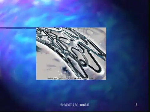
覆膜金属支架治疗CDF幻灯
- 格式:ppt
- 大小:20.12 MB
- 文档页数:40


Vol. 1 No. 3 2010 1Xu meidong, Yao liqing, Zhong yunshi et al.Department of Endoscopy Center, Zhongshan Hospital, Fudan University, Shanghai, China. 200032ǏAbstract ǐObjective Methods ResultsConclusionTo evaluate the clinical value of treatment of advanced esophageal carcinoma by using membrane-covered metallic stent.The clinical data of 106 patients with advanced esophageal carcinoma treated with endoscopy from June 2003 to June 2006 were analyzed retrospectively.122 membrane-covered metallic stents were placed in 106 cases, the success rate was 100%.The degree of dysphagia was reduced from 3.17 grade to 1.01 grade. The cure rate of 11 patients with esophagotracheal fi stula was 100%. The mean survival was 8.2 months.The placement of membrane-covered metallic stent is a safe, economical, effective and well-tolerated method for relieving esophageal stricture of advanced esophageal carcinoma.Esophageal neoplasms; Esophageal stenosis; Stent; EndoscopyǏKey words ǐThe malignant stenosis and esophageal obstruction caused by advanced esophageal carcinoma and recurrence of postoperative anastomotic stoma of esophageal carcinoma are difficult problems in esophageal surgery. It is because most of the patients lose their chances of surgical operation or could not bear the operation. Dysphagia would seriously affect the survival quality and survival time of patients. In the recent years, with the incessant development of endoscopic intervention technique, a kind of palliative treatment method which is safe, effective and reasonable concerns is provided for that category of patient. From June, 2003 to June, 2006, 106 patients of advanced esophageal carcinoma were treated by endoscopic dilatation and placement of membrane-covered metallic stent, the curative effect was satisfactory and the report is as follows.Clinical evaluation of treating an advancedesophageal carcinoma by using membrane-covered metallic stent125 1 Data and method1.1 General dataFrom June, 2003 to June, 2006, 106 patients with advanced esophageal carcinoma were treated by using endoscopic dilatation and placement of membrane-covered metallic stent, including 79 male and 27 female, who were 42~89 years old. Their average age was 61.7. Among them, 85 cases were pathologically affi rmed to be squamous cell carcinoma, 19 cases glandular cancer, 1 case adenosquamous carcinoma, and 1 case lymphatic sarcoma. The stenosis length was between 3~10 cm and was 5.49 cm on average. The patients had symptom of dysphagia in varying degrees before operation, and it was confirmed to be malignant esophageal stenosis by gastroscope or GI, refer to Table 1 for details. The stooler classifi cation criterion is adopted for appraising the extentRadioactive Esophageal Stent with 125I Particles2 Vol. 1 No.3 2010of dysphagia of patients: Grade 0 means no dysphagia. Grade 1 means obstruction in eating solid food. Grade 2 means obstruction in eating semi-liquid diet. Grade 3 means obstruction in eating liquid diet. Grade 4 means difficulty in drinking water (including those with esophago-tracheal fistula), refer to Table 2 for details.1.2 Method1.2.1 PreoperativeGIF-240 or 260 Video-gastroscope or model GIF-XP260 ultrafine gastroscope (Olympus), Balloon- type CRE dilatation catheter (Boston) or Savary dilating bougie (Cook) and 400 cm long φ0.89 mm Jagwire guide wire orφ0.97 mm guide wire with metallic soft head. The patients shall be starved for 6 hours before operation, accepted an intramuscular injection of 5 mg Diazepam 15 minutes before operation, and accepted an intravenous injection of 10mg Anisodamine hydrobromide for sedation, spasmolysis and reduce excretion.1.2.2 Stenotic dilatationGastroscopy and biopsy shall be conventionally performed to confirm the cause, region, level and length of stricture. The guide wire was inserted into the distal-end of stricture through endoscope under X-ray fluoroscopy. The ERCP tube was inserted along the guide wire for injecting cardiografin. The region, form and length of stenosis affection were confirmed under the contrast medium. A lso, it provided the information whether the stenosis was correlated to esophago-tracheal fistula. For the relatively serious stricture, where the endoscope and even stent pushing system could not pass though, bougie or balloon dilation shall be used in order to dilate the stricture region. Savary dilating bougie could dilate the stricture step by step up to 13 mm. In case of balloon dilating, the dilating balloon shall be inserted into the stricture part by the help of guide wire. When the region in balloon is located at the thinnest part of stricture, the balloon was inflated by contrast medium or aseptic physiological saline with pressure pump. The pressure ranged 304.0~810.6kPa was applied according to different needs, the balloon diameter shall be maintained at 12mm~15mm, and the dilatation time shall be kept for 2~5min. The endoscope was inserted again to observe the dilation result at distal-end of stricture. The guide wire was inserted as deep as possible again. The stricture location and length was measured at the time of withdrawing endoscope. The metallic markings were made for locating the distal end and proximal end of stricture.1.2.3 Placement of internal stentA n appropriate membrane-covered stent of nickel-titanium memory alloy with individualized shape (Nanjing Micro-tech or Boston company), its diameter shall be 18~20 mm, and its length shall be the length of stricture segment plus additional about 4cm, was used. Under the X-ray guiding, the stent delivering system shall be inserted into the stricture part through guide wire, the position of distal-end was confirmed with the help of metallic label or contrast medium, and the stent was deployed so that both sides of stent projected beyond the stricture segment by 2 cm. With the combination of endoscope and X-ray fluoroscopy, the positioning of stent was closely monitoring. If the length or position of stent has not reached the requirements, it shall be corrected by endoscope immediately.1.2.4 Postoperative treatment and follow-upRestoring the nutrition by gradually drinking of warm water, eating liquid diet with no residue and low residue were recommended till the regular eating was achieved. However, chewing and swallowing was an important issue. Eating thick, sticky and high-fiber food, glutinous kind or hard type was not recommended. Meanwhile, if there was any symptom of pectoralgia, short breath, abdominal pain, hematemesis or hemafecia, consulting doctor was recommended. On the third day after operation, conventionally X-ray follow-up examination would be performed, by taking water soluble contrast medium orally or radiography directly, to observe the positioning of stent. The dietary habits and survival circumstance of patients by periodically follow-up in outpatient service or by telephone.2 Result2.1 Rate of success and curative effect122 times of stents placement through endoscope for the 106 patients in this group under X-ray guidance, with success in one time without exception, and the overall success rate of operation is 100%. Among them, 2 times of stents were placed for 14 cases, and 3 times of stents were placed for 2 cases. The stricture of whole group of patients was immediately alleviated after stent placement, the irritating cough disappeared, and they were gradually able to eat liquid, semi-liquid and even regular diet, the score for classification of dysphagia was reduced to grade 1.01 from grade 3.17 two weeks later on the average (refer to Table 2), and a significant differenceDigestive SystemDigestive SystemR a d i o a c t i v e E s o p h a g e a l S t e n t w i t h 125I P a r t i c l e sVol. 1 No. 3 2010 3between before and after treatment were found (P<0.01). 11 patients, who had esophago-tracheal fistula, orally took cardiografin for fluoroscopy after operation, no contrast medium was leaked and there was no symptom of irritating cough, etc. It indicated the esophageal fi stula orifi ce was all fully blocked after stent placement, and the blocking rate is 100%. One patient was died of respiratory failure in two days after operation. Postoperative follow-up was performed to the other 105 cases. The follow-up time was between 3 weeks to 12 months, 12 cases did not go back for follow-up and the follow-up rate was 88.57% (93/105). The reason of missing follow-up included a lack of accurate telephone number and rejection by family members of dead patients. The follow-up period was equal to survival period, and the postoperative living standard of patients who could eat food was obviously improved. A mong them, the longest survival period reached 12 months, the shortest survival period was 2 months, the stent was smooth during the survival period, and the average survival period was 8.2 months.2.2 ComplicationAll patients had retrosternal pain and epigastric pain after operation and alleviated by themselves within one week. 8 patients had relatively severe pain, narcotic analgesic was needed to be injected repeatedly for relieving pain, such as bucinnazine or dolantin, etc., and the occurrence rate was 6.56% (8/122). In the process of dilatation and stent placement, there was mild bleeding in all cases because of laceration of tumor tissue at the stricture part, and bleeding could be stopped for most patients after haemostatic medication was locally nebulized. 5 patients had symptom of hemorrhage after operation, such as hematemesis and passing tarry stools, etc., the occurrence rate was 4.10% (5/122), and the hemorrhage was stopped after antacid, homeostasis and heteropathy treatment for 3~5d. One patient (0.81%) had diffi culty in breathing after operation, and died because of respiratory failure 2 days later. No perforation was occurred. It was found through postoperative follow-up that 35 patients had symptom of reflux esophagitis, such as food reflux, regurgitation and retrosternal burning sensation, etc., the occurrence rate was 28.69% (35/122), the symptom could partly be alleviated through application of antacid and dynamic medicine. Three cases (2.46%) had a stent migration at the distal-end and caused restenosis, the stent was taken out by gastroscope after dilating the stenosis lumen, and stent was re-placed again. Stent blocking occurred in 14cases with occurrence rate of 11.48% (14/122). Among them, one patient’s stent was blocked by food. Stent blockages by tumor-overgrowing (TG) were occurred in 7 cases. Granulation and fi brosis (GF) occurred in 6 cases. For the food impaction, massive food was taken out by gastroscope, and it was dredged after repeated rinsing; 2 cases of GF accepted repeated APC treatment, and liquid diet could be maintained till patient passed away. Stents were re-placed for the other 9 cases of GF or TG, and the symptom of blocking was relieved. Stenosis and obstruction occurred in two cases within the follow-up period, and 3 times of stent placement were carried out to them individually.3 DiscussionPatient, suffered an advanced esophageal carcinoma, including advanced primary esophageal carcinoma, cancerous esophago-tracheal fistula and recurrence of anastomotic stoma after operation, etc., loses the opportunity of operation. An expected result could not be reached even if operation is performed. When the curative effect is poor and the trauma is increased in size, the occurrence rate of postoperative complication and the mortality rate is high, surgical resection is not a preferable method. Placement of self-expandable metallic stent can effectively alleviate the obstruction symptom of patients, increase food intake and improve the quality of life. It has been more extensively applied clinically, especially the clinical application effect of membrane-covered metallic stent is relatively ideal [1, 2]. It was found by Saranovic (et al.) [3] in a research on treating advanced esophageal carcinoma by using metallic stent with membrane-covered and without membrane-covered: the symptom of dysphagia of patient could be effectively alleviated after placement of stent into esophagus, the score for the average dysphagia of 98 patients in the membrane-covered group was reduced from 2.67 to 0.05, and that of those in group without membrane-covered was reduced from 2.73 to 0.15. It was considered by the report of Yang (et al.) [4] that membrane-covered stent would be adopted for the patients of advanced esophageal carcinoma accompanied by esophago-tracheal fistula, and the blocking rate of fi stula orifi ce could reach 100%. 122 times of stents were placed in the 106 cases of this group, with the success rate of operation being 100%. The score for dysphagia classification in two weeks after operation was reduced from grade 3.17 to grade 1.014 Vol. 1 No. 3 2010Radioactive Esophageal Stent with 125I ParticlesTable 1 Affection category and regionafter operation; the esophageal fi stula orifi ce of 11 cases of esophago-tracheal fi stula could be fully blocked after operation, and the blocking rate was 100%. The average survival period after operation was 8.2 months, being similar to the report in literature.The common complication of placing metallic stent into esophagus includes retrosternal and epigastric pain,hemorrhage, perforation, respiratory diffi culty or asphyxia, stent blockage (food impaction, GF or TG), stent migration and reflux esophagitis, and the report on occurrence rate differs [5~8]. The occurrence rate of severe pain in this group after operation was 6.56%, that of hemorrhage 4.10%, that of respiratory difficulty 0.81%, that of reflux esophagitis 28.69%, that of stent migration 2.46%, that of stent blocking 11.48%, and no perforation occurred. All complications could be alleviated by medication or endoscope treatment.How to improve the success rate of placement of esophageal stent and reduce the occurrence rate of complication? A ccording to our experience, the following measures will be taken. (1) Accuracy of stent deployment could be increased by the combinationof endoscopy and X-ray fluoroscopy. (2) The metal marking is an important guidance for locating the sides of stricture and the stent deployment. (3) The occurrence of postoperative pain and hemorrhage can be reduced by selecting a membrane-covered stent with appropriate diameter, shape and length. A skillful stent placement is necessary. An appropriate shape of upper rim of stent can reduce the tension between the stent and pipe wall and reduce the irritation to esophageal mucosa and ingrowth of granulation. Membrane-covered stent can prevent ingrowth of tumor into stent cavity. Both sides of stent shall go beyond the affection by 2cm. (4) the 1st week after stent placement, the patient shall take liquid or semi-liquid diet. Then, the composition of solid food shall be gradually increased till regular diet restores, patients shall chew carefully and swallow slowly, especially, avoid from eating glutinous or hard food. (5) Appropriate radiotherapy and chemotherapy shall be adopted after stent placement in order to suppress the ingrowth of tumor and granulation tissue and reduce the occurrence rate of stent blockage.RegionAffection category Case numberUpper segment Middle segmentLower segment and cardiac orifi cePrimary esophageal carcinoma stenosis 6343623Recurrence of postoperative anastomotic stoma 13265esophageal carcinoma stenosis after radiotherapy 196112Esophago-tracheal (esophagobronchial) fi stula 11281Total 105146131Digestive SystemVol. 1 No. 3 2010 5Digestive SystemR a d i o a c t i v e E s o p h a g e a l S t e n t w i t h 125I P a r t i c l e sReferences1. Morgan R, Adam A. Use of metallic stents and balloons in the esophageal and gastrointestinal tract [J]. J Vasc Interv Radiol, 2001, 12(3):283-2972. Yao Liqin, Xu Meidong. The clinical application and prospect of alimentary canal stent [J]. Chinese Journal of Practical Surgery, 2002, 22(10): 583-585.3. Saranovic Dj, Djuric-Stefanovic A , Ivanovic A , et al. Fluoroscopically guided insertion of self-expandable metal esophageal stents for palliative treatment of patients with malignant stenosis of esophagus and cardia: comparison of uncovered and covered stent types [J]. Dis Esophagus, 2005,18(4):230-238.4. Yang HS, Zhang LB, Wang TW, et al. Clinical application of metallic stents in treatment of esophageal carcinoma [J]. World J Gastroenterol, 2005, 11(3):451-453.Table 2 Contrast of classifi cation for dysphagia before and after stent treatment*P<0.01 The group of those after treatment includes 105 cases, and 1 case died within 2 days after operation.Classifi cation for dysphagiaBefore treatment (n)After treatment (n)001910662172035404350Average number3.171.015. A cunas B, Rozanes I, A kpinar S, et al. Palliation of malignant esophageal strictures with self-expanding nitinol stents: drawbacks and complications [J]. Radiology, 1996, 199(3): 648-652.6. Bartelsman JF, Bruno MJ, Jensema AJ, et al. Palliation of patients with esophagogastric neoplasms by insertion of a covered expandable modified Gianturco-Z endoprosthesis: experiences in 153 patients [J]. Gastrointestinal Endosc, 2000, 51(2): 134–138.7. Han YM, Song HY, Lee JM, et al. Esophagotracheal fistulae due to esophageal carcinoma: palliation with a covered Gianturco stent [J]. Radiology, 1996, 199(3): 65–70.8. Li ZS, Wan XJ, Xu GM, et al. Clinical study of esophageal stent restenosis [J]. Chinese Journal of Dig Endo, 2000, 17(4): 217-219.。