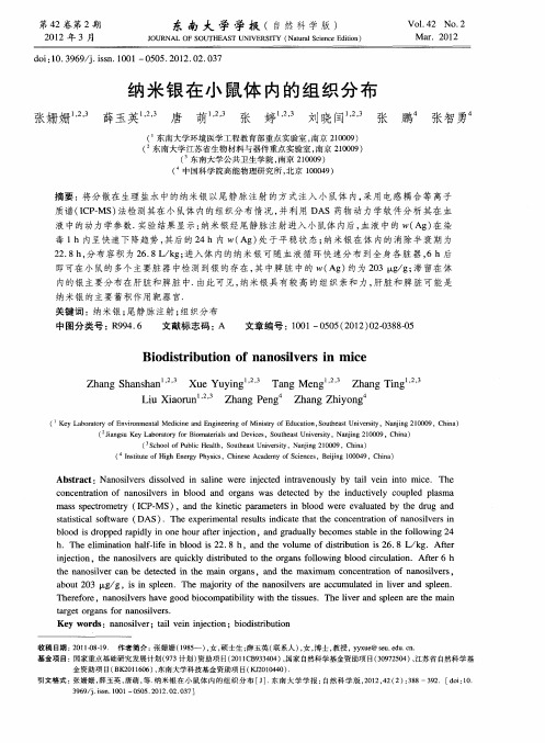
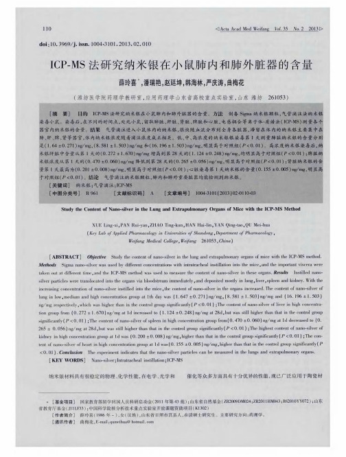
纳米银在小鼠体内的组织分布
- 格式:pdf
- 大小:323.19 KB
- 文档页数:5

纳米银冻干粉小鼠经口最大耐受剂量实验王醒;江筱莉;周昱薇;陈明祺;吕海;顾宁【期刊名称】《东南大学学报(医学版)》【年(卷),期】2014(033)001【摘要】目的:测定纳米银冻干粉小鼠经口给药的最大耐受剂量.方法:按照最大耐受剂量测定法,将ICR小鼠40只随机分为对照组和实验组,每组20只,实验组采用最大浓度、最大容积的纳米银冻干粉一次经口给药,对照组给予等量生理盐水,观察小鼠给药后2周内的行为、外观、进食、排泄物、主要脏器的大体和病理学改变以及死亡情况,并计算小鼠对纳米银的最大耐受量.结果:纳米银冻干粉小鼠经口染毒后,行为无异常,摄食量、体重等与对照组比较差异无统计学意义(P>0.05),各主要脏器无明显异常改变,2周内无死亡.结论:采用最大浓度、最大容积的纳米银冻干粉行小鼠经口给药最大耐受量试验,经折算最大耐受剂量为临床成人日用量的3 592倍.【总页数】4页(P31-34)【作者】王醒;江筱莉;周昱薇;陈明祺;吕海;顾宁【作者单位】东南大学生物科学与医学工程学院,江苏南京210009;东南大学生物科学与医学工程学院,江苏南京210009;东南大学生物科学与医学工程学院,江苏南京210009;江苏省中医院重症医学科,江苏南京210029;江苏省中医院重症医学科,江苏南京210029;东南大学生物科学与医学工程学院,江苏南京210009【正文语种】中文【中图分类】R-33;R114【相关文献】1.蜂王浆冻干粉对HepG2细胞葡萄糖消耗作用及对糖尿病小鼠降糖作用的实验研究 [J], 汪宁;朱荃;周义维;童黄锦2.牛初乳冻干粉改善小鼠睡眠状况的实验研究 [J], 江海涛;任源浩;陈勇;陈舒泛;吴京燕;杨云峰;蒋新陵3.纳米银涂层可吸收线吻合小鼠肠壁近期抗炎疗效的实验研究 [J], 刘雪来;宋岩彪;张创;曹学会;靳晓次;费川;张永婷;李索林4.荟生源两种组方对小鼠经口急性毒性实验 [J], 姚淮育;吴虹谚;何昊;付满玲;吴显和5.干、鲜壁虎冻干粉对S180荷瘤小鼠的抑瘤作用及其急性毒性实验研究 [J], 杨金霞;杨国生;朱伟;富宏;刘庚信;王学美因版权原因,仅展示原文概要,查看原文内容请购买。


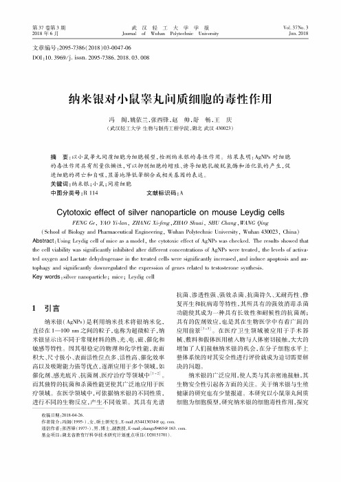
第!7卷第!期2018年6月武汉轻工大学学报Journal of Wuhan Polytechnic UniversityVol.37No.3Jun.2018文章编号:2095-7386(2018)03-0047-06DOI ;10. 3969/j. issn. 2095-7386. 2018.03.008纳米银对小鼠睾丸间质细胞的毒性作用冯阁,姚依兰,张西锋,赵帅,舒畅,王庆(武汉轻工大学生物与制药工程学院,湖北武汉430023)摘要:以小鼠睾丸间质细胞为细胞模型,检测纳米银的毒性作用。
结果表明:AgNPs对细胞的毒性作用具有剂量依懒性,可以抑制细胞的增殖、诱导细胞乳酸脱氢酶和活化氧的产生,促进细胞的凋亡和自噬,显著地降低睾酮合成相关基因的表达。
关键词:纳米银;小鼠;间质细胞中图分类号:K114 文献标识码:ACytotoxic effect of silver nanoparticleon mouseLeydig cellsFENG $,YAO Yi-lcm,ZHANG Xi-feng,ZHA0 Shuai,SHU Chang,WANG Qing(School of Biology and Pharmaceutical Engineering,Wuhan Polyteclmic University,Wuhan 430023, China)AbstfclCt ;Using Leydig cell of mice as a model,the cytotoxic effect of AgNPs was checked. The results showed thatthe cell viability was significantly inhibited after diferent concentrations of AgNPs were treated,the levels of activated oxygen and Lactate dehydrogenase in the treated cells were significantly increased,and induce apoptosis tophagy and significantly downregulated the expression of genes related to testosterone synthesis.Key words ; silver nanoparticle %mice %Leydig celll引言纳米银(AgNPs)是利用纳米技术将银纳米化,直径在1一100nm之间的粒子,也称为超微粒子,纳米银显示出不同于常规材料的热、光、电、磁、催化和 敏感等特性。
纳米银对小鼠流感治疗作用的研究李秀景;尹俭俭;郑丛龙【摘要】目的探讨不同浓度的纳米银对小鼠流感的治疗作用.方法选择18~22 g 健康昆明小鼠48只,随机分成6组:正常对照组、模型组、治疗阳性对照组、高剂量组(纳米银400 μg/mL)、中剂量组(纳米银100 μg/mL)和低剂量组(纳米银25 μg/mL).小鼠感染H1N1流感病毒A/PR/8/34(PR8 H1N1),4 h后鼻腔给予不同浓度纳米银,连续治疗3 d.记录各组小鼠每天的一般生理状况以及生存和死亡情况,观察14 d;通过比较各组小鼠的死亡率、生存天数、肺指数和肺组织病毒滴度,观察各组小鼠肺组织病理变化,测定各组小鼠肺组织的细菌感染情况,研究不同浓度纳米银对小鼠流感的疗效.结果高剂量组、中剂量组和低剂量组的死亡率均明显减低,其平均生存天数均明显延长,与模型组比较差异有显著性意义,P<0.01;高剂量组、中剂量组和低剂量组的肺指数均有所降低,且高剂量组和中剂量组与模型组比较差异有显著性意义,P<0.01;模型组小鼠肺组织的病毒滴度为1:512,有的小鼠高达1:1024,高剂量组、中剂量组和低剂量组的大部分小鼠的肺组织病毒滴度<1:2.模型组小鼠肺组织的形态结构消失,肺泡壁增厚,组织间可见大量红细胞,肺泡腔见水肿液;高剂量组小鼠肺组织的病变明显减轻,肺组织形态结构较为完整,肺泡腔中无水肿液.模型组携菌量较多,高剂量组、中剂量组和低剂量组的携菌量减少,与模型组相比差异有显著性意义,P<0.01.结论纳米银对小鼠流感具有明显的治疗作用,其治疗效果与纳米银的剂量有关.%Objective To discuss the therapeutical effect of different concentrations of silver nanoparticles against influenza in mice. Methods Forty - eight Kun Ming mice, which were healthy and weigh 18 -22 g, were randomly divided into normal control group, model group, positive control group , high dose group (the concentration of silver nanoparticles is 400μg/mL) , middle dose group (100 μg/mL) and low dose group (25 μg/mL). Mice were infected with the influenza virus A/PR/8/34 H1N1 (PR8 H1N1) and intranasal administration of silver nanoparticles at different concentrations after 4 hours, and treated continuously 3 days. Record the general physical condition , the condition of life and death count of each group every day for 14 days. To study the curative effect of silver nanoparticles at different concentrations against influenza in mice, the indexes that mortality rate , survival days , lung index, lung virus titer, lung histopathological examination and lung bacterial infections of each group were used . Results Compared with model group , the mortality of high, middle and low dose group reduced obviously , and monse survival days in these gvoup were significantly longer (P <0.01). The lung index of high, middle and low dose group reduced and the high and middle dose group reduced significantly (P <0.01). The lung virus titer of model group was 1:512, and some mice were as high as 1: 1024; the with hist in the high, middle and low dose group was < 1: 2. In model group, the morphological structure of lung tissue was disappeared , the alveolar walls were thickened , a large number of red blood cells wlhe found , and the edema fluid was found in alveolar cavity . In high dose group, the pathological changes of lung tissue were obviously alleviated , and it is no edema fluid in alveolar cavi -ty. There were large numbers of bacteria in model group , the bacterial content in high , middle and low dose group reduced significantly ( P < 0.01). Conclusion Two silver nanoparticles wouldhmte therapeutic effect on influenza in mice , and the curative effect was dose dependent.【期刊名称】《大连医科大学学报》【年(卷),期】2013(035)003【总页数】6页(P223-228)【关键词】纳米银;流感;小鼠【作者】李秀景;尹俭俭;郑丛龙【作者单位】大连大学医学院,病原生物学教研室,辽宁,大连,116622;大连大学医学院,病原生物学教研室,辽宁,大连,116622;大连大学医学院,病原生物学教研室,辽宁,大连,116622【正文语种】中文【中图分类】R373.1+3流感病毒能够引起个体的严重感染甚至导致死亡,对人类的健康造成威胁。
纳米银植入大鼠皮下组织亚慢性毒性及在不同组织中的分布陈丹丹;奚廷斐;白净;王谨【期刊名称】《中国组织工程研究》【年(卷),期】2009(013)016【摘要】背景:有实验报道纳米银的生物材料直接与人体接触或植入体内,能够产生不良的生物学效应.目的:验证纳米银对人体是否存在潜在的不良生物学反应,评价其生物安全性.设计、时间及地点:动物实验观察,2005-06/2006 06在中国药品生物制品检定所以来器械检测中心完成.材料:纳米银粉和微米银粉为美国Sigma化学试剂公司产品.方法:将大鼠随机分为3组:纳米银组,微米银组,空白对照组,10只/组,雌雄各半.纳米银粉组和微米银粉组皮下植入进行亚慢性毒性试验.实验开始时植入1次,植入剂昔为0.33 g/kg.对照组只做手术,不给药.同时,每组随机选择4只大鼠,对12只大鼠的血清和部分脏器使用等离子体质谱分析仪进行银在大鼠各脏器中分布的测定.主要观察指标:血液生化学指标,脏器系数,银含量.结果:与对照组比较,给药组大鼠的个别脏器系数与对照组比较有显著性差异,但各给药组脏器的绝对质量与对照组比较没有湿著性差异,结合起来分析脏器系数的异常不具有临床意义.其它各脏器系数均在正常值范围内,与对照组比较尤显著性差芹.末见与受试物毒性作用有关的病理组织学改变,给予纳米银粉大鼠末见毒件反应.结论:纳米银对人体可能存在潜在的不良生物学效应.%BACKGROUND: It has been reported that nanosilver-containing biomaterials produce bad biological effects after they directly contact with or are implanted into human body.OBJECTIVE: To investigate whether nanosilver yields potential adverse biological effects on human body and to evaluate its bioiogical safety.DESIGN, TIME AND SETTING: Ananimal experiment observation was performed at the Medical Device Center of National Institute for the Control of Pharmaceutical and Biological Products from June 2005 to August 2006.MATERIALS: Nanosilver particles and microsilver particles were purchased from Sigma Company, USA.METHODS: Thirty rats were randomly divided into 3 groups, with 5 male and 5 female rats per group: nanosilver, microsilver, and blank control. Nanosilver and microsilver particles were respectively and subcutaneously implanted for subchronic toxicity test.The nanosilver and microsilver groups were given 0.33 g/kg nanosilver and microsilver, respectively. Rats from the blank control group received identical procedure, with the exception of drug application. Four rats were selected from each group for determination of silver content in serum and some organs by plasma mass spectrometry.MAIN OUTCOME MEASURES: serum biochemical indices, organ coefficient, and silver content.RESULTS: There was significant difference in individual organ coefficient between each drug application group and blank control group. But no significant difference in absolute mass was found between each drug application and the blank control group.These findings suggested no clinical significance of organ coefficient. Other organ coefficients were in the normal range, and there was no significant difference between each drug application group and the blank control group. Patho-histological changes related to toxicity were not found. Rats from the nanosilver group did not show toxic reaction.CONCLUSION: Nanosilver produces potential adverse biological effects after implanted into human body.【总页数】4页(P3181-3184)【作者】陈丹丹;奚廷斐;白净;王谨【作者单位】中国药品生物制品检定所,北京市,100050;中国药品生物制品检定所,北京市,100050;清华大学医学院生物医学工程系,北京市,100084;中国药品生物制品检定所,北京市,100050【正文语种】中文【中图分类】R318【相关文献】1.巴戟天不同炮制品中水晶兰苷的大鼠体内血药浓度及组织分布研究 [J], 史辑;景海漪;黄玉秋;范亚楠;贾天柱2.纳米银和微米银在大鼠组织器官中的分布 [J], 陈丹丹;奚廷斐;白净;王谨3.钙调神经磷酸酶在大鼠不同组织中的分布及活性 [J], 符民桂;陈亚红;刘秀华;庞永政;唐朝枢4.胱硫醚β-合酶在大鼠不同组织中的分布及其在As时的表达变化 [J], 郑斌;韩梅;温进坤5.羟基磷灰石植入皮下组织不同阶段弹性模量的变化 [J], 徐莲云;侯振德;赵巍;毕平;王泓因版权原因,仅展示原文概要,查看原文内容请购买。