超微型显微成像系统(中英文版)
- 格式:docx
- 大小:203.84 KB
- 文档页数:2
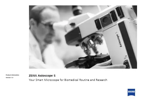
ZEISS Axioscope 5Your Smart Microscope forBiomedical Routine and Research2ZEISS Axioscope 5Your Smart Microscope forBiomedical Routine and ResearchThis smart microscope system teams Axioscope 5 with a high-performance LED light source and a sensitive, standalone microscope camera to document your samples in transmitted light or fluorescence – all in one go. In particular, it’s never been easier to acquire fluorescent images consisting of up to four different channels. Now, instead of going through that tedious process of at least 15 steps with a manual microscope, all you have to do is push a button.Axioscope 5’s high quality optics deliver brilliant images of your sample. In addition, this smart, encoded microscope facilitates digital documentation enormously, andSnap images without taking your hands off the microscopeMink Uterus Endometrium Epithelial Cells, vimentin – red, F-actin – green, nucleus – blue; acquired with ZEISS Axioscope 5, Colibri 3 and Axiocam 202 mono in stand-alone mode, objective: Plan-Apochromat 40× /0.95without getting in the way of your established workflow in the lab. You’ll work more efficiently, save time and produce high contrast images with the very best resolution.Enjoy Fast Digital Documentation with Smart MicroscopyAxioscope 5 makes documenting your specimens very efficient. The color impression shows up in the camera image exactly the same as it appears through the eyepieces. The smart Axioscope 5 system makes automatic adjustments for illumination and white balance to keep digital documentation easy. All you have to do is focus on your sample, press the ergonomic Snap button on the microscope, and that’s it. Acquiring high quality images with high color fidelity has never been easier – and quicker.Visualize Your Sample Clearlyand EffortlesslyAxioscope 5 uses its transmittedwhite light LED to provide powerful illumination with high color reproductivity. You will clearly seethe subtle differences in your sample. And experience all the advantagesof LED illumination properties suchas stable color temperature, low energy consumption and long lifetime. Axioscope 5 comes with a light intensity manager that produces uniform brightness at all magnifications so adjusting lamp brightness whenyou change magnification is a thingof the past. That saves you time and reduces eye fatigue, too.Histological specimen (human). CDx immuno-histological stain; Red: immunoreactive antigens in cytoplasm; Blue: nuclear counterstainingZ iehl-Neelsen-Färbung,objective: EC Plan-Neo fl uar 63× / 0.95 Korr.Chromosome specimen (human). Giemsa stain,objective: Plan-Apochromat 63× / 1.4Renal tissue (human). Trichrome stain,objective: Plan-Apochromat 40× / 0.95Capture Multichannel FluorescenceImages with Just One ClickAcquiring fluorescent images has neverbeen so easy. Using Axioscope 5 inconjunction with its LED light sourceColibri 3, you have the perfect setupfor easy multichannel fluorescencedocumentation. Switch effortlesslybetween the channels for UV, blue,green and red excitation. Just selectthe relevant channels and press Snap.The system then takes over andautomatically adjusts the exposuretime, acquires the image, switchesthe channel and starts again. So thereyou have it in one go: your overlayedmultichannel fluorescence image,complete with a scale bar – evenwithout a PC.3N o t a l l p r o d u c t s a r e a v a i l a b l e i n e v e r y c o u n t r y . U s e o f p r o d u c t s f o r m e d i c a l d i a g n o s t i c , t h e r a p e u t i c o r t r e a t m e n t p u r p o s e s m a y b e l i m i t e d b y l o c a l r e g u l a t i o n s . C o n t a c t y o u r l o c a l Z E I S S r e p r e s e n t a t i v e f o r m o r e i n f o r m a t i o n .E N _41_012_193 | C Z 04-2019 | D e s i g n , s c o p e o f d e l i v e r y , a n d t e c h n i c a l p r o g r e s s s u b j e c t t o c h a n g e w i t h o u t n o t i c e . | © C a r l Z e i s s M i c r o s c o p y G m b HYour Flexible Choice of ComponentsCarl Zeiss Microscopy GmbH 07745 Jena, Germany ******************** /axioscopeThis is Smart Microscopy –Digital Documentation Made Easy Used in combination with themicroscope cameras Axiocam 202mono or Axiocam 208 color, you have the full advantage of a smart standalone m icroscope solution.Camera settings such as white balance, contrast and exposure time are done automatically.W ithout needingadditional imaging software or even acomputer, you can:• Snap images and record videosdirectly from your stand• Use mouse (and optionally keyboard) to control your c amera via OSD (on screen display)• Save settings• Store images with all metadata of them icroscope and camera as well as scaling i nformation• Predefine the name or rename your imageStand-alone for Basic Routine Imaging ZEISS Axioscope 5 operatesi ndependently of a computer system.VZEISS Labscope for Advanced Routine ImagingOperating ZEISS Axioscope 5 with ZEISS Labscope imaging software is ideal forc onnected microscopy and standard multichannel fluorescence imaging.VVZEISS ZEN for Research ApplicationsUse ZEN imaging software to perform advanced imaging tasks with ZEISSA xioscope 5.VVBoost your Efficiency –with Smart MicroscopyEfficiency and quality are key in your lab, but it can take a lot of time to acquire detail-rich, true-color images. You know the drill: place the sample, focus your region of interest, switch to the computer, a djust settings such as white balance, exposure time and gain, then acquire an image, insert a scale bar, switch back to the microscope … and so on. That’s what a typical documentation workflow looks like. Now, with the Axioscope 5 system, you can stay focused on your sample at all times, thanks to smart microscopy. Digital documentation is inherent in the system design. Just press the e rgonomic Snap button on the microscope and you’re done. The procedure integrates perfectly with your established microscopy workflow and boosts your efficiency tremendously.。

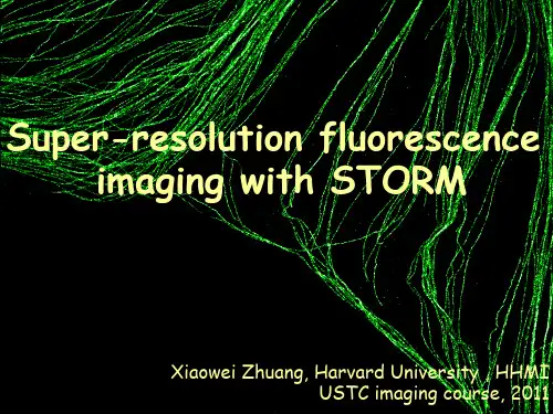
单分子定位显微技术(Single Molecule Localization Microscopy,简称SMLM)是一种基于荧光显微技术的高分辨率成像方法,它的原理是通过利用单个荧光分子的闪烁信号来定位样品中的单个分子,从而实现高分辨率成像。
SMLM技术可以实现分子级别的可视化,其分辨率可以达到10纳米以下,远高于传统显微技术。
由于其高分辨率和高灵敏度,SMLM 技术已经成为生物学、化学、材料科学等多个领域中重要的研究工具。
一、原理SMLM的原理基于单个荧光标记的瞬时发光特性。
当激光或其他光源激发荧光标记时,标记会发射荧光光子。
这些光子在相机或光探测器上被捕获,并转化为电子信号。
在SMLM 中,通过分析这些光子的时间和空间分布,可以确定每个荧光标记的位置。
SMLM的关键在于利用光学显微镜的成像系统将荧光标记限制在光学焦点内,使得每次只有单个标记被激发发光。
因此,可以记录每个标记的光子发射时间和位置信息,并将这些信息用于精确定位标记的位置。
SMLM技术包括多种方法,其中最常用的是单分子激发的STORM(stochastic optical reconstruction microscopy)和PALM(photoactivated localization microscopy)。
在SMLM过程中,需要解决许多技术挑战,包括背景噪声的影响、荧光标记的失活和漂移等问题。
为了解决这些问题,研究者们开发了各种算法和图像处理技术,如滤波、校正和重建方法等。
这些技术的应用可以提高SMLM的空间分辨率和信噪比,并为更深入的细胞生物学研究提供了新的工具。
图 1 STORM其中,STORM是最常用的SMLM技术,其基本原理是利用亚波长分辨率下荧光发光的闪烁特性,对单个发光染料的位置进行定位,进而构建出超分辨率的图像。
STORM技术将样品在一定范围内随机激发,从而使样品中的荧光染料分子以随机的方式发光。
利用图像处理算法,可以对发光点的位置进行高精度的计算,得到超分辨率图像。

中国科学 G 辑: 物理学 力学 天文学 2007年 第37卷 增刊: 138~145收稿日期: 2007-05-20; 接受日期: 2007-09-03Australia Research Council 和国家自然科学基金(批准号: 60410131, 90508003)资助项目* E-mail:《中国科学》杂志社SCIENCE IN CHINA PRESS 光纤非线性光学显微成像付 玲①②* M. Gu ①②(① 华中科技大学武汉光电国家实验室(等), 武汉 430074; ② Centre for Micro-Photonics, Faculty of En-gineering and Industrial Sciences, Swinburne University of Technology, Hawthorn, Victoria 3122, Australia)摘要 在光学显微成像领域, 基于光纤的小型非线性光学显微镜和内窥镜作为传统显微镜和其他光学成像方法的一种重要补充形式, 近几年来受到人们的关注. 该文介绍和总结了光纤非线性光学显微成像技术及其在生物医学中的应用.首先介绍了结合非线性光学显微技术和单模光纤耦合器获得小型非线性光学显微镜的方法; 特别对基于双包层光子晶体光纤和微电机系统扫描镜的光纤非线性内窥成像系统进行了分析; 最后通过消化器官的组织成像实验说明了光纤非线性光学显微镜的重要应用. 研究证明了基于光纤和微电机系统MEMS 扫描镜的非线性内窥镜的新概念, 并用于生物组织的成像.关键词 非线性光学显微成像 光纤耦合器 双包层光子晶体光纤 微电机系统扫描镜 组织成像显微镜改变了人类认识世界的方法, 是人类历史上最重要的发明之一. 经过300多年的发展, 光学显微成像已成为自然科学研究领域中的重要分支, 为生命和信息等科学领域不断提供新方法和新概念[1]. 特别是上世纪末期出现的非线性光学显微镜具有光学层析能力并能在厚组织(毫米量级)内实现高空间分辨率(亚微米)成像, 是现代光学显微成像技术的革新, 为人们研究厚组织中的分子和细胞功能提供了有力的工具[2,3]. 非线性光学成像技术主要基于多光子吸收(multiphoton absorption)[4]、高次谐波(higher harmonic)[5]和相干反斯托克斯拉曼散射(coherent anti-Stokes Raman scattering, CARS)[6]等光学非线性效应, 其中以双光子荧光成像的应用最为广泛. 自非线性光学显微镜诞生以来[4], 科学家们一直致力于将其用于活体(in vivo )成像. 然而, 复杂的光学系统和沉重的光学器件使非线性光学显微成像的研究局限于庞大的显微镜系统, 无法应用到活体的内部器官(internal organs)和整体动物(intact animals)成像.近年来, 随着新型光纤和微制造技术的迅猛发展, 光纤非线性显微镜和内窥镜(fiber-optic nonlinear optical microscopy/endoscopy)的研究正在扭转这一局面[7]. 光纤非线性显微镜和内窥镜是小型的非线性光学显微成像系统, 由光纤传输激发光或者非线性光信号, 并利用微型扫增刊付玲等: 光纤非线性光学显微成像139描机制构建光学图像. 光纤的超小尺寸(一般只有数百个微米的直径)与良好的机械和光学性能增加了成像系统的灵活性并减小了系统的尺寸, 使非线性光学成像技术在内部器官和活体动物中的研究成为可能. 自Denk研究小组和Gu研究小组先后研制出基于单模光纤的双光子荧光显微镜之后[8,9], 光纤非线性显微镜和内窥镜的研究在短短6年之内进展迅速[10~19]. 光纤非线性显微镜和内窥镜研究中的关键问题是: (ⅰ) 近红外波段超短脉冲激光的传输和非线性光信号的收集. 光纤的色散和自相位调制等非线性效应会导致脉冲宽度和光谱的展宽, 降低激发效率[20]; 光纤的低数值孔径和小芯径限制了可见光信号的高效收集、系统的信噪比和成像深度. (ⅱ) 扫描机制. 扫描器件应该具有毫米量级的尺寸, 并能实现快速扫描和高图像分辨率来实时监测生物过程. (ⅲ) 系统设计. 系统设计是非线性内窥镜研究的关键, 直接决定了系统的灵活性, 功能和应用范围. 该文研究了基于单模光纤耦合器(single-mode fibre coupler)和双包层光子晶体光纤(double-clad photonic crystal fibre, DCPCF)的非线性光学显微成像系统, 首次实现了基于光纤耦合器的双光子荧光和二次谐波显微成像技术, 并证明了基于DCPCF和微电机系统(microelectromechanical system, MEMS)扫描镜的非线性内窥镜的新概念, 使光纤成像系统的信噪比提高了两阶.1基于单模光纤耦合器的非线性光学显微镜Bird和Gu[10,11]首次将单模光纤耦合器引入双光子荧光显微成像, 证明了三端口的单模光纤耦合器能传输近红外波段的超短脉冲激发光并收集荧光. 与基于单模光纤的非线性光学显微镜相比[9], 使用单模光纤耦合器能将非线性显微镜的纵向分辨率提高30%, 且使系统更紧凑并具有自准直的特性. 然而, 二次谐波比双光子荧光更远离单模光纤耦合器的工作波长; 二次谐波源于相干过程, 其偏振各向异性的测量(polarisation anisotropy measurement)要求光纤耦合器能同时传输偏振的激发光和谐波信号[15]. 因此, 我们首先研究了单模光纤耦合器在不同波长的传输特性, 结果如图1(a). 实验中端口3单模光纤耦合器(Newport, F-CPL-S12785)的工作波长为780 nm, 分束比(Splitting ratio)是50:50, 纤芯与包层的直径分别为5和125 μm. 当波长范围在770~870 nm时, 耦合器中端口3~1的耦合效率为20%~41%; 端口2~1在435~532 nm 波长范围内的耦合效率为29%~41%. 当长度为2 m的光纤耦合器传输80~150 mW的激发光至样品端时, 尽管光纤中的群速度色散和自相位调制使脉冲展宽到数皮秒, 系统仍有足够的激发光功率用于非线性成像. 实验结果还表明, 单模光纤耦合器在可见光波段的功能类似于普通光学显微镜中的二向色镜(Dichroic mirror). 当435 nm的激光耦合入端口1, 端口2和端口3的分束比为99.6:0.4, 而且两个端口的输出模式分别表现出单模和多模的特征(图1(b), (c)). 532 nm的激光在单模光纤耦合器中传播时, 测量得到的分束比和输出模式也具有相同特征(图1(d)). 因此, 为了优化在基于单模光纤耦合器的非线性显微镜系统中激发光与非线性信号的传输, 超短脉冲激光通过端口3(激发臂)传递到端口1(样品臂), 后向散射的双光子荧光和二次谐波则由端口1通过端口2(信号臂)被探测器收集(图1). 光纤纤芯能消除焦点以外的杂散光, 在成像系统中具有共聚焦针孔(Pinhole)的功能.除了单模光纤耦合器传输特性的研究, 二次谐波光谱的研究也是验证光纤成像系统的关键. 在图1的实验系统中, 我们以AF-50为样品, 用光谱仪(Acton Research Corporation)测量了经过单模光纤耦合器的双光子荧光和二次谐波的光谱. 当激发波长是800 nm时, 通过单模光纤耦合器的辐射谱包含400 nm的二次谐波谱线和430~600 nm的双光子荧光(图2). 二次谐波140中国科学G辑物理学力学天文学第37卷的光谱宽度约为9 nm, 约为激发光带宽的. 当激发光在780~870 nm范围内调谐时, 二次谐波光谱的中心波长则随激发光波长改变, 始终展示出倍频的特性.图1 基于单模光纤耦合器的双光子荧光和二次谐波成像系统(a) 单模光纤耦合器在可见光和近红外波段的耦合效率. (b)~(d) 可见光在激发臂和信号臂的输出模式; O1和O2为 0.25 NA10×显微物镜, O3为0.85 NA 40×显微物镜双光子荧光和二次谐波显微镜中的光学层析能力源于信号光强度与激发光强度的非线性关系, 纵向分辨率是非线性显微成像系统的重要参数. 通过在纵向位置扫描薄层AF-50, 我们得到了基于单模光纤耦合器的显微镜系统对双光子荧光和二次谐波的响应曲线(图2中的插图). 此结果表明, 在激发波长为800 nm时, 光纤显微镜对二次谐波和双光子荧光成像的纵向分辨率分别为1.8和2.1 μm. 可见二次谐波较短的信号波长使其成像的纵向分辨率比双光子荧光成像的纵向分辨率提高了14%. 当激发光波长由800 nm调谐到860 nm时, 二次谐波成像的纵向分辨率增加到 1.9 μm. 通过三维相干传递函数的理论计算, 我们发现与光纤参数和光学系统参数相关的归一化光纤尺寸对基于单模光纤的二次谐波成像系统的纵向分辨率起决定性的作用, 光纤中二次谐波波长的增加使归一化光纤尺寸增大, 纵向分辨率降低[21].图2 基于单模光纤耦合器的非线性显微镜的双光子荧光(TPEF)和二次谐波(SHG)光谱与纵向响应曲线增刊付玲等: 光纤非线性光学显微成像141用偏振的二次谐波信号来反应结构蛋白的形态是二次谐波显微镜的特性之一. 为了证明基于单模光纤耦合器的成像系统具有探测二次谐波偏振各向异性的能力, 我们研究了单模光纤耦合器对偏振光传输和成像的影响. 测量结果表明, 在整个近红外和可见光波段, 单模光纤耦合器中的材料双折射特性使其能传播特定偏振方向的线性偏振光[22]. 因此单模光纤耦合器能传递线性偏振的激发光与高度有序的蛋白纤维(例如鱼鳞, black tetra fish scale)相互作用, 二次谐波经过光纤耦合器后的偏振状态可通过转动探测器前的偏振片并测量收集到的二次谐波强度来确定. 图3(a)和(b)是在探测器前偏振片(Newport, 10GT04)转角正交情况下光纤系统收集到的鱼鳞的二次谐波图像, 激发波长为800 nm, 探测器前的滤光片为400/9 nm. 当偏振片转角改变时, 收集到的二次谐波强度与转角θ 满足cos2θ 的关系(图3(c)). 此结果证明, 基于单模光纤耦合器的显微镜系统能保持二次谐波信号的线性偏振态, 并对结构蛋白成像.图3 二次谐波偏振各向异性的测量(a)和(b)偏振方向正交的二次谐波成像, 箭头方向为探测器前偏振片的转角方向. 标尺为20 μm. (c) 二次谐波的强度与探测器前偏振片转角的cos2θ 关系2基于双包层光子晶体光纤的非线性光学内窥镜由于单模光纤能传输高质量的激光束并改善成像系统的层析能力, 因而在光纤成像系统中被普遍采用. 但是单模光纤的低数值孔径(约0.1~0.2)和较小的纤芯尺寸极大地限制了对生物样品中微弱光信号的收集和探测, 使非线性光学显微镜系统的灵敏度较低. 作为光纤领域的革新产物, 光子晶体光纤能通过设计光纤中二维光子晶体的结构实现普通光纤不能具备的功能, 彻底地改变了光纤传输光束的方式. 我们首次改造了有源的DCPCF, 并将其用于光学显微成像来提高系统的灵敏度. 由于MEMS器件具有小尺寸、低功耗、利于建造“芯片显微镜”等优点, 显微内窥成像系统中的扫描机制则由MEMS扫描镜来实现. 基于DCPCF和MEMS 扫描镜的非线性内窥成像系统如图4. 飞秒脉冲光经过光栅对(Newport, 1200 grooves/mm)和一个显微物镜(Melles Griot, 4×/0.12NA)后被耦合到DCPCF. 光纤中出射的光经过MEMS扫描镜后被渐变折射率(Gradient index, GRIN)透镜聚焦到样品上. 非线性光信号则通过MEMS扫描镜后被DCPCF收集到探测器.142 中国科学 G 辑 物理学 力学 天文学 第37卷图4 基于DCPCF 和MEMS 扫描镜的非线性光学内窥镜系统右上角是双包层光子晶体光纤的扫描电子显微镜图像与800 nm 激发光输出模式的叠加, 右下角是能实现两维扫描的MEMS 扫描镜非线性光学内窥成像系统中的DCPCF 是由用于光纤激光器的有源DCPCF 演化而来. 我们除去了有源光纤纤芯中的掺杂元素, 其目的是使近红外波段的激发光能在纤芯进行单模传输, 而位于可见光波段的非线性光信号则能被具有高数值孔径的内包层收集[16]. 成像系统中DCPCF(Crystal Fiber A/S)的纤芯和内包层直径分别为20和165 μm(图4). 纤芯被孔间距比(Hole to hole pitch ratio)为0.26的空气小孔包围. DCPCF 的内包层和外包层则被由一圈空气小孔分开, 使内包层拥有高数值孔径(~ 0.6). 需要指出的是, 这种大面积和高数值孔径的光纤只能利用光子晶体的结构实现, 而非普通光纤的制造技术. 通过对DCPCF 光传输特性的研究, 我们发现如果选择与纤芯数值孔径接近的耦合物镜, 改造过后的DCPCF 在410~870 nm 的耦合效率可达到80%~90%(图5). 而使用高数值孔径(0.65)的耦合物镜则会导致光泄露到内包层, 使耦合效率降低20%. 因此, 低数值孔径的耦合物镜能使近红外光耦合到纤芯中的效率为28%, 可见光耦合到内包层的效率为90%, 此数值是单模光纤对可见光耦合效率的两倍.GRIN 透镜是一种利用特定折射率分布使光线聚焦的小型透镜. 当光纤和GRIN 透镜结合用于非线性光学成像系统, GRIN 透镜的有效数值孔径依赖于光纤和GRIN 透镜之间的距离, 因而直接影响到光纤成像系统的效果[23]. 例如,光束的耦合效率与有效数值孔径有关; 成像的分辨率与有效数值孔径的平方成正比; 双光子荧光的强度与有效数值孔径的4次方成正比. 因此, 研究光纤系统的纵向分辨率和信号水平随光纤和GRIN 透镜距离的变化是优化系统设计的重要途径. 实验采用了直径为0.5 mm, 节距(pitch)为0.2, 数值孔径为0.5的GRIN 透镜(GRINTECH)和AF-50为样品. 实验结果表明, 当DCPCF 与GRIN 透镜之间的距离增大时, 双光子荧光成像的纵向分辨率逐渐得到改善, 而且探测到的荧光强度逐渐增大(图6(a)).如果光纤与透镜之间图5 双包层光子晶体光纤在可见至近红外波段的耦合效率增刊付玲等: 光纤非线性光学显微成像143的距离为5 mm, 激发光束充满GRIN透镜的后孔径, 系统能得到最佳的纵向分辨率, 即双光子荧光和二次谐波的纵向分辨率分别为6和5.4 μm (图6(b)). 与基于单模光纤耦合器和GRIN透镜的非线性显微镜系统相比, 由于DCPCF具有高数值孔径和大截面面积, 使用DCPCF取代单模光纤能实现纵向分辨率和非线性光信号的同时最优化, 而使用单模光纤的成像系统在探测到的荧光信号最强时却不具备最高的纵向分辨率[17,23].图6(a) 双光子荧光强度和纵向分辨率随GRIN透镜和光纤间距的变化, 荧光强度和纵向分辨率能在间距较大时被同时优化;(b) 双光子荧光与二次谐波的纵向响应曲线MEMS扫描镜是成像系统中实现二维光束扫描的器件. 我们使用的MEMS扫描镜是基于电热驱动(Electrothermal actuation)的原理, 在驱动电压少于12 V的情况下其光学扫描角度可达到30°[24]. 镜面的尺寸为0.5 mm, 表面铝膜对800 nm激发光的发射率约为80%(图4). 通过在MEMS扫描镜的快、慢轴应用特殊设计的波形和2.5~7.5 V的电压, 激光束的反射角被改变从而实现二维的线扫描(raster scanning), 其扫描速率为7线/s. 图7(a)是由基于DCPCF、MEMS 扫描镜、和GRIN透镜的显微镜系统获得的荧光微球的双光子荧光图像. 此结果证明了MEMS 扫描镜不仅能传递近红外超短脉冲用于非线性效应的激发, 而且能收集位于可见波段的荧光信号; 飞秒脉冲经过MEMS镜面之后, 脉冲大约展宽了25%, 故可通过镀膜技术来改善这一现象. 更重要的是, 以MEMS扫描镜作为扫描机制的非线性光学成像系统所产生的图像与通过普通扫描台获得的图像高度一致(图7(b)).图7 基于DCPCF, MEMS扫描镜、GRIN透镜(a)和基于单模光纤耦合器、扫描台、GRIN透镜(b)的显微镜系统对微球的双光子荧光成像标尺为10 μm144中国科学G辑物理学力学天文学第37卷DCPCF的独特光学性质使非线性显微镜成像系统的灵敏度得到极大改善. 通过比较由DCPCF和单模光纤耦合器得到的双光子荧光图像(图7(a)和(b)), 我们发现DCPCF的应用使成像系统的信号水平提高了两个量级. 如果使用光栅对补偿脉冲光在DCPCF中经历的群速度色散, 成像系统的信号水平将会在此基础上再提高一个量级. 成像系统灵敏度的改善主要是因为DCPCF比单模光纤具有更高的数值孔径和更大截面面积, 减少了飞秒脉冲在光纤中传输的自相位调制效应, 使飞秒脉冲具有最小的畸变, 而且对GRIN透镜的色差有更强的兼容性.3基于光纤非线性光学显微镜的组织成像利用基于DCPCF, MEMS扫描镜和GRIN透镜的非线性光学内窥镜系统(图4), 我们对大鼠的大肠和胃部组织进行了成像, 结果如图8. 为了增加荧光成像的对比度, 大肠和胃部组织从大鼠(Sprague-Dawley)中剥离后, 用浓度为1%的Acridine Orange(Sigma)对其内表面上皮组织染色, 成像实验在大鼠牺牲后两小时之内完成. 大肠组织双光子荧光成像的穿透深度约为100 μm, 图8(a)是双光子荧光成像的截面之一. 光纤显微镜系统能清晰地显示表面上皮细胞和肠道孔穴(intestinal crypts, 箭头)的形态信息. 从大鼠胃部组织获取的双光子荧光图像(图8(b))展示出与大肠截然不同的形态结构, 显微系统的分辨率亦能将胃部的坑穴(gastric pits, 箭头)从上皮细胞中区分出来.图8 使用基于DCPCF, MEMS扫描镜和GRIN透镜的光纤内窥镜系统对大鼠大肠组织(a)和大鼠胃部组织(b)的双光子荧光成像标尺为20 μm4总结和展望本文介绍和总结了基于单模光纤耦合器和DCPCF的非线性显微镜系统及其生物医学应用. 单模光纤耦合器能传递近红外波段的激发光和收集可见光波段的双光子荧光和二次谐波, 并在非线性显微镜系统中展示出二向色镜的特性. 基于单模光纤耦合器的成像系统能保持激发光和二次谐波的线性偏振态并用于研究结构蛋白的形态. 通过引入新型的DCPCF, 光纤非线性光学显微镜的灵敏度得到极大改善. MEMS扫描镜和GRIN透镜的结合使基于DCPCF的成像系统更加小型化, 大鼠大肠和胃部的组织成像也证明了非线性光学内窥镜系统的可行性. 在将来的研究中, DCPCF耦合器[25]与内窥镜系统将会实现整个成像系统的全光纤化, 这对光纤非线性光学显微镜的发展有着重要意义. 另外, 此光纤成像技术也可与显微成像领域中新的扫描技术[26,27]结合, 拓展多光子荧光成像的应用. 随着光纤器件和微制造技术的不断发展, 光纤非线性光学显微镜系统将日趋小型化, 为传统的光学显微成像技术提供重要的补充并在生物医学领域发挥作用.增刊付玲等: 光纤非线性光学显微成像145参考文献1 Yuste R. Fluorescence microscopy today. Nat Methods, 2005, 2: 902—904[DOI]2 Zipfel W R, Williams R M, Webb W W. Nonlinear magic: multiphoton microscopy in the biosciences. Nat Biotech, 2003, 21:1369—1377[DOI]3 Helmchen F, Denk W. Deep tissue two-photon microscopy. Nat Methods, 2005, 2: 932—940[DOI]4 Denk W, Strickler J H, Webb W W. Two-photon laser scanning fluorescence microscopy. Science, 1990, 248: 73—75[DOI]5 Campagnola P J, Loew L M. Second harmonic imaging microscopy for visualizing biomolecular arrays in cells, tissues andorganisms. Nat Biotech, 2003, 21: 1356—1360[DOI]6 Cheng J X, Xie X S. Coherent anti-Stokes Raman scattering microscopy: instrumentation, theory, and applications. J PhysChem B, 2004, 108: 827—840[DOI]7 Flusberg B A, Cocker E D, Piyawattanametha W, et al. Fiber-optic fluorescence imaging. Nat Methods, 2005, 2: 941—950[DOI]8 Helmchen F, Fee M S, Tank D W, et al. A miniature head-mounted two-photon microscope: high-resolution brain imaging infreely moving animals. Neuron, 2001, 31: 903—912[DOI]9 Bird D, Gu M. Resolution improvement in two-photon fluorescence microscopy with a single-mode fiber. Appl Opt, 2002,41: 1852—1857[DOI]10 Bird D, Gu M. Compact two-photon fluorescence microscope based on a single-mode fiber coupler. Opt Lett, 2002, 27:1031—1033[DOI]11 Bird D, Gu M. Two-photon fluorescence endoscopy with a micro-optic scanning head. Opt Lett, 2003, 28: 1552—1554[DOI]12 Jung J C, Schnitzer M J. Multiphoton endoscopy. Opt Lett, 2003, 28: 902—904[DOI]13 GÖbel W, Kerr J N D, Nimmerjahn A, et al. Miniaturized two-photon microscope based on a flexible coherent fiber bundleand a gradient-index lens objective. Opt Lett, 2004, 29: 2521—2523[DOI]14 Levene M J, Dombeck D A, Kasischke K A, et al. In vivo multiphoton microscopy of deep brain tissue. J Neurophysiol,2004, 91: 1908—1912[DOI]15 Fu L, Gan X, Gu M. Use of a single-mode fiber coupler for second-harmonic-generation microscopy. Opt Lett, 2005, 30:385—387[DOI]16 Fu L, Gan X, Gu M. Nonlinear optical microscopy based on double-clad photonic crystal fibers. Opt Express, 2005, 13:5528—5534[DOI]17 Fu L, Jain A, Xie H, et al. Nonlinear optical endoscopy based on a double-clad photonic crystal fiber and a MEMS mirror.Opt Express, 2006, 14: 1027—1032[DOI]18 Myaing M T, MacDonald D J, Li X. Fiber-optic scanning two-photon fluorescence endoscope. Opt Lett, 2006, 31: 1076—1078[DOI]19 Fu L, Gu M. Fibre-optic nonlinear optical endoscopy. J Microscopy, 2007, 226: 195—206[DOI]20 Agrawal G P. Nonlinear Fiber Optics. San Diego: Academic. 198921 Gu M, Fu L. Three-dimensional image formation in fiber-optical second harmonic generation microscopy. Opt Express,2006, 14: 1175—1181[DOI]22 Fu L, Gan X, Bird D, et al. Polarisation characteristics of a 1×2 fiber coupler under femtosecond pulsed and continuouswave illumination. Opt Laser Technol, 2005, 37: 494—497[DOI]23 Fu L, Gan X, Gu M. Characterization of the GRIN lens-fiber spacing toward applications in two-photon fluorescence endo-scopy. Appl Opt, 2005, 44: 7270—7274[DOI]24 Jain A, Xie H. An electrothermal SCS micromirror for large bi-directional 2D scanning. 13th International conference onSolid-State Sensor, Actuators and Microsystems. Seoul: IEEE Computer Society Press, 2005. 988—99125 Fu L, Gu M. A double-clad photonic crystal fiber coupler for compact nonlinear optical microscopy imaging. Opt Lett, 2006,31: 1471—1473[DOI]26 Zeng S, Lv X, Zhan C, et al. Simultaneous compensation for spatial and temporal dispersion of acousto-optical deflectorsfor two-dimensional scanning with a single prism. Opt Lett, 2006, 31: 1091—1093[DOI]27 Li D, Zeng S, Lv X, et al. Dispersion characteristics of acousto-optic deflector for scanning Gaussian laser beam of femto-second pulses. Opt express, 2007, 15: 4726—4734[DOI]。
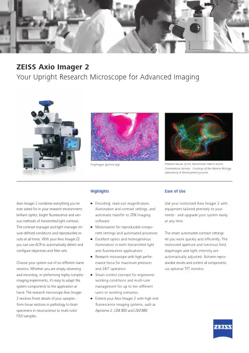
Pilidium larvae of the Nemertean ribbon worm,Cerebratulus lacteus. Courtesy of the Marine BiologyLaboratory & Development journalEsophagus (guinea pig)ZEISS Axio Imager 2Your Upright Research Microscope for Advanced ImagingAxio Imager 2 combines everything you’veever asked for in your research environment:brilliant optics, bright fl uorescence and vari-ous methods of transmitted light contrast.The contrast manager and light manager en-sure defi ned conditions and reproducible re-sults at all times. With your Axio Imager.Z2you can use ACR to automatically detect andconfi gure objectives and fi lter sets.Choose your system out of six different standversions. Whether you are simply observingand recording, or performing highly compleximaging experiments, it’s easy to adapt thesystem components to the application athand. The research microscope Axio Imager2 resolves fi nest details of your samples -from tissue sections in pathology to brainspecimens in neuroscience to multi-colorFISH samples.Highlights• Encoding: read-out magnifi cation,illumination and contrast settings, andautomatic transfer to ZEN Imagingsoftware• Motorization for reproducible compo-nent settings and automated processes• Excellent optics and homogeneousillumination in both transmitted lightand fl uorescence applications• Research microscope with high perfor-mance focus for maximum precisionand 24/7 operation• Smart control concept for ergonomicworking conditions and multi-usermanagement for up to ten differentusers or working scenarios.• Extend your Axio Imager 2 with high endfl uorescence imaging systems, such asApotome.2, LSM 800 and LSM 880Ease of UseUse your motorized Axio Imager 2 withequipment tailored precisely to yourneeds - and upgrade your system easilyat any time.The smart automated contrast settingslet you work quickly and effi ciently. Themotorized aperture and luminous fi elddiaphragm and light intensity areautomatically adjusted. Achieve repro-ducible results and control all componentsvia optional TFT monitor.***************/axioimagerN o t a l l p r o d u c t s a r e a v a i l a b l e i n e v e r y c o u n t r y . U s e o f p r o d u c t s f o r m e d i c a l d i a g n o s t i c , t h e r a p e u t i c o r t r e a t m e n t p u r p o s e s m a y b e l i m i t e d b y l o c a l r e g u l a t i o n s . C o n t a c t y o u r l o c a l Z E I S S r e p r e s e n t a t i v e f o r m o r e i n f o r m a t i o n .E N _41_012_113 | C Z 08-2015 | D e s i g n , s c o p e o f d e l i v e r y a n d t e c h n i c a l p r o g r e s s s u b j e c t t o c h a n g e w i t h o u t n o t i c e . | © C a r l Z e i s s M i c r o s c o p y G m bHPerformance:The motorized reflector turret accommo-dates either six or ten Push & Click filtermodules. Auto-configure all motorized components with Smart Setup of the ZEN imaging software.Acquire fluorescence images with an excellent signal-to-noise ratio. The fluorescence beam path and high efficiency fluorescence filter sets of this research microscope deliver exposure times that are up to 50 percent shorter.Stand Versions:• Axio Imager.A2 LED • Axio Imager.A2• Axio Imager.D2• Axio Imager.M2p • Axio Imager.M2• Axio Imager.Z2 Suitable Applications:• Cell biology • Neuroscience • Molecular genetics • PathologyNote: This product is primarily for research use. Only Axio Imager.M2p is for use in diagnostic procedures or patient management.ZEISS Axio Imager 2Your Upright Research Microscope for Advanced Imaging。

/OPMI-picoZEISS OPMI picoSeeing without compromise.S 100 / O P M I p i c o23ZEISS OPMI picoSimple, compact, provenOPMI ® pico from ZEISS, the most frequently sold ZEISS surgical microscope worldwide, is the trusted choice among doctors. It makes details and fine structures clearly visible. The system enables you to better visualize regions of interest and consistently provide your patients with high-quality examinations and treatments.ZEISS OPMI pico’s cost-effective LED illumination meets your standards and needs. The complete integration of the light source makes it easier to clean the system.Restorative dentistry Quickly detect enamel and dentine fractures as well as approximal caries. High-precision views enable accurate assessment of crown edges, preparation levels, and veneers.EndodonticsVisualize fine anatomical structures and details of root canals and isthmuses for a clear view right down to the apex.Implantology Conduct high-precision examinations and implant treatments quickly and confidently. Reliably detect important anatomical structures.Periodontics Benefit of support for soft-tissue evaluation and management to assist healing, low scarring, and improved cosmetic outcomes.The compact and easy-to-use ZEISS OPMI pico offers solid support for even the most demanding applications – whether in restorative dentistry, endodontics, implantology, or periodontics:Ergonomic seating enables users to conveniently maintain an upright working posture.ZEISS OPMI pico also features camera options to facilitate patient consultation and documentation, depending on thespecific needs in your practice.4Better viewLong-lasting LEDFor color rendition and light that strongly resembles natural daylight. It is a very cost-effective and virtually maintenance-free light source. The low heat output requires minimal ventilation and generates little noise.Full overview of fine detailsZEISS OPMI pico delivers high-quality images at every magnification level.ZEISS OPMI pico enables you to visualize high-contrast, true-color images – the key to improving the quality of examination and treatment. With ZEISS OPMI pico, optics and illumination go hand in hand.View the whole mouth The Varioskop ® 100 objective lens allows you to adjust the focal length over a range of 200 mm to 300 mm to focus on the whole oral cavity in the vertical axis – without moving themicroscope.5While treating your patients you can sit comfortably in an upright, ergonomically correct position for a more relaxed working day.Comfortable working conditions and improved visionOver 75% of dentists found that using a dental microscope had a positive effect on their neck and back pain.5 Working in a comfortable posture allows you to fully focus on your patient and their procedure, and increases your productivity.6A dental microscope can offer other ergonomic benefits, such as improved vision and reduced eye fatigue, common in dental professionals and corresponding to increasing age.7–9Better ergonomicsAdjust the ZEISS OPMI pico to your demands and not vice versa Reduce or increase the distance for the required treatment field as needed with the patented multi-link design of Foldable Tube f170 / f 260. With its long reach, this highly flexible system easily accommodates the needs of the user and different positions of the patient.6Better integrationBenefits of an integrated full HD camera • Complete function integration • Co-observation and documentation • Simplified cleaning of the housing • Images and videos recorded onto a shared network drive or a USB device• Capture of full HD images during recording or from a recorded videoBenefits of live viewing and streamings • Video livestream into the network• Smart Recording: retroactively record video that occurred in the previous 30 secondsComplete integration of technology and design in the suspension arm for a well-balanced architectureFunctional elements such as the video control console, HD video camera, cables, light sources, and light guides are completely integrated into the stand to avoid workspace clutter.Better educationReal-life pictures are very convincing. ZEISS OPMI pico can accommodate a full HD video camera with recording and streaming, allowing you to present patients with high-definition material to explain procedures.A highly compact instrument with a small footprint, ZEISS OPMI pico fits seamlessly into virtually any practice workflow environment. Simply plug in the cable, switch on the power, and the ZEISS OPMI pico isready for use.7Basic configurationPackage optionsAdd-onsSuspension system optionsFloor standCeiling mountWall mount (wallplate)Workplace integrationFurther space-saving suspension system options are available for the Centro carrier system and specified treatment units.Carl Zeiss Meditec AG Goeschwitzer Strasse 51–52 07745 JenaGermany/opmi-pico /med/contacts en-INT_3_1_12IPrintedinGermany.CZ-II/223Internationaledition:Onlyforsaleinselectedcountries.Thecontentsofthebrochuremaydifferfromthecurrentstatusofapprovaloftheproductorserviceofferinginyourcountry.Pleasecontactourregionalrepresentativesformoreinformation.Subjecttochangesindesignandscopeofdeliveryandduetoongoingtechnicaldevelopment.OPMI,VarioskopandVisionGuardareeithertrademarksorregisteredtrademarksofCarlZeissMeditecAGorothercompaniesoftheZEISSGroupinGermanyand/orothercountries.©CarlZeissMeditecAG,223.Allrightsreserved.Application images courtesy of:1 Dr. Claudia Cia Worschech, Sao Paulo, Brazil2 Dr. José Aranguren Cangas, Madrid, Spain3 Dr. Behnam Shakibaie, Tehran, Iran4 Dr. Rino Burkhardt, Zurich, Switzerland5Z augg B et al. Influence of magnification tools on the recognition of artificaila preparation and restoration defects (in German).Schweiz Monatsschr Zahnmed 2004;114:890-896. [Abstract]6L inger W. Advantages for patients under the dental microscope.Available from: https:///blog/dental-microscope7P errin P, et al. Visual acuities of dentists in their respective clinical conditions. Clin Oral Investig 2014;19:2055-2058.8E ichenberger M, et al. Visual acuity of dentists under simulated clinical conditions. Clin Oral Investig 2013;17:725-729.9Y adav VS, et al. Periodontal microsurgery: Reaching new heights of precision.J Indian Soc Periodontol 2018;22(1):5-11.。

质臻至简蔡司Axiovert 5用于细胞培养和研究的智能显微镜/axiovert20 µm HeLa Kyoto细胞,物镜:LD Plan-Neofluar 63×。
双通道荧光成像:细胞核为蓝色,微管蛋白为红色。
正在为您的实验室寻找一款功能强大的显微镜?想要一款成像时间短、图像质量优的显微镜?这很有必要!拥有一款高质易用的显微镜,对于需要在实验室进行长时间工作的您来说显得十分重要。
智能的倒置细胞培养显微镜蔡司Axiovert 5是您明智的选择:您仅需专注于样品和工作流,按下拍照按钮,即可获得用于数据记录的清晰图像。
该设备将透射光配备的各种观察方式与多通道荧光相结合,用于研究您的细胞或组织培养。
不仅如此,当实验室空间紧张时,您甚至可以将该智能显微镜作为单机使用,将图像保存在U盘上,而无需使用额外的计算机或软件。
用于细胞培养和研究的智能显微镜› 简介› 优势› 应用› 系统› 技术参数›售后服务更简单、更智能、更高度集成使用智能显微技术,让工作更智能蔡司Axiovert 5显微镜十分智能,且成像快速、结果出众。
您只需专注于样品,按下按钮,即可保存细胞或组织培养的清晰图像。
这款智能显微镜还会自动为您调整透射光以及多通道荧光图像的设置及参数。
自动叠加的多通道荧光图像包含标尺信息,该信息将自动保存在图像文件的元数据中。
轻松自如,享受您的日常工作Axiovert 5让您不用再时时刻刻焦急地等待实验结果。
其设计符合人体工程学,功能巧妙,可为您全天候的工作提供支持。
您只需专注于样品本身,使用单手便能完成包括拍照、移动载物台、调焦和控制亮度在内的各种主要操作。
光强管理功能可在所有放大倍率下提供统一的亮度,让您无需在更换物镜时手动调节灯泡亮度。
为了进一步提高细胞分析流程的速度和数据可靠性,您可以选择使用Labscope 中的AI 细胞融合度和AI 细胞计数分析功能,实时获得可重复的结果。
放眼未来,选择一款立足前沿的活细胞 显微镜从常规细胞培养到研究,Axiovert 5可无缝融入您的实验室和工作流。
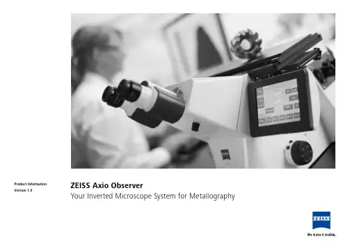
ZEISS Axio ObserverYour Inverted Microscope System for MetallographyProduct InformationVersion 1.050 µmSpherulitic graphite in nodular cast iron seen in C-DIC contrast.Your Inverted Microscope System for MetallographyFast, flexible, economic: Take advantage of Axio Observer’s inverted construction to investigate a large number of samples in no time at all – or to explore heavy ones, just as efficiently. There’s no need to refocus, even when changing magnifi-cation or switching samples. Axio Observer combines the proven quality of ZEISS optics with automated components to give you reliable, reproducible results. Using dedicated software modules you can analyze, for example, non-metallic inclusions, grain sizes and phases – it’s fully automatic. Axio Observer is your open imaging platform: invest in only the features you need today.As requirements change, a simple upgrade keeps your system ready for all materials applications.› In Brief › The Advantages › The Applications › The System› Technology and Details ›ServiceAnimationSimpler. More intelligent. More integrated.Save Time in Metallographic Investigations As an inverted microscope platform, Axio Observer makes work so much more enjoyable. Whether investigating a large number or even heavy samples, you’ll save time in both sample preparation and investigation. Meanwhile, its inverted design facilitates parallel alignment to the objective lens. Observe more samples in less time: simply put your specimen on the stage, focus once and keep the focus for all further magnifications and samples.Upgrade Your SystemKeep an eye on your budget as well as your samples.With Axio Observer, you invest only in the featuresyou need now. You can always upgrade your system,simply and economically, any time you need to.Choose between encoded or motorized compo-nents and a range of accessories – you can dependon having any relevant contrasting techniques yourapplication requires.Count on Reliable Results and Brilliant ImagesYou will appreciate the stable imaging conditionsof Axio Observer, especially when working withhigh magnifications. Homogeneous illuminationacross the entire field of view produces brilliantimages. And you will get reliable, reproducibleresults every time, thanks to the proven opticalquality of ZEISS combined with automated com-ponents. Profit from short time-to-image for yourmetallographic structure analysis with dedicatedsoftware modules, e.g. NMI, Grains, Multiphase.› In Brief› The Advantages› The Applications› The System› Technology and Details › ServiceExpand Your PossibilitiesChoose Between Three Different Stands• Control all motorized components of yourAxio Observer 7 materials with its touchscreendisplay. Automatic Component Recognition(ACR) means it will always recognize the settingsfor objectives and filtersets you have chosen.• Axio Observer 5 materials – nearly all compo-nents can be read out or even motorized• Axio Observer 3 materials with an encodednosepiece, light manager, CAN and USB inter-face that enables a read-out of the magnificationGet Crisp Images with Polarization ContrastInvestigate your samples with polarization contrastusing fixed analyzers, a measuring analyzer rotatingthrough 360° and a rotating analyzer with rotatingfull-wave plate.Now, you can also use a rotatable polarizer tochange the direction of incidence of the polarizedlight. This also makes bireflection and pleochroismvisible on anisotropic samples. In addition, someore phases display anisotropy in the polarized re-flected light, whereby a color change is generateddepending on the placement of the polarizer a fewdegrees +/- from the marked position.Take Advantage of a Variety of Stage InsertsSelect from a variety of stage inserts to tailor thesystem to your needs. The high-grade spring steelwill not yield under loads, even when examiningmany samples. Thus you can be sure that the opticalreference plane is maintained. Stage inserts comewith different inside apertures to match standardspecimen diameters, plus a 10 mm aperture forvery small specimens.› In Brief› The Advantages› The Applications› The System› Technology and Details› ServiceTailored Precisely to Your Applications› The Advantages› The Applications› The System› Technology and Details› Service50 µm50 µm50 µm50 µmBrightfieldDarkfieldPolarization ContrastPolarization with Additional Lambda PlateZEISS Axio Observer at WorkSpherulitic graphite in nodular grey cast iron, spheruliths with ferrite envelope and perlitic ground mass, same position acquired in reflected light with different contrasting techniques, objective: EC Epiplan-NEOFLUAR 50×/0.80 HD DIC› In Brief › The Advantages › The Applications › The System› Technology and Details › Service50 µm50 µm50 µm50 µm50 µmCast aluminum-silicon, reflected light, brightfield, objective: EC Epiplan-NEOFLUAR 20×/0.50 HD DICZEISS Axio Observer at WorkCast aluminum-silicon, reflected light, darkfield, objective: EC Epiplan-NEOFLUAR 20×/0.50 HD DICNiccolite, reflected light, polarization contrast with lambda plate, objective: EC Epiplan-NEOFLUAR 20×/0.50 HD DICZinc, reflected light, polarization contrast with lambda plate, objective: EC Epiplan-NEOFLUAR 20×/0.50 HD DICNiccolite, reflected light, polarization contrast with slightly twisted polarizers, objective: EC Epiplan-NEOFLUAR 20×/0.50 HD DIC› In Brief › The Advantages › The Applications › The System› Technology and Details › Service500 µm500 µm500 µm500 µmBarker-etched aluminum, reflected light, polarization contrast, objective: EC Epiplan-NEOFLUAR 5×/0.13 HD DICZEISS Axio Observer at WorkBarker-etched aluminum, reflected light, polarization contrast with lambda plate, objective: EC Epiplan-NEOFLUAR 5×/0.13 HD DICBarker-etched aluminum, reflected light, circular polarization contrast, objective: EC Epiplan-NEOFLUAR 5×/0.13 HD DICBarker-etched aluminum, reflected light, differential interference contrast with circular polarized light (C-DIC), objective: EC Epiplan-NEOFLUAR 5×/0.13 HD DIC› In Brief › The Advantages › The Applications › The System› Technology and Details › Service1234561 Microscope• Axio Observer 3 materials (encoded)• Axio Observer 5 materials (encoded, partly motorized)• Axio Observer 7 materials (motorized)2 Objectives • EC Epiplan• EC Epiplan-NEOFLUAR • EC Epiplan-APOCHROMATYour Flexible Choice of Components3 Illumination Reflected light:• microLED • HAL 100• HBOTransmitted light:• HAL 100• microLED4 Cameras • Axiocam HRc • Axiocam MRc 5• Axiocam MRc • Axiocam 506 color • Axiocam 503 color • Axiocam ICc 5• Axiocam ICc 1• Axiocam 105 color5 Software • AxioVision • AxioVision LE • ZEN 2 core • ZEN 2 starter6 Accessories• Correlative Microscopy • Fixed, measuring,rotating analyzer and polarizers • Gliding stage, scanning stages› In Brief › The Advantages › The Applications › The System› Technology and Details › ServiceSystem Overview› The Advantages› The Applications› The System› Technology and Details› ServiceSystem Overview› The Advantages› The Applications› The System› Technology and Details› ServiceTechnical Specifications› The Advantages› The Applications› The System› Technology and Details› ServiceTechnical Specifications› The Advantages› The Applications› The System› Technology and Details› ServiceTechnical Specifications› The Advantages› The Applications› The System› Technology and Details› ServiceTechnical Specifications› The Advantages› The Applications› The System› Technology and Details› ServiceTechnical Specifications› The Advantages› The Applications› The System› Technology and Details› ServiceTechnical Specifications› The Advantages› The Applications› The System› Technology and Details› ServiceTechnical Specifications› The Advantages› The Applications› The System› Technology and Details› ServiceBecause the ZEISS microscope system is one of your most important tools, we make sure it is always ready to perform. What’s more, we’ll see to it that you are employing all the options that get the best from your microscope. You can choose from a range of service products, each delivered by highly qualified ZEISS specialists who will support you long beyond the purchase of your system. Our aim is to enable you to experience those special moments that inspire your work.Repair. Maintain. Optimize.Attain maximum uptime with your microscope. A ZEISS Protect Service Agreement lets you budget for operating costs, all the while reducing costly downtime and achieving the best results through the improved performance of your system. Choose from service agreements designed to give you a range of options and control levels. We’ll work with you to select the service program that addresses your system needs and usage requirements, in line with your organization’s standard practices.Our service on-demand also brings you distinct advantages. ZEISS service staff will analyze issues at hand and resolve them – whether using remote maintenance software or working on site. Enhance Your Microscope System.Your ZEISS microscope system is designed for a variety of updates: open interfaces allow you to maintain a high technological level at all times. As a result you’ll work more efficiently now, while extending the productive lifetime of your microscope as new update possibilities come on stream.Profit from the optimized performance of your microscope system with services from ZEISS – now and for years to come.Count on Service in the True Sense of the Word› In Brief › The Advantages › The Applications › The System› Technology and Details › ServiceE N _42_011_176 | C Z 10-2015 | D e s i g n , s c o p e o f d e l i v e r y , a n d t e c h n i c a l p r o g r e s s s u b j e c t t o c h a n g e w i t h o u t n o t i c e . | © C a r l Z e i s s M i c r o s c o p y G m b HCarl Zeiss Microscopy GmbH 07745 Jena, Germany ********************。
ZEISS Axioscope 5Your Smart Microscope for Biomedical Routine and ResearchProduct InformationVersion 1.0In the past, documenting samples with multiple fluorescent labels in your routine lab could be time consuming. To get best image quality, you needed to manually switch filters, adjust illumination intensities and exposure times and to snap each single channel image. For three different channels, this could sum up to 15 steps and clicks. With Smart Microscopy from ZEISS, this is a thing of the past.Your Axioscope 5 with Axiocam 202 mono and Colibri 3 LED illumination takes this workload from you. You don't even need to move your hands from them icroscope stand anymore. All you have to do is focus and press Snap – and you're done! You can now concentrate on the essence of your job and let your Axioscope 5 work for you. You'll work more efficiently, save time and produce high contrast images with best image quality. What's more: this even works without any PC involved.Your Smart Microscope for Biomedical Routine and ResearchClick here to explore all features in an interactive infographic.J› In Brief › The Advantages › The Applications › The System› Technology and Details ›ServiceAnimationSimpler. More Intelligent. More Integrated.Capture Four Fluorescence Channelswith Just One ClickAcquiring fluorescent images has never been so easy. Combine Axioscope 5 with the high perfor-mance LED light source Colibri 3 and the sensitive, standalone microscope camera Axiocam 202 mono to have the perfect setup for easy multi-channel fluorescence d ocumentation. Switche ffortlessly between the channels for UV, blue, green and red excitation. Just select the relevant channels and press Snap. The system then takes over and automatically a djusts the exposure time, acquires the image, switches the channel and starts again. That's it: you get your overlayedm ultichannel fluorescence image including scale bar – even without a PC.Benefit from Smart LED IlluminationAxioscope 5 uses its transmitted white light LEDto provide powerful illumination with high colorfidelity. You will clearly see the subtle d ifferencesin your sample. And experience all the advantagesof LED illumination such as stable color tempera-ture, low energy consumption and long lifetime.Axioscope 5 comes with a light intensity managerthat produces uniform brightness at all magnifica-tions. Adjusting lamp brightness when you changemagnification is a thing of the past. That savesyou time and reduces eye fatigue, too.Smart Microscopy Makes Your DigitalD ocumentation FasterAxioscope 5 makes documenting your specimensvery efficient. The color impression shows up inthe camera image exactly the same as it appearsthrough the eyepieces. The smart Axioscope 5s ystem makes automatic adjustments for bright-ness and white balance to keep digital documen-tation easy. All you have to do is focus on yoursample, press the ergonomic Snap buttonon the microscope, and that's it. Acquiring highquality images with high color fidelity has neverbeen easier – and faster.› In Brief› The Advantages› The Applications› The System› Technology and Details ›ServiceExpand Your PossibilitiesUsed in combination with the microscope cameras Axiocam 202 mono or Axiocam 208 color, you have the full advantage of a smart standalonem icroscope solution.Camera settings such as white balance, contrast and exposure time are done automatically.W ithout needing additional imaging software or even a computer, you can:Stand-alone for Basic Routine Imaging ZEISS Axioscope 5 operates i ndependently of acomputer system.VZEISS Labscope for Advanced Routine Imaging Operating ZEISS Axioscope 5 with ZEISS Labscope imaging software is ideal for c onnected microscopyand standard multichannel fluorescence imaging.VZEISS ZEN for Research ApplicationsUse ZEN imaging software to perform advanced imaging tasks with ZEISS Axioscope 5.VVV• Snap images and record videos directly from your stand• Use mouse (and optionally keyboard) to control your c amera via OSD (on screen display)• Save settings• Store images with all metadata of the m icroscope and camera as well as scaling i nformation • Predefine the name or rename your imageThis is Smart Microscopy – Digital Documentation Made Easy › In Brief› The Advantages › The Applications › The System› Technology and Details › ServiceExpand Your PossibilitiesBoost your Efficiency – with Smart MicroscopyEfficiency and quality are key in your lab, but it can take a lot of time to acquire detail-rich, true-color images. You know the drill: place the sample, focus your region of interest, switch to the computer,a djust settings such as white balance, exposure time and gain, then acquire an image, insert a scale bar, switch back to the microscope … and so on. That's what a typical documentation workflowlooks like. Now, with the Axioscope 5 system, you can stay focused on your sample at all times, thanks to smart microscopy. Digital documentation is inherent in the system design. Just press thee rgonomic Snap button on the microscope and you're done. The procedure integrates perfectly with your established microscopy workflow and boosts your efficiency tremendously.› In Brief› The Advantages › The Applications › The System› Technology and Details › ServiceExpand Your PossibilitiesRat kidney, acquired in transmitted light brightfield,objective: Plan-Apochromat 20× / 0.8Rabbit muscle, acquired in DIC contrast,objective: Plan-Apochromat 63× / 1.4Trout cartilage acquired in phase c ontrast, objective: Plan-Apo-chromat 63× / 1.4Crystal, acquired in polarization c ontrast,objective: Plan-Neofluar 20×Whether unstained cells, histologically staineds ections, or other samples: transmitted light tech-niques continue to be the standard for manye xaminations.With Axioscope 5 you can use a sheer variety ofc ontrasting techniques for your applications:the classical methods of brightfield, darkfield,phase contrast, but also Differential I nterferenceContrast (DIC) and polarization c ontrast.A xioscope 5 can also be equipped with P lasDIC,the cost effective interference contrasting tech-nique.› In Brief› The Advantages› The Applications› The System› Technology and Details› ServiceExpand Your PossibilitiesMink Uterus Endometrium Epithelial Cells, vimentin – red, F-actin – green, nucleus – blue; acquired with ZEISS Axioscope 5, Colibri 3 andAxiocam 202 mono in stand-alone mode, objective: Plan-Apochromat 40× / 0.95ZEISS Colibri 3 LED IlluminationComplement your Axioscope 5 with the optional fluorescence LED illumination Colibri 3, and acquire brilliant fluorescence images with ease. Colibri 3 delivers the right wavelength and intensity to ex -cite fluorescent dyes and proteins in a gentle way.• Save time and money thanks to the long LED lifetime and adjustment-free operation. • Choose up to four configurable wavelengths to fit your needs. Upgrade anytime you need to.• Individually control and switch between c hannels for UV, blue, green and red excitation – or use selected wavelengths simultaneously.• With direct visual status feedback, you area lways sure which FL-LED is in use.• The integrated design saves space and makesfor easy and ergonomic operation.100 µm50 μmIndian muntiac, fibroblasts, F-actin – red, nucleus – green objective: Plan-Apochromat 20× / 0.8Mouse kidney in fluorescence, cryosection, AF 488 – WGA, AF 568 Phalloidin, DAPI, objective: Plan-Apochromat 20× / 0.8› In Brief› The Advantages › The Applications › The System› Technology and Details › ServiceHistological specimen, CDx immunohistological stain;Red: immunoreactive antigens in cytoplasm;Blue: nuclear counterstaining Ziehl-Neelsen-Färbung, objective: EC Plan-Neofluar 63× / 0.95 Korr.Chromosome specimen, Giemsa stain, objective: Plan-Apochromat 63× / 1.4 Renal tissue, Trichrome stain,objective: Plan-Apochromat 40× / 0.95Tailored Precisely to Your Applications› In Brief › The Advantages › The Applications › The System› Technology and Details › Service1235341 Microscope• ZEISS Axioscope 5, transmitted light, LED • ZEISS Axioscope 5, transmitted light, Hal 50• ZEISS Axioscope 5, fluorescence 2 Recommended Objectives • Plan-Apochromat • Plan-Neofluar • N-Achroplan5 Software • Stand-alone• Labscope imaging app • ZEN imaging softwareYour Flexible Choice of Components3 Illumination Transmitted light:• LED 10W, Hal 50, Hal 100Reflected light, fluorescence: • Colibri 3, HXP 120, and other4 Recommended Microscope Cameras • ZEISS Axiocam 202 mono • ZEISS Axiocam 208 color› In Brief › The Advantages › The Applications › The System› Technology and Details › ServiceSystem Overview› The Advantages› The Applications› The System› Technology and Details› ServiceSystem Overview› The Advantages› The Applications› The System› Technology and Details› ServiceTechnical Specifications› In Brief› The Advantages› The Applications› The System› Technology and Details› ServiceTechnical Specifications› The Advantages› The Applications› The System› Technology and Details› ServiceTechnical Specifications› The Advantages› The Applications› The System› Technology and Details› Service>> /microserviceBecause the ZEISS microscope system is one of your most important tools, we make sure it is always ready to perform. What’s more, we’ll see to it that you are employing all the options that get the best from your microscope. You can choose from a range of service products, each delivered by highly qualified ZEISS specialists who will support you long beyond the purchase of your system. Our aim is to enable you to experience those special moments that inspire your work.Repair. Maintain. Optimize.Attain maximum uptime with your microscope. A ZEISS Protect Service Agreement lets you budget for operating costs, all the while reducing costly downtime and achieving the best results through the improved performance of your system. Choose from service agreements designed to give you a range of options and control levels. We’ll work with you to select the service program that addresses your system needs and usage requirements, in line with your organization’s standard practices.Our service on-demand also brings you distinct advantages. ZEISS service staff will analyze issues at hand and resolve them – whether using remote maintenance software or working on site. Enhance Your Microscope System.Your ZEISS microscope system is designed for a variety of updates: open interfaces allow you to maintain a high technological level at all times. As a result you’ll work more efficiently now, while extending the productive lifetime of your microscope as new update possibilities come on stream.Profit from the optimized performance of your microscope system with services from ZEISS – now and for years to come.Count on Service in the True Sense of the Word› In Brief › The Advantages › The Applications › The System› Technology and Details › ServiceN o t a l l p r o d u c t s a r e a v a i l a b l e i n e v e r y c o u n t r y . U s e o f p r o d u c t s f o r m e d i c a l d i a g n o s t i c , t h e r a p e u t i c o r t r e a t m e n t p u r p o s e s m a y b e l i m i t e d b y l o c a l r e g u l a t i o n s .C o n t a c t y o u r l o c a l Z E I S S r e p r e s e n t a t i v e f o r m o r e i n f o r m a t i o n .E N _41_011_205 | C Z 05-2019 | D e s i g n , s c o p e o f d e l i v e r y , a n d t e c h n i c a l p r o g r e s s s u b j e c t t o c h a n g e w i t h o u t n o t i c e . | © C a r l Z e i s s M i c r o s c o p y G m b HCarl Zeiss Microscopy GmbH 07745 Jena, Germany ******************** /axioscope。
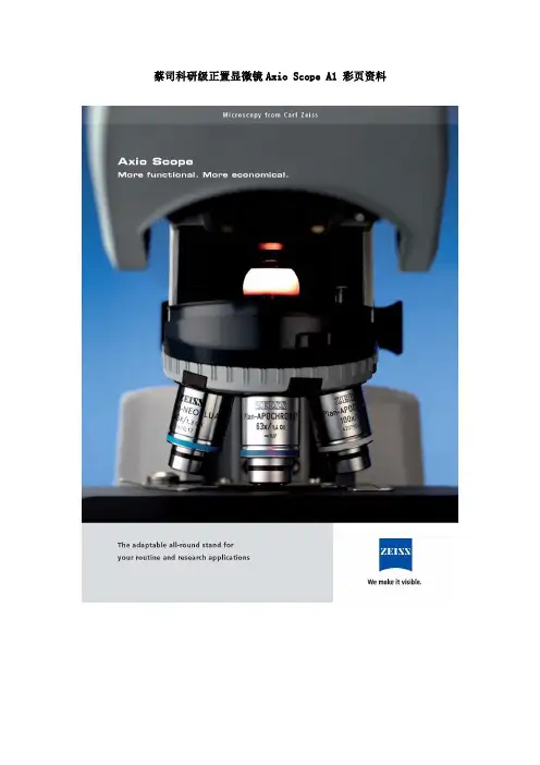
蔡司科研级正置显微镜Axio Scope A1 彩页资料一、灵活性前所未有的模组化Axio Scope——卡尔•蔡司的这款新型多功能显微镜在各方面都是您的不二选择。
您将被这款系统的性能深深震撼,与此同时,您的财务部门也将为其价格优势而叫好。
您只需买您所需,因此各个配置方案都十分经济。
购买之后您可以在任何时间按照您的需要来升级。
1. 高度弹性设计,23种配置,灵活升级,满足您不同需求2. 可以定制5种显微镜上部、3个下部和2个可变柱子,非常灵活I:用于纯粹的透射光。
6个用于明场的物镜孔(6xBF)II:用于标准的荧光应用。
含3xDIC/3xBF,用于HBO50,HBO100和HXP120,Colibri 的标准界面。
III:用于LED荧光应用。
3xDIC/3xBF,可接受4种不同LED模块的综合照明,长寿命,经济。
IV: 用于反射光和荧光。
6xBF/DF,用于HAL100或HBO的标准界面,物镜转盘上的DF插件。
V:用于反射光和荧光。
6xBF/DF,用于HAL100或HBO的标准界面,物镜转盘上的DF插件。
可调的apertA:常规应用。
带LED照明B:标准应用:50W的反射光。
带有视场光栏和孔径光栏的科勒光路、带滤块滑片和6位滤块转轮。
C:用于高强度照明的透射光应用。
100W的卤素灯。
带有视场光栏和孔径光栏的科勒光路、带滤块滑片和6位滤块转轮。
D: 380mm高的Vario柱,用于反射光和荧光应用。
E: 560mm高的Vario柱,用于反射光和荧光应用。
二、方便升级Axio Scope模块界面的概念使得将来升级变得更简单,您不需要等待昂贵的服务支持自己就可以安装组件。
三、多样化的用途Axio Scope在研究所和实验室提供多样的用途,从简单的日常应用到更复杂的研究。
从解剖到细胞学,从透射光应用到多种荧光标记,从最薄的组织切片到380mm厚的样品。
高达560mm大样品空间可选多人共览成像可选;23mm大视野观察;多种光学变倍可选;适合显微镜操作使用透射光照明成像LED透视光可选,无需色温转换滤片以及减光片,无需库勒照明调节,方便使用;明场、暗场、相差、DIC、PlasDIC、偏光等多种观察方式;专利PlasDIC设计,允许塑料器皿DIC成像。
The Next GenerationMADE BY ZEISS 23ZEISS OPMI PENTERO 900The Next GenerationEvolution in ExcellenceOPMI ® PENTERO ® 900 from ZEISS represents the next generation visualization system. Building on the ground b reaking innovations introduced in 2004, it combines unique design concepts and new functionalities in a proven, fully-integrated platform. Key functions have been enhanced and new visualization methods integrated, raising ZEISS OPMI PENTERO 900 to a new level of performance. ZEISS OPMI PENTERO 900 continues the evolutionary process, turning technological progress into medical innovations effectively advancing what is possible in modern microsurgery.Experience the new OPMI PENTERO 900.• Brilliant VisualizationExperience optical immersion with state-of-the-art apochromatic optics, razor-sharp video images in full HD quality and innovative fluorescence methods.• Superior PerformanceSmooth, intuitive system handling and superior functionality ensure efficient surgery and fast system set-up.• Beyond VisualizationZEISS OPMI PENTERO 900 interacts with current and emerging workplace technologiesand workflow based solutions to create a better OR experience.1953First surgical microscope:OPMI 11976Contraves ®suspension:OPMI 1 / NC 11993First microscopeintegrated navigation system:OPMI ES MKM autopilot1998First microscopeintegrated fluorescence:OPMI Neuro FLmicroscopes:OPMI Pentero2011Anotherdefining momentExperience Optical ImmersionZEISS OPMI PENTERO 900 delivers state-of-the-art apochromatic optics providing crystal-clear images, sharp details and natural color rendition. Whether viewing through the eyepieces or on its flexible and integrated HD touchscreen display, ZEISS OPMI PENTERO 900 elevates visualization of the surgical field to the next level. The entire HD video chain – camera, recorder, editor and monitor – is fully integrated into the system without the need for external components, exposed cabling or the use of multiple control interfaces. The HD video system can be configured and controlled via the central HD touchscreen, handgrip or foot switchfor maximum flexibility and surgical performance. ZEISS OPMI PENTERO 900 offers a unique HD experience with visual brilliance for live demonstrations, teaching presentations and patient documentation.4Uniquely Designed Apochromatic OpticsThe distinctive design concept with ZEISS apochromatic optics throughout the entire optical pathway allows the system to deliver unmatched optical clarity, detail resolution and color reproduction, both through the eyepieces and the video image.Highquality Data Injection The advanced MultiVision ™ display significantly enhances image quality and enables efficient data injection during procedures. Higher resolution, enhanced contrast and better color rendition ensure outstanding quality leading to improved outcomes.Brilliant High Definition Video DisplayThe large touchscreen display delivers impressively, crisp images in HD quality. The extendable suspension arm allows the display to be rotated, tilted or moved into different viewing positions.Fully Integrated HD VideoThe fully integrated HD video camera, recorder and editor enable surgeons to capture razor-sharp images for teaching, documentation and presentation purposes. All video functions can be centrally controlled from the intuitive graphical user interface.MADE BY ZEISSAdvancing fluorescence – Continuation of Scientific ProgressZEISS has partnered with visionary thought leaders in the development of innovative intraoperative fluorescence technologies. The first fluorescence module, INFRARED 800™, successfully established intraoperative fluorescence as a key visualization technology. FLOW® 800 is a unique tool enabling the visual analysisof blood flow dynamics, further establishing ZEISS as a leader in intraoperative fluorescence. With BLUE400 and now, with the introduction of YELLOW 560™, we further broaden the capability for fluorescencebased research applications. With its intuitive workflow, automated functions and unmatched performance, theZEISS OPMI PENTERO 900 platform supports fluorescence-based surgeries like no other system.5 INFRARED 800¹Intraoperative visual assessmentof blood flow and vessel patencyduring AVM, bypass and aneurysmsurgery. INFRARED 800 is indicatedfor use in neurosurgery, plasticand reconstructive procedures andcoronary artery bypass graft surgery.YELLOW 5603Visualizes in the 540 to 690 nmwavelength range for supportingresearch applications. It is the firstZEISS fluorescence module capableof highlighting fluorescent structureswhile viewing surrounding structuresin their natural color.BLUE 4002Capable of supporting fluorescence-based surgery for researchapplications by providingvisualization in the 620-710 nmrange. BLUE 400 is a completelyintegrated module with no externalcomponents or cables.Optionally available in HD quality.FLOW 800¹Unique fluorescence applicationenabling visual analysis of vascularblood flow dynamics. It compilesINFRARED 800 video sequences intovisual maps, diagrams or side-by-side images, enabling an in-depthinterpretation of fluorescence videos.¹)For the complete 510(k) summary for INFRARED 800 with FLOW 800 option, visit /cdrh_docs/pdf10/k100468.pdf.²)Image obtained with investigational new drug.3) Image obtained with drug under investigation for new intended use.6Maximum EfficiencyZEISS OPMI PENTERO 900 is a unique surgical visualization platform specifically conceived and designed for even the most demanding microsurgical applications. Extended system ergonomics and functions provide increased con v enience, streamlining the surgical workflow. All relevant functions are combined into a cohesive system that can be controlled from the intuitive touchscreen user interface. Smooth system handling and superior performance are delivered through proven functions like mouth switch control, a unique depth of field, AutoFocus ™ and many more. The Foldable Tube f170/f260 and wireless foot control panel enhance ergonomics, improving surgeon comfort and performance.// S UPERIOR PERFORMANCEMADE BY ZEISSUnparalleled Depth of Field Depending on preference or application, the integrated, electronically controlled double-iris diaphragm enables surgeons to choose between maximum light and resolution or depth of field.Dynamic Foldable Tube The highly flexible Foldable Tube f170/f260 offers increased positioning capability, magnification and user comfort. With a quick dial turn, the integrated PROMAG ™functionality provides an additional50 percent magnification gain.Wireless Foot ControlThe wireless foot control, designedto manage multiple microscope functions, can be freely positioned for rapid set-up and user convenienceduring surgery. The intelligent power management function ensures superior long-lasting operability.Efficient Light SettingsThe patented, two-channe lillumination design reduces shadowing in deep cavities. The Automatic Iris Control ™limits illumination to the field of interest and the new Focus Light Link ™automatically limits brightness, both in the aim of preventing inadvertent light exposure.7AutoBalanceAutomatic balancing of the microscope with the press of a single button for fast system setup. AutoBalance can even be performed while the system is draped ensuring optimum system mobility. A unique fine balance function also allows precise system control via the mouth switch.AutoDrapeAutoDrape allows for easy and fast system draping by automatically evacuating air from the sterile drape. The drape clings tightly to the system in a matter of seconds without obstructing the microscope’s mobility.OR Turn-Around SimplifiedDesigned as much for the OR staff as for the surgeon, ZEISS OPMI PENTERO 900 incorporates workflow-conducive features that reduce prep time for the nursing staff before each surgical case. AutoBalance ™ quickly balances the microscope at the touch of a button, AutoDrape ® facilitates a quick and easy draping process, and the unique FlexiTrak ™ enables the OR staff to easily maneuver the system in the clinical environment. Additionally, the intuitive user interface allows for easy access to all microscope functions through a large, HD touchscreen monitor including patient data, pre-configured surgeon settings and video recording. More than any other surgical microscope system, ZEISS OPMI PENTERO 900 streamlines the surgical workflow and maximizes OR efficiency.VisionGuard Drape Technology ZEISS drapes are manufactured with VisionGuard ®, a unique, optical lensthat works with the microscope’s objective as a single optical unit for unparalleled optical clarity. The lens can be replaced with a new sterile lens during surgery without compromising sterility.Rapid Remote DiagnosisFast internet access via VPN enables remote diagnostic capability, resulting in better service support and improved system uptime. System log files can be accessed online by ZEISS service specialists for rapid analysis and support.8Workflow-based Solutions ZEISS OPMI PENTERO 900 provides tailored, workflow-based solutions that are accessible through a common interface. The modules were designed with surgeons for surgeons to meet the requirements of clinical applications and are seamlessly integrated into the surgical workflow.Applicationdriven Technologies Focused on enhancing clinical outcomes, ZEISS OPMI PENTERO 900 provides surgeons with application-driven solutions like MultiVision and fluorescence-based visualization. Each integrated module introduces a new level of simplicity, speed and accuracy to the surgical procedure.Universal ConnectivityZEISS OPMI PENTERO 900 offers comprehensive connectivity with workplace technologies and data management functionality. Integrated modules like MultiVision and DICOM ensure a seamless connection with other visualization systems and into the hospital’s communication infrastructure, respectively.Intelligent Design From complete cable and component integration to overall intuitive system design, ZEISS OPMI PENTERO 900 creates the optimal surgical environment. One cohesive touchscreen interface serves all system configurations and functions.Workplace InnovationsClose cooperation with leading surgeons across the globe led to the development of a ground-breaking visualization platform. ZEISS OPMI PENTERO 900 offers advanced functionality in an elegantly designed workplace. The thoughtfully designed workflow based solutions, specifically tailored to meet the demands of clinical applications, differentiate this system from any other. The complete integration of workplace components as well as the ability to adapt emerging technologies present the surgeon with a wide variety of product solutions to meet their individual requirements. Now more than ever, ZEISS OPMI PENTERO 900 provides an experience that goes far beyond visualization.// B EYOND VISUALIZATIONMADE BY ZEISSTechnical DataRated Voltage(115V): 100 V – 125 V(230V): 220 V – 240 Vmax. 1.200 VACurrentConsumptionRated Frequency50 Hz – 60 HzElectrical Standard Complying with IEC 60601-1:2005;UL 60601-1; CAN/CSA-C22.2No. 601.1-M90Protection class I, degree of protection IP20Type B equipmentClass 2 laser product as perEN 60 825-1:2002Weight Weight approx. 358 kgWeight of system incl. transport container:approx. 610 kg910OptionsVideoIntegrated 3-CMOS HD video camera Integrated SD or HD video recording and editingAdaptation of consumer (SLR) photo/video camera Intraoperative FluorescenceBLUE 400INFRARED 800INFRARED 800 with FLOW 800YELLOW 560Connectivity / Data Management DICOM module for patient data transfer from/to PACSAccessories12.5x magnetic widefieldeyepieces with integrated eyecups Straight tube, focal length f = 170mm Stereo co-observation tube Foldable Tube f170/f260,including the PROMAG function for additional 50% magnification and integrated rotate function Wired foot control panel Wireless foot control panelStandard ConfigurationApochromatic OpticsMotorized focus; Varioskop ® with working distance 200 – 500 mmMotorized zoom; 1:6 zoom ratio10x magnetic widefield eyepieces with integrated eyecupsAutofocus with 2 visible laser dots, automatic mode with magnetic brakes IlluminationSuperlux ® 330 light source with 2 x 300 W xenonAutomatic Iris Control for adjusting the illumination to the field of view Individual light threshold setting Focus Light Link: working distance controlled light intensityDisplay of remaining lamp life timeSystem OperationMultifunctional programmable handgrips Magnetic clutches for all system axes Central user interfaceXY robotic movement in 3 axes (variable speed)System SetupAutoBalanceAutoDrape – air evacuation system Mouthswitch fine balance Video22" HD video touchscreen on extendable armIntegrated 3-CMOS SD video camera Integrated video still image capturing on HDD and USB-mediaConnectivity / Data ManagementVideo-in for external SD video sources Navigation interfaceInterface for micromanipulator Remote diagnosis via internet/VPN11Image Courtesy of:BrainLAB AG, Feldkirchen, Germany (p. 4, 8)Barrow Neurological Institute, Phoenix, Arizona, USA (p. 1, 4, 5, 8)Michael Buchfelder, MD, PhD, Neurosurgery Department, Erlangen University Hospital, Erlangen, Germany (p. 5)Walter Stummer, MD, PhD, Department of Neurosurgery, University Hospital Münster, Münster, Germany (p. 5, 8)Yasuo Murai, MD, PhD, Department of Neurosurgery, Nippon Medical School, Tokyo, Japan (p. 8)Yasushi Takagi, MD, PhD, Department of Neurosurgery, Kyoto University Graduate School of Medicine, Kyoto, Japan (p. 5)Carl Zeiss Meditec AG Goeschwitzer Strasse 51–5207745 Jena Germany//contactsYour local contact:ArgentinaCarl Zeiss Argentina S.A.Calle Nahuel Huapi 4015 / 25C1430 BCO Buenos Aires ArgentinaPhone: +54 11 45 45 66 61****************.arAustraliaCarl Zeiss Pty. Ltd.Unit 13, 2 Eden Park DriveNorth Ryde, New South Wales 2113AustraliaPhone: +61 2 9020 1333*************.au AustriaCarl Zeiss GmbH Laxenburger Str. 21100 Vienna AustriaPhone: +43 1 79 51 80*****************BelgiumCarl Zeiss NV-SA Ikaroslaan 491930 Zaventem BelgiumPhone: +32 2 719 39 11*************BrazilCarl Zeiss do Brasil Ltda.Av. Naçoes Unidas, 21711CEP04795-100 São Paulo BrazilPhone: +55 11 5693 5521*******************CanadaCarl Zeiss Canada Ltd.45 Valleybrook Drive Toronto, ON M3B 2S6CanadaPhone: +1 800 387 8037***************ChinaCarl Zeiss Shanghai Co. Ltd.1/f., Ke Yuan Building 11 Ri Yin Nan RoadWaigaoqiao Free Trade Zone 2005 Yang Gao Bei Road Shanghai 200131ChinaPhone: +86 21 5048 17 17*************.cnCzech Republic Carl Zeiss spol. s.r.o.Radlická 14/3201150 00 Prague 5Czech RepublicPhone: +420 233 101 221**************FranceCarl Zeiss Meditec France SAS 60, route de Sartrouville 78230 Le Pecq FrancePhone: +33 1 34 80 21 00************GermanyCarl Zeiss Meditec VG mbH Carl-Zeiss-Strasse 2273447 Oberkochen GermanyPhone: +49 7364 20 6000**********************.com Surgical Ophthalmology:Phone: +49 800 470 50 30***********************.com Hong KongCarl Zeiss Far East Co. Ltd.Units 11-12. 25/FTower 2, Ever Gain Plaza No. 88 Container Port Road Kwai Chung Hong KongPhone: +852 2332 0402**************.hk IndiaCarl Zeiss India Pvt. Ltd.22. Kensington Road UlsoorBangalore 560 008IndiaPhone: +91 80 2557 88 88*************.in ItalyCarl Zeiss S.p.A.Viale delle Industrie 2020020 Arese (Milan)ItalyPhone: +39 02 93773 1*************JapanCarl Zeiss Meditec Japan Co. Ltd.Shinjuku Ku Tokyo 160-000322 Honchio-Cho JapanOphthalmic instruments:Phone: +81 3 33 55 0331*****************.jp Surgical instruments:Phone: +81 3 33 55 0341****************.jpMalaysiaCarl Zeiss Sdn Bhd.Lot2, Jalan 243/51 A 46100 Petaling Jaya Selangor Darul Ehsan MalaysiaPhone: +60 3 7877 50 58******************.sgMexicoCarl Zeiss de México S.A. de C.V.Avenida Miguel Angel de Quevedo 49604010 Mexico City MexicoPhone: +52 55 59 99 0200*******************Netherlands Carl Zeiss B.V.Trapezium 300Postbus 3103364 DL Sliedrecht NetherlandsPhone: +31 184 43 34 00*************New ZealandCarl Zeiss (N.Z.) Ltd.15B Paramount Drive P.O. Box 121 - 1001Henderson, Auckland 0650New ZealandPhone: +64 9 838 5626*************.au PolandCarl Zeiss sp. Z o.o.ul. Lopuszanska 3202-220 Warsaw PolandPhone: +48 22 858 2343*****************SingaporeCarl Zeiss Ptd. Ltd.50 Kaki Bukit Place Singapore 415926SingaporePhone: +65 6741 9600**************.sg South AfricaCarl Zeiss (Pty.) Ltd.363 Oak Avenue FerndaleRandburg 2194South AfricaPhone: +27 11 886 9510*************.zaSouth Korea Carl Zeiss Co. Ltd.Seoul 121-828Mapo-gu141-1, Sangsu-dong 2F, BR Elitel Bldg.South KoreaPhone: +82 2 3140 2600**************.krSpainCarl Zeiss Meditec Iberia S.A.Ronda de Poniente, 15Tres Cantos 28760 Madrid SpainPhone: +34 91 203 37 00*************Sweden Carl Zeiss ABTegeluddsvaegen 7610254 Stockholm SwedenPhone: +46 84 59 25 00*************Switzerland Carl Zeiss AGFeldbachstrasse 818714 Feldbach SwitzerlandPhone: +41 55 254 7534************ThailandCarl Zeiss ThailandFloor 8, Thosapol Land Building 2230 Ratchadapisek Road Huaykwang, Bangkok 10310ThailandPhone: +66 2 2 74 06 43******************.sg United Kingdom Carl Zeiss Ltd.15-20 Woodfield Road Welwyn Garden City Hertfordshire, AL7 1JQ United KingdomPhone: +44 1707 871200*************.ukUnited States of America Carl Zeiss Meditec USA, Inc.5300 Central Parkway Dublin, CA 94568USAPhone: +1 925 557 /us/medC A P -e n -U S _30_020_0011I C Z -V I I /2021T h e c o n t e n t s o f t h e b r o c h u r e m a y d i f f e r f r o m t h e c u r r e n t s t a t u s o f a p p r o v a l o f t h e p r o d u c t i n y o u r c o u n t r y . P l e a s e c o n t a c t o u r r e g i o n a l r e p r e s e n t a t i v e f o r m o r e i n f o r m a t i o n . S u b j e c t t o c h a n g e i n d e s i g n a n d s c o p e o f d e l i v e r y a n d a s a r e s u l t o f o n g o i n g t e c h n i c a l d e v e l o p m e n t . O P M I , P E N T E R O , F L O W , V i s i o n G u a r d , V a r i o s k o p a n d A u t oD r a p e , M u l t i V i s i o n , I N F A RE D 800, B L U E 400, Y E L L O W 560, P R O M A G , A u t o B a l a n c e ,F l e x i T r a k , A u t o m a t i c I r i s C o n t r o l a n d F o c u s L i g h t L i n k a r e e i t h e r t r a d e m a r k s o r r e g i s t e r e d t r a d e m a r k s o f C a r l Z e i s s M e d i t e c AG o r o t h e r c o m p a n i e s o f t h e Z E I S S G r o u p i n G e r m a n y a n d /o r o t h e r c o u n t r i e s . C o n t r a v e s ® i s a r e g i s t e r e d t r a d e m a r k o f t h e C o n t r a v e s A G .© C a r l Z e i s s M e d i t e c U S A , I n c ., 2021. A l l r i g h t s r e s e r v e d .0297Carl Zeiss Meditec USA, Inc. 5300 Central Parkway Dublin, CA 94568USA/us/med。
F O C U S E D Q U A L I TYMantis - ergonomic stereo microscopesMantis are unique ergonomic high performance stereo microscopes, offering 3D optical imaging with magnification options up to 20x.Large fields of view and long working distances allow for a wide range of inspection, manipulation and re-working tasks, all with exceptional hand-eye coordination.Patented optical technology allows operators freedom of head movement for superb ergonomics and hand-eye co-ordination, with the ability to wear glasses if required. All Mantis systems aid with productivity and quality improvements.Key Features92x to 20x magnification options9Superior ergonomics for improved productivity9Quick change turret for flexible magnification9Optimised for long working distance & large field of view9Bright true colour, LED illumination9Optional factory-integrated HD digital camera9Inspect and document with ease (Mantis Elite-Cam HD only)9Choice of stands and accessories to suitMantis CompactMantis Compact a high value stereo microscope which excels in the low magnificationrange for inspection or manipulation tasks where bench magnifiers have traditionallybeen used.Patented optical technology allows operators freedom of head movement for superbergonomics and hand-eye co-ordination, with the ability to wear glasses if required.All Mantis systems aid with productivity and quality improvements.92x, 4x, 6x and 8x quick change objectives9Patented eyepiece-less optics maximise head freedom providing unrivalledergonomic performance9Small footprintFind out more:/mantis-compact Mantis EliteMantis Elite has enhanced optical performance, comparing to Mantis Compact,including higher magnification, a large field of view and long working distances.Mantis Elite is a perfect alternative to traditional stereo microscopes for a wide rangeof inspection, preparation and manipulation tasks requiring hand-eye co-ordination.92x - 20x magnification options with quick change turret allows users to switchbetween low and high magnification9Superb hand-eye co-ordination for inspection and re-working tasks9Bright true colour LED illumination provides up to 10,000 hours of shadow-freeviewingFind out more:/mantis-elite Mantis Elite-Cam HDMantis Elite-Cam HD is a variant of the successful Mantis Elite stereo microscope,with an internally integrated USB digital camera, bringing image capture capabilitiesto the outstanding optical performance of Mantis.By adding an HD camera to Mantis Elite, Vision Engineering has created asupremely capable inspection solution, providing flexibility and simplicity for anyprecision magnification task.9Quickly and simply add annotations / mark-up to captured images using theViCapture software supplied9Video recording (.avi), ideal for training purposesFind out more:/mantis-elite-camImproving microscopy ergonomics...Around 62%of microscope operators declared suffering fromMUSCULOSKETLETAL PROBLEMS 262%85%Around 55%of microscope users have experiencedFREQUENT HEADACHES 155%94%Common locations were neck and back. Other problematic areas included shoulder, wrist and hand.78%Unrivalled Mantis advantage...Businesses choose Vision Engineering’s ergonomic stereo microscopes because operators are more comfortable during inspection, so more efficient, more accurate and more productive.Give your stereo microscope a health check!Ergonomic working positionAn ergonomic body position makes the Mantis more comfortable, less fatiguing and, more importantly, much easier to use. Additionally, optimal operator ergonomics minimises the risk of repetitive strain-related injuries. A happy worker is a productive worker.Freedom of head movementAn additional benefit of Vision Engineering’s patented eyepiece-less design is that users do not need to align their eyes with eyepieces. This freedom of movement reduces associated neck and back strain associated with the fixed body position of conventional microscope eyepieces.A natural view of the subjectWith conventional microscope eyepieces, operators must position their eyes very close to the eyepieces, blocking out ambient light. The intense light exiting the eyepieces causes the pupils to contract. Constant contraction and expansion of the pupils is the main cause of eye fatigue with microscopes.With the patented eyepieces of Mantis, users sit back from the viewer, allowing ambient light into the eyes. Additionally, the light exiting the ‘viewing lens’ is spread overa larger area, proving a more natural view of the subject.Easy hand-eye co-ordinationEasy hand-eye co-ordination is possible with the Mantis – critical for re-work, repair, dissection and other manipulation tasks. Sitting back from the viewer provides userswith much better peripheral vision, so they can co-ordinate hands in a natural manner.Ability to wear glassesWith Mantis, operators do not need to remove their glasses (or safety glasses) to use the microscope./ergonomicsDisclaimer - Vision Engineering Ltd. has a policy of continuous development and reserves the right to change or update, without notice, the design, materials or specification of any products, the information contained within this brochure/datasheet and to discontinue production or distribution of any of the products described.EO&E: Errors and omissions accepted.Mantis_LIT5429EN_10.1| Copyright ©2022 Vision Engineering Ltd. | All rights reserved.To see our focused quality , please contact your Vision Engineering branch, local authorised distributor, or visit our website: Vision Engineering Ltd.(UK Manufacturing & Commercial)The Freeman Building, Galileo Drive, Send, Surrey, GU23 7ER, UK T +44 (0) 1483 248300E ************************.uk Vision Engineering Ltd. (Italia)Via G. Paisiello 10620092 Cinisello Balsamo MI, Italia T +39 02 6129 3518E *****************Vision Engineering (South East Asia)P-03A-20, Impian Meridian,Jalan Subang 1, USJ 1, 47600 Subang Jaya,Selangor Darul Ehsan, Malaysia T +604-619 2622E *******************Vision Engineering (Mexico)T +01 800 099 5325E ********************Vision Engineering Inc.(NA Manufacturing & Commercial)570 Danbury Road,New Milford, CT 06776, USA T +1 (860) 355 3776E ******************Vision Engineering Ltd. (France)ZAC de la Tremblaie, Av. de la Tremblaie91220 Le Plessis Paté, France T +33 (0) 160 76 60 00E *****************Vision Engineering (China)Room 904B, Building B, No.970, Nanning Road, Xuhui Vanke Center Shanghai, 200235, P .R. ChinaT +86 (0) 21 5036 7556 E ******************.cn Vision Engineering (Brazil)E ******************.br Vision Engineering (Latin America)E ********************Vision Engineering Ltd. (Central Europe)Anton-Pendele-Str . 3,82275 Emmering, Deutschland T +49 (0) 8141 40167-0 E *****************Nippon Vision Engineering (Japan)272-2 Saedo-cho, Tsuduki-ku, Yokohama-shi, Kanagawa 224-0054, Japan T +81 (45) 935 1117 E *****************Vision Engineering (India)T + 91 (0) 80-5555-33-60E *****************.inFM 557119Vision Engineering Ltd. has been certified for the quality management systemISO 9001:2015.Vision Engineering Ltd. has been designing and manufacturing high quality ergonomic microscopes, digital instruments, inspection and non-contact measuring systems for over 60 years.InnovationWith a philosophy of design innovation, Vision Engineering holds world patents for a number of optical / digital techniques, significantly improvingviewing ergonomics and enabling customer quality and productivity improvements.QualityVision Engineering prides itself on quality products, electronics, mechanics and optics and is certified for the quality management system ISO 9001:2015. Quality is as important to us as it is to our customers. Our systems have proved themselves many times over and are chosen by the world’s leading companies.GlobalVision Engineering has manufacturing and design facilities in the UK and USA, plus sales and support offices throughout Europe, the Americas, the Far East, and Asia. We support our customers with close technical and service support anywhere in the world.VISION ENGINEERINGOUR DIFFERENCE。
EVOS ™ M7000 Imaging SystemFor fluorescence and transmitted light applicationsCatalog Number AMF7000Pub. No. MAN0018380 Doc. Part No. 710246 Rev. A.0Note: For safety and biohazard guidelines, see the “Safety” appendix in the EVOS ™ M7000 User Guide (Pub. No. MAN0018326). Read the Safety Data Sheets (SDSs) and follow the handling instructions. Wear appropriate protective eyewear, clothing, and gloves.This document is intended as a benchtop reference for the users of the EVOS ™ M7000 Imaging System (Cat. No. AMF7000). See the EVOS ™ M7000 User Guide for detailed instructions and troubleshooting. The user guide is provided on the USB flash drive and is also available for download at .Capture imagesa. Turn the instrument power switch located in back of the instrument base to the ON position.b. Turn the computer and monitor ON.c. When the computer shows the Windows ™ desktop and the X-Y stage of the instrument has stopped moving, click the M7000 icon to start the EVOS ™ M7000 software.d. When the Capture tab is displayed, the EVOS ™M7000 Imaging System is ready to use.1Turn onthe EVOS ™M7000Imaging System a. Place the vessel containing your sample on the stage using the appropriate vessel holder. b. On the Capture tab, click the Vessel button to open the Vessel Selection dialog.c. Select the Vessel category that corresponds to your sample vessel,then select the Holder and Vessel type from the dropdown menus.d. Click Done to complete your selection and close the dialog.The Vessel map on the Capture tab displays your selected vessel.2Selectsample vessel a. Click the desired Objective button to select the corresponding magnification. b. Select the desired Light source for which you wish to adjust brightness and set focus.3Select objective andlight sourceb. For Mode, select Simple or Actual.• Simple mode allows you to control Brightness as a single parameter.• Actual mode allows you to adjust Light (i.e., LED intensity), Exposure, and Gain individually.c. For Camera, select Mono (monochrome) or Color.• Mono is used for image capture in fluorescence and transmitted light (brightfield) channels• Color is used for image capture in the brightfield channel onlyd. Optional: Choose Phase options.Note: Phase options are available only for the transmitted light (brightfield) channel; they are not availablefor fluorescence channels.e. Click the Brightness and camera settings button again to collapse the controls.and cameraoptionsa. Click the Light button to turn on the excitation light for the selected light source and enterthe instrument in the Live mode.b. Adjust brightness using the Brightness controls.Note: For best results, optimize the brightness parameters as follows:• When searching for sample: Increase Gain for a brighter signal and decrease Exposure for faster framerate during navigation around the vessel.• When capturing image: Decrease Gain to reduce background noise and increase Exposure to regainsignal intensity, as needed.• For brighter signal: Increase Light intensity for brighter illumination. If needed, follow by increasingGain.• For time lapse imaging: Increase Gain and Exposure, and decrease Light intensity to reducephotobleaching and phototoxicity.a. Choose the channels you wish display in the Viewing Area by selecting the appropriate channel checkboxon the corresponding Light source button.You can select multiple channels to display in the Viewing Area. In the following example, DAPI, RFP, andtransmitted light (brightfield) channels have been selected.b. Click the Image Display settings button to expand the image display controls for the selected channels.c.Adjust the Brightness , Contrast , and Gamma settings for each of the selected channels usingthe corresponding sliders.d.Optional: To remove a channel from displaying in the Viewing Area, unselect the corresponding Displaycheckbox.To display an available channel not shown in the Viewing Area, re-select the checkbox.e.Click the Image color display button to display the sample pseudo-colored in the default emissioncolor of the selected channels. By default, color display option is on.f.Click the Image Display settings button again to collapse the controls.Note: Adjustments made to Image display settings only affect how the image is displayed in the ViewingArea; they do not change how the image is captured.5AdjustbrightnessBrightness controls in Simple mode Brightness controls in Actual mode6Set imagedisplayoptionsa. Click Autofocus to run the autofocus procedure to find the best focal plane for thecurrent channel and sample.Alternatively, use the Coarse and Fine focus sliders to manually find the best focal position.b. To choose a different autofocus method, click Advanced focus settings button, then select from theAutoFocus Method dropdown.Available options for AutoFocus Method are:• Small Structure: Choose this option for samples with many fine, hair-like structures (e.g., filaments or structural stains).• Large Structure: Choose this option when your sample contains large structures (e.g., whole cell stains).• Small Bright Objects: Choose this option when capturing samples with localized staining (e.g., nuclei).Z-Offsets allow you to specify the optimal focus position in each channel relative to the focus position in other channels.a. Verify that the Lock Z-Offsets option is checked in the Advanced focus settings window, then click theAdvanced focus settings button again to close the window.b. Select the Objective and the Light source (i.e., channel) you wish to capture.c. Click the Light button to enter the Live mode, and focus on the sample manually using the Coarse and Finefocus sliders or automatically by clicking the AutoFocus button.d. After you have found the optimal focus position in the first channel, click the A dvanced focus settingsbutton, then uncheck the Lock Z-Offsets option.e. Without changing the objective, select the next Light source (i.e., channel) you wish to capture.f. If the light is off, click the Light button to turn it on, then focus on the sample manually using the Coarseand Fine focus sliders or automatically by clicking AutoFocus.g. Repeat this procedure for every channel you wish to capture.h. When you have found the optimal focus position in all the additional channels you wish to capture, clickthe Advanced focus settings button, then check the Lock Z-Offsets option.When you have completed this procedure, the focus position in each selected channel will be offset relative to each other.7 8Focus on the sample Optional: SetZ-Offsetsa. While in Field View , click the Light button and enter the instrument in the Live mode.b. To go to a specific location on the sample vessel, click and dragthe crosshair to the corresponding location on the virtual vessel.If needed, click on the Zoom button to open a larger view of the Vessel map for easier navigation to the desired location.Click the Zoom button again to close the zoom window.c. While in the Live mode, use the Jog Control to move the stage at an intermediate pace to thedesired location as you view the sample until you find the field of view you wish to capture.Alternatively, click on the navigation arrows (up, down, left, right) on the Viewing area to move in the corresponding direction exactly one field of view. You can also click and drag the field of view itself to move around the sample vessel.d. Click the Lightbutton again to exit the Live mode in Field View.9Find theregion ofinterest inthe Livemode 10Select fieldand capture images a. In Area View , position the capture crosshair over the region of interest and click to select the field you wish to capture. Alternatively, find the field you wish to capture in the Live mode.b. Click Capture to acquire an image of the selected field using the current capture settings.A thumbnail of the captured image is displayed above the Light source button for the specific channel in which the image was captured (in this example, DAPI).The Viewing are displays the captured image at the location of its capture when the zoom level is greaterthan one field of view.c. To capture the same field in another channel, select the desired channel using the corresponding Lightsource button.d. Readjust the brightness and focus (if needed), then click Capture.A thumbnail of the captured image is displayed above the Light source button for the new channel in whichthe image was captured.The Viewing area displays a multicolor overlay of the images at the location of their capture.e. To capture another field, position the capture crosshair over the new region of interest, then click to selectthe new field.f. Readjust the brightness and focus (if needed), then click Capture.g. To capture a field using a different magnification, select the desired Objective, then repeat the captureprocedure with the new magnification as described.h. To capture a selected field in multiple channels simultaneously, select the channels you wish to capture bychecking the small circular checkboxes on the the corresponding light source buttons.In the following example, DAPI, RFP, and transmitted light (brightfield) channels have been selected.i.Readjust the brightness and focus (if needed), then click Capture Channels.The instrument captures an image of the selected field in each of the selected channels using the current capture settings. After capture, the Viewing area displays a multicolor overlay of the captured images at the location of their capture.Save captured imagesa. When finished capturing images, click Save… to open the Save widialogndow.b. Select the captured fields you wish save. Available options are:• Currently selected field: Saves images only from the currently selected field.• All newly captured fields: Saves images that have been captured and stored in the image cache, but notyet saved. This option is available only if you have previously saved images from the same session.• All captured fields: Saves images from all captured fields that are held in the image cache.This is typically all of the images that you captured during an imaging session.a. Click Browse , navigate to the desired folder, then click Select .To create a save new folder, navigate to the desired location, click New , type in the name of the newly creat-ed folder, then click Select .Note: We recommend that you save your captured images to an external hard drive.b. If desired, type the prefix you wish to use for your save images in the File name prefix textbox.a. Select File types to save by checking the corresponding checkboxes. You can choose multiple file types foryour captured images. Available options for file type are Raw images and Displayed images .• Raw images: Saves images captured in different channels individually as 16-bit raw images.This is the recommended format for image analysis.Available raw image options are:–Single field, individual channels: Saves images capturedin each field and each channel individually.–Z stack planes, individual channels: Saves the z-stackprojection and each z-stack plane for each field and each channel as a separate image.Available File format for raw images are: TIF , PNG , C01, and DIB .• Displayed images: Saves images in a format that can be viewed in most image display applications.Available displayed image options are:–Single field, individual channels: Saves imagescaptured in each field and each channel individually.–Merged image: Saves the images of a field captured indifferent channels as a multi-channel overlay.–Tiled image, merged channels: Merges the imagescaptured in each channel, then aligns them close together into a tiled format.Available sizes for tiled images are Small (2000 × 2000 pixels), Medium (4000 × 4000 pixels), Large (10,000 × 10,000 pixels),or Maximum (26,000 × 26,000 pixels)Available Color options for displayed images are:–Grayscale: 16-bit–Pseudocolor:24-bit RGB (8-bit per RGB channel).123Select captured fieldto saveSelect savelocation Select file types to saveAvailable Brightness options for displayed images are:–High: Limits pixel depth and reduces exposure time and dynamic range.–Full: Provides more detail for quantitative imaging.Available File format for displayed images are: TIF, PNG, and JPEG.Note: While pseudocolors help differentiate the channels used in multi-channel overlays, grayscale images usually show more detail.24-bit images (8-bit per RGB channel) are NOT recommended for image analysis as not all channels will display in many image analysis applications.b. If desired, check the Include Grid option and select the grid size.When checked, this option superimposes a grid on the displayed images.You have the following options for the grid size (in pixels): Auto, 10 × 10, 50 × 50, 100 × 100, 200 × 200, and 500 × 500.c. To see more file type options in a table format, click More options…Full menu of save options are displayed in a convenient table format, allowing you to save your captured images in a number of formats simultaneously.d. After you selected save options for your captured images, click Save.The instrument will save the images in the designated folder based on your specifications.Documentation and supportTechnical supportFor the latest services and support information for all locations, go to /support.At the website, you can:• Access worldwide telephone and fax numbers to contact Technical Support and Sales facilities• Search through frequently asked questions (FAQs)• Submit a question directly to Technical Support (/support)• Search for user documents, SDSs, vector maps and sequences, application notes, formulations, handbooks, certificates of analysis, citations, and other product support documents• Obtain information about customer training• Download software updates and patchesSDSSafety Data Sheets (SDSs) are available at /support.Limited Product WarrantyLife Technologies Corporation and/or its affiliate(s) warrant their products as set forth in the Life Technologies’ General Terms and Conditions of Sale found on Life Technologies’ website at /us/en/home/global/terms-and-conditions.html. If you have any questions, please contact Life Technologies at /support.For Research Use Only. Not for use in diagnostic procedures.The information in this guide is subject to change without notice.DisclaimerTO THE EXTENT ALLOWED BY LAW, LIFE TECHNOLOGIES AND/OR ITS AFFILIATE(S) WILL NOT BE LIABLE FOR SPECIAL, INCIDENTAL, INDIRECT, PUNITIVE, MULTIPLE OR CONSEQUENTIAL DAMAGES IN CONNECTION WITH OR ARISING FROM THIS DOCUMENT, INCLUDING YOUR USE OF IT.Revision history: Pub. No. MAN0018380Important Licensing InformationThese products may be covered by one or more Limited Use Label Licenses. By use of these products, you accept the terms and conditions of all applicable Limited Use Label Licenses.Manufacturer: Life Technologies Corporation | 22025 20th Ave SE St #100 | Bothell, WA 98021All trademarks are the property of Thermo Fisher Scientific and its subsidiaries, unless otherwise specified.©2018 Thermo Fisher Scientific Inc. All rights reserved.。
SPECIFIERS’ GUIDE Optem® FUSION Micro-Imaging Lens System Visible to SWIR (400-1700nm)Covering the optical, mechanical,and operational parameters ofFUSION system components.Now Supports Key Visible, NIR and SWIR wavebands8th EditionTable of ContentsHOW TO USE THIS GUIDEConfiguring your Optem FUSION Lens System now with compatibility (3)LENS SYSTEM COMPONENT OPTIONSOptem FUSION Component Matrix ..........................................................................................4-5OPTICAL PERFORMANCE CHARTSFixed Magnification .................................................................................................................6-7Fixed w/Mini Tube Magnification .............................................................................................8-9Fixed Magnification w/ LWD Objectives................................................................................10-11Fixed Magnification w/Mini Tube w/ LWD Objectives ............................................................12-137:1 Zoom with Lower Lens .................................................................................................14-157:1 Zoom w/Mini Tube with Lower Lens .............................................................................16-177:1 Zoom w/ LWD Objectives ...............................................................................................18-197:1 Zoom w/Mini Tube w/ LWD Objectives ...........................................................................20-2112.5:1 Zoom and Fetura+ with Lower Lens ..........................................................................22-2312.5:1 Zoom and Fetura+ w/Mini Tube with Lower Lens ......................................................24-2512.5:1 Zoom and Fetura+ w/ LWD Objectives ......................................................................26-2712.5:1 Zoom and Fetura+ w/Mini Tube w/ LWD Objectives ..................................................28-29Wide Angle Lens with Lower Lens .......................................................................................30-31FIxed Zoom and 7:1 Zoom with compatibility..............................................................32-33FUSION Flexibility and Notes ................................................................................................34-352How to use this guideO ptem FUSIO N is a modular Lens System, which can be modified to affect form, function and performance across the Visible (400-700nm), the Near Infrared (NIR – 700-1100nm), and the Short-Wave Infrared (SWIR – 900-1700nm) wavebands. Follow the steps outlined herein to configure an Optem FUSION Lens System to meet your specific optical, functional and physical configuration requirements. Refer to the Optical Performance Chart that matches your chosen Fixed-magnification or Zoom configuration (e.g., with or without Objectives, with Standard or Mini Camera Tubes). Following the flow lines in the FUSION Component Matrix (p. 4-5), you can identify the components you will require to configure the lens performance you desire.The first step in configuring your O ptem FUSIO N Lens System is to determine whether you require a fixed field-of-view or a zoom system and what your desired operating wavelength will be (from Visible to SWIR). If you require zoom, determine how much zoom range you require, 7:1 or 12.5:1. This decision will direct you to the correct FUSION Optical Performance chart (p. 06 - 33).Your second choice is determining what working distance you require for your system. Specify the correct Lower Lens to best fit your working distance requirements. Working distance is the mechanical space between the bottom of the lens system and your specimen. The addition of a ringlight or right-angle module to the bottom of the system will further reduce this working distance. Using the shortest working distance will provide the best resolution for all systems. Choose longer working distances when necessary to increase your desired field-of-view. Working distances for each Lower Lens is listed just below the Lower Lenses shown across the top of each Optical Performance chart. You need to know your intended camera format (chip size) to determine the field-of-view for your lens system. If you do not have a particular camera format in mind, use the numbers in the chart to help you determine the best camera chip size selection for your Optem FUSION Lens System.Navigate from the top of the Component Matrix down to the Lower Lenses to identify the components needed to complete your FUSION Lens System. Fixed Magnification systems require the Aperture Module 35-07-20-000 for best performance. The correct aperture diameter is provided in the Fixed Optical Performance Chart (p. 07 - 13 and p.32).These steps above are a general guide in configuring your Lens System. Your application may require choosing components in a different order than detailed herein. In this case, refer to the appropriate Optical Performance chart and identify your desired parameters (typically field-of-view at high- and low-end zoom) as it relates to your intended camera format.NOTE:•Now including Optical PerformanceCharts for FUSION systems (p. 32-33).• With the Optem FUSION System,the opticalmagnification is the product of themagnification of the Lower Lens, Zoom dialsetting (if Zoom and the magnification of theCamera Tube. This formula works even whenusing Qioptiq Microscope Objectives.Contact your Qioptiq representative or your Qioptiq dealer for additional help determining your specific requirements.13234®5Optical Performance - Fixed Magnification with Lower Chart 1A-- Useable FOV Limited by Coax illumination (>50% Illumination)-- FOV Limited by vignetting6Lenses7Optical Performance - Fixed Magnification with Lower Chart 1B-- Useable FOV Limited by Coax illumination (>50% Illumination)*Also known as Half Frame 35mm format8Lenses - Mini Camera Tubes9Optical Performance - Fixed Magnification with LWD Chart 1C-- Useable FOV Limited by Coax illumination (>50% Illumination)-- FOV Limited by vignetting10ObjectivesNote: Hi Res Objectives are not flat-field and therefore limited byfield curvature at lower magnifications with both fixed andzoom systems.Optical Performance - Fixed Magnification with LWD Chart 1D-- Useable FOV Limited by Coax illumination (>50% Illumination)Objectives - Mini Camera TubesNote: Hi Res Objectives are not flat-field and therefore limited byfield curvature at lower magnifications with both fixed andzoom systems.Optical Performance - OPTEM 7:1 Zoom with LowerChart 2ANR = Not recommended for Coax illumination-- FOV Limited by vignetting-- Useable FOV Limited by Coax illumination (>50% Illumination)LensOptical Performance - OPTEM 7:1 Zoom with LowerChart 2BNR = Not recommended for Coax illumination-- FOV Limited by vignetting-- Useable FOV Limited by Coax illumination (>50% Illumination)*Also known as Half Frame 35mm formatLens - Mini Camera TubesOptical Performance - OPTEM 7:1 Zoom with LWDChart 2C50X M Plan Apo is NOT recommended for use with a 7:1 Zoom 0.4X and 0.5X Camera Tubes are not recommended for use with a 7:1 Zoom and Microscope Objectives.-- FOV Limited by vignetting-- Useable FOV Limited by Coax illumination (>50% Illumination)ObjectivesNote: Hi Res Objectives are not flat-field and therefore limited byfield curvature at lower magnifications with both fixed andzoom systems.Optical Performance - OPTEM 7:1 Zoom with LWDChart 2D50X M Plan Apo is NOT recommended for use with a 7:1 Zoom 0.4X and 0.5X Camera Tubes are not recommended for use with a 7:1 Zoom and Microscope Objectives.-- FOV Limited by vignetting-- Useable FOV Limited by Coax illumination (>50% Illumination)*Also known as Half Frame 35mm formatObjectives - Mini Camera TubesNote: Hi Res Objectives are not flat-field and therefore limited byfield curvature at lower magnifications with both fixed andzoom systems.Optical Performance - OPTEM 12.5:1 Zoom andChart 3ANR = Not recommended for Coax illumination-- FOV Limited by vignetting-- Useable FOV Limited by Coax illumination (>50% Illumination)Fetura+ with Lower LensOptical Performance - OPTEM 12.5:1 Zoom andChart 3BNR = Not recommended for Coax illumination-- FOV Limited by vignetting-- Useable FOV Limited by Coax illumination (>50% Illumination)Fetura+ with Lower Lens - Mini Camera TubesOptical Performance - OPTEM 12.5:1 Zoom andChart 3C-- FOV Limited by vignetting50X M Plan Apo is NOT recommended for use with a 12.5:1 Zoom0.4X and 0.5X Camera Tubes are not recommended for use witha 12.5:1 Zoom and Microscope Objectives.Fetura+ with LWD ObjectivesNote: Hi Res Objectives are not flat-field and therefore limited byfield curvature at lower magnifications with both fixed andzoom systems.Optical Performance - OPTEM 12.5:1 Zoom andChart 3D-- Useable FOV Limited by Coax illumination (>50% Illumination) Array -- FOV Limited by vignetting50X M Plan Apo is NOT recommended for use with a 12.5:1 Zoom0.4X and 0.5X Camera Tubes are not recommended for use witha 12.5:1 Zoom and Microscope Objectives.*Also known as Half Frame 35mm formatFetura+ with LWD Objectives - Mini Camera TubesNote: Hi Res Objectives are not flat-field and therefore limited byfield curvature at lower magnifications with both fixed andzoom systems.Optical Performance - Infinity Macro Lens with 2X Chart 4-- FOV Limited by vignetting1X Camera Tube shown. For other Camera tubes, multiply the magnification in the chart by the Camera tube mag., and divide the FOVvalues by the Camera tube mag. NA, Resolution, and DOF are not affected by the Camera Tube.NOTE: Wide Angle Objective is not recommended for use with coaxial Illumination.The Wide Angle Objective can be focused out to infinity. At WDs greater than 1m, it can be thought of as a video lens.NOTE: The Camera Tube Magnification will affect the EFL and the f/# of the video lens.Fixed Mag:EFL:73 *CT magf/# :200/Stop Diameter*CT mag 7:1 Zoom:EFL (low mag):30*CT magEFL (high mag):210*CT magf/#: (low mag):18*CT magf/#: (high mag):38*CT mag42-35-41-00031Lower Lens12.5:1 Zoom:EFL (low mag):20*CT mag EFL (high mag):250*CT mag f/#: (low mag):15.5*CT mag f/#: (high mag):36*CT mag35-00-03-000Optical Performance - Fixed Magnification SWIR Chart 5A-- Useable FOV Limited by Coax illumination (>50% Illumination) Array -- FOV Limited by vignettingNote:• SWIR Specifications based on primary wavelength of 1177nm• FUSION SWIR supports operating waveband from 700 - 1700 nm32Optical Performance - OPTEM 7:1 Zoom SWIRChart 5B-- Useable FOV Limited by Coax illumination (>50% Illumination)-- FOV Limited by vignetting33FUSION Flexibility...At the heart of FUSION‘s extreme versatility, is the simplicity of stacking a variety of modules to affect magnification, function, form and wavelength of the lens system.Following are the basic required components of a functional FUSION lens assembly. From here, features and accessories are easily swapped in to meet your requirements.>Modular design allows FUSIONto evolve quickly and easilyWith a simple change-out of afew modules, you can modify FUSION performance, function, form and wavelength to meet the evolving requirements of your system. This streamlines prototyping stages and aids the evolution of your system.Your Key to Photonic Innovation...Qioptiq serves a wide variety of applications across theindustrial manufacturing sector. With a rich history ofdelivering innovative photonic components, modulesand instruments to integrators, engineers and OEMs,we also offer custom-tailored photonic solutions tomaximize the potential and capabilities of tomorrow’smany processes and industrial manufacturing systems....and Manufacturing AdvancementFor technical information************************North AmericaToll free: 800-724-4274EuropeTel: +49 551 6935-0Asia PacificTel: +65 6499 7766Copyright©2023 Qioptiq, Inc. All rights reserved.O-OI_MN-FUSION Specifiers Guide-ed 8.0_2023.07 • 2424-304-003-02Due to our on-going commitment to excellence in serving our customers, all specifications herein are subject to change without notice.。
ZEISS ConfoMapSurface Imaging and Analysis Software for ZEISS MicroscopesProduct Information Version1.0› In Brief› The Advantages › The Features› TheModules The standard ConfoMap® ST package, based on MountiansMap® softwarefrom Digital Surf, includes numerous analytical studies. Geometric studiescalculate distances, angles, areas, volumes and step heights on 2D profilesand 3D surfaces. Functional studies, including the bearing ratio curve andheight distribution, facilitate the assessment of friction and wear onengineering surfaces. The roughness and waviness components of a surfaceare separated using the latest ISO advanced filtering techniques and 3Dsurface texture parameters are calculated in accordance with ISO 25178standard (see modules for selected parameters). Additionally a layer orregion of interest on a micro-mechanical or electronic component can beextracted and analyzed in exactly the same way as a full surface.ConfoMap® ST software can be extended by adding modules for advancedsurface texture analysis, dimensional analysis, grain and particle analysis,3D Fourier analysis, the analysis of surface evolution, and statistics.Working in ConfoMap®, a visual surface analysis report is built straightforward frame by frame in accordance with latest international and nationalmetrology standards and methods. Every analysis step is recorded in ananalysis workflow to assure full metrological traceability.This powerful software package works in conjunction with both ZEISSconfocal and widefield microscope systems providing ease of use forcomplexanalyses.ZEISS ConfoMap: Surface Imaging and Analysis Software for ZEISS Light Microscopes› In Brief› The Advantages › The Features › The ModulesExpand Your PossibilitiesFor use with ZEISS microscope systems:•Smartproof 5 confocal microscope •LSM 800 MAT laser scanning confocal microscope•Smartzoom 5 digital microscope•Many other ZEISS microscopes using ZEN 2 core imagingsoftwareConfoMapConfocalWidefieldZEN 2 core softwareZEISS Smartproof 5ZEISS LSM 800MATA variety of Stereo, Zoom, Inverted, and Upright Light Microscopes ZEISS Smartzoom 52Dimensional3Dimensional› In Brief› The Advantages › The Features › TheModulesVisualize. Analyze.Report.See your sample in a new wayConfoMap® ST provides the highest quality surface imaging. You can visualize a surface in 3D, zoom in and rotate in real time, apply different renderings, select the height amplification and control the lighting type. A standard oruser-defined palette can be selected for the vertical scale and the palette can be fine tuned automatically or interactively to highlight surface features. In addition true color overly is available for a real life view.Provide fast reportingFast, automated, traceable 2D, 3D and surface analysis report creation is simple and quick.Documents are built visually frame by frame. Every step (e.g. 3D surface view, application of filter, geometric study and parameter table) is shown in a graphical analysis workflow that assures full metrological traceability. Once a document has been created it can be applied as a template to automate the analysis of all similar data sets. The results of analyses can be exported in a format compatible with third party software.All the tools at your finger tipsModern look, easy to use with total GUI (Graphical User Interface) flexibility to configure your workspace.Simplify your GUI.Panels can be moved, stacked, docked, and combined for optimal use of screen space. All functions are organized in groups and sub-groups that are clearly labeled and in logical order.The standard version software contains a wide array of imaging and analytical tools. Depending on the application requirements modules can be added to expand the tool set.› In Brief› The Advantages › The Features› The Modules Work Made EasyOrganized to fit your needs.Easy and simple navigation. Page View provides an overview of your document Report Pages. Shortcuts allow quick access to often used tools. Thevaluable Analysis Workflow records step by step processes preformed during analysis which can be reused on subsequent images with the click of the mouse. Results Manager tracks the critical data generated as a result of analysis. All analysis results can be exported in Excel format for interfacing withthird party software, for example quality management systems.1.Configurable tool bars.Context Sensitive Text2.Analysis Workflow3.Page View4.Shortcuts5.Report Pages6.ResultsManager23654ConfoMap at WorkTrue Color Overlay with surface topography making surface features more 3D Surface Texture Measurement. User selectable standards and parameters.analysis of tribological studiesISO 4287Amplitude parameters - Roughness profile Rp 5.37µm Gaussian filter, 0.25 mm Rv 5.03µm Gaussian filter, 0.25 mm Rz 10.4µm Gaussian filter, 0.25 mmRc 10.3µm Gaussian filter, 0.25 mm, ISO 4287 w/o amendme …Rt 10.4µm Gaussian filter, 0.25 mm Ra 3.00µm Gaussian filter, 0.25 mm Rq 3.41µm Gaussian filter, 0.25 mmVolumetric measurement of a hole / valley or peak. Parameters such as volume, surface, Threshold slicing of 2D and 3D images for % area (porosity) and volumetric measurement.Motifs Analysis is a segmentation method (new for ISO 25178) fallowing detection of the hills and dales on a surface with morphological parameters.› In Brief› The Advantages › The Features › The ModulesConfoMap at Work12345501001502002503003504004501.01.5Mean values on 5 steps Value Unit› In Brief› The Advantages › The Features › TheModulesConfoMap at WorkContour Extraction extracts a horizontal profile following the edges of a segmented imageresulting in a parametric profile.Advanced Contour Analysis provides metrology on 2D images and surface profiles with automatic contourdetection.Tolerance limits can be set on measurement parameters and surface roughness measurements. Comparison to CAD files as well.Statistics module is used to monitor numerical results and present in control charts , tables and charts with user defined control limits.› In Brief› The Advantages › The Features › The ModulesConfoMap at WorkAdvanced surface leveling by plane fit, subtraction or defined multipoint while ignoring surface structures.Form removal function -mathematically removing the general form such as of cylinders,spheres and more complex shapes.Outlier removal function. Under certain optical conditions “spikes” or non-measured pointsmay be created in the image. The outlier removal function handles this issue in a elegantand efficient way.Standard filter (Cut-off) used to separate the roughness and waviness phenomena of the profile. Selectable filter types (e.g.ISO).Spectral Analysis -Interactive representation and filtering of frequency spectrum(FFT). In addition avg. power spectrum density (PSD) and wavelet transform are available.Texture Direction and Texture Isotropy for analysis of surfaces having main directions and / or periodic structures in two directions.› In Brief› The Advantages› The Features › The ModulesConfoMap at Work› In Brief› The Advantages› The Features› TheModules ConfoMap Applications Milled aluminum surface with height profile...Smartproof 5with C Epiplan Apochromat 20x/0.7 objective lens.Metalized finger on solar cell surface 3D view with color overlay... Smartzoom 5 with Plan Apochromat D 5x/0.3 objective lens.Wear mark on polymer surface of a medical device…LSM 800 with C Epiplan Apochromat 50x/0.95 objective lens.Typical Areas of Use Machining and Micromechanics Automobile& Aerospace MedicalDevicesElectronicsMicro-optics / MicroReplication Materials ScienceForensicsTasks Measure surface texture/roughness (2D/3D) Measure 3D geometric features Measure wear (tribology) Measure geometical features Measure porosity Measure volume Measure form And much more!› In Brief› The Advantages › The Features› TheModulesFunctions and› In Brief› The Advantages › The Features› TheModules Functions and Modules› In Brief› The Advantages › The Features› The Modules Functions and* Distance between the highest profile peak and the intersection line of the surface ratio Mr1 or distance between the intersection line of the surface ratio Mr2 and the deepest valley respectively.› In Brief› The Advantages › The Features› The Modules Functions and› In Brief› The Advantages › The Features› The Modules Functions andModules。
一、超微型显微成像系统产品介绍如下所示:
1.功能和用途
1.1功能
1.1.1系统组件包括显微镜镜体、固定板、GRIN透镜、CMOS、图像采集卡及采集软件等。
1.1.2在单细胞分辨水平,记录一群神经元的钙信号。
1.1.3适用于自由活动动物的在体实验。
1.1.4通过植入GRIN透镜,可以实现深脑成像。
1.1.5系统体积小、重量轻,不影响小鼠自由运动和行为实验。
2.1用途:
2.1.1用于行为动物在体钙成像的超微型显微成像系统。
2.1.2检测新型可遗传编码的乙酰胆碱和多巴胺等探针的荧光变化,即可实时监测乙酰胆碱、多巴胺等浓度的动态变化情况。
二、产品彩图:
Miniature Fluorescent Microscope
1.1 function
1.1.1 System Components include Miniscope body、Base Plate、GRIN Lens、CMOS、DAQ card and software;
1.1.2 Record the calcium signal of a group of neurons at the single cell resolution level;
1.1.3 experiments for freely moving animals;
1.1.4 Deep brain imaging can be achieved by implanting a GRIN lens;
1.1.5 The system is small in size and light in weight, and does not affect the free movement and behavioral experiments of mice.
2.1 Uses:
2.1.1 Ultra-microscopic microscopic imaging system for in vivo calcium imaging of behavioral animals.
2.1.2 To detect the changes in the fluorescence of new genetically-encoded probes such as acetylcholine and dopamine, the dynamic changes of concentrations of acetylcholine and dopamine can be monitored in real time.。