2014 plos one AMPK
- 格式:pdf
- 大小:1.07 MB
- 文档页数:8

Synaptopodin与肾脏疾病的研究进展佚名【期刊名称】《浙江临床医学》【年(卷),期】2019(021)001【总页数】3页(P133-135)【正文语种】中文Synaptopodin(SP)又名突触极蛋白,是足细胞裂隙膜(SD)的重要分子结构。
被认为是足细胞分化成熟的特异性标志物,其损伤缺失会导致裂孔隔膜的疏松、消失,滤过屏障的破坏进而出现蛋白尿。
目前研究证实SP与肌动蛋白紧密相连,可影响足突内的细胞骨架成分,肌动蛋白纤维的聚集和解聚,从而调节足细胞的可塑性,在稳定足细胞应力纤维、维持足突细胞结构及功能、调节和维持肾小球滤过功能等方面均具有重要作用。
1 Synaptopodin的结构与分布SP是一种与肌动蛋白微丝紧密相连的富含脯氨酸的线状蛋白质,存在3中异构体,即神经元Synpo-short、肾Synpo-long和Synpo-T,Synpo-long的C末端与和Synpo-T的C末端相同。
这3种同型特异性的与α-actinin-4相互作用并延长α-actinin-4诱导的肌动蛋白丝[1]。
SP与除myopodin以外的任何已知蛋白均无显著同源性,是myopodin基因家族的第二个成员,也是肾小球足细胞分化成熟的标志[2]。
SP在人类的分子量为73.7KD,等电点是9.38;而在小鼠的分子量为74.0KD,等电点是9.27。
人和大鼠的SP有84%的同源性。
SP多肽链富含脯氨酸,占总量的20%左右,且均匀分布于多肽链中,这使SP不可能组成任何的球状结构域。
SP含有两个PPXY结构域或几个潜在的磷酸激酶的作用位点,可通过其PPXY结构域和其他蛋白的WW结构域相连接。
人类SP的cDNA源于一个2071bp的阅读框和1.5kb的3端非编码区序列,其编码SP多肽链含有690个氨基酸[3]。
SP在海马、大脑皮质、纹状体、嗅觉的远端神经元和分化的足细胞中高度表达[4]。
SP存在于树突棘中,其中棘器是必须装置,棘器由光滑内质网组成。

脂肪细胞因子与骨关节炎研究进展郑洁;郭海英;潘思京;张鹏飞【摘要】脂肪细胞因子是一类由白色脂肪组织分泌的内源性活性多肽.近年来发现,瘦素、脂联素、内脂素及抵抗素等脂肪细胞因子在骨关节炎病理生理过程中发挥了重要作用,既可作为促炎性细胞因子参与骨关节炎性反应过程;也可独立地或与一些炎性因子协同作用,通过MMPs等软骨基质降解酶加速骨关节炎后期软骨退变过程.【期刊名称】《基础医学与临床》【年(卷),期】2014(034)004【总页数】4页(P562-565)【关键词】脂肪细胞因子;骨性关节炎;瘦素;脂联素;内脂素;抵抗素【作者】郑洁;郭海英;潘思京;张鹏飞【作者单位】南京中医药大学第二临床医学院,江苏南京210023;陕西中医学院针灸推拿系,陕西咸阳712046;南京中医药大学第二临床医学院,江苏南京210023;南京中医药大学第二临床医学院,江苏南京210023;南京中医药大学第二临床医学院,江苏南京210023【正文语种】中文【中图分类】R683骨关节炎(osteoarthritis, OA)是由多因素造成的关节退行性疾病,以关节软骨破坏、软骨下骨变化、骨赘形成以及滑膜炎性反应为主要特征。
OA通常被认为是一种非炎性疾病,炎性反应过程只是加重OA临床症状及进程的诱发因素。
但研究发现,炎性反应在OA的发生发展过程中起到了不可忽视的作用,炎性反应可能是OA的原发事件,也可能是关节软骨生化变化的继发事件[1]。
脂肪细胞因子(adipokines)是一类由白色脂肪组织分泌的内源性活性多肽,被认为与肥胖、胰岛素抵抗、糖尿病等多种代谢性疾病密切相关。
近年来发现,脂肪细胞因子除参与调节糖脂代谢,在免疫反应、炎性反应过程以及OA发病中均发挥了重要的调节作用[2]。
一些脂肪细胞因子也因而被视为肥胖、炎性反应及OA发生的潜在因素。
本文将近年来瘦素、脂联素、内脂素和抵抗素等脂肪细胞因子在OA发病中的研究进展作一综述。
瘦素(leptin)是由肥胖基因编码的肽类激素,主要由白色脂肪组织分泌。

待探索。
3.4 Cu/Zn 超氧化物歧化酶(Cu/Zn superoxide dis‐mutase ; superoxide dismutase 1, SOD1)突变小鼠对肌肉和认知的双重损伤 SOD1是AD 患者大脑氧化损伤的主要靶标之一,细胞内Aβ与SOD1相互作用可降低SOD1活性和增加Aβ聚集,SOD1基因敲除小鼠会造成骨骼肌减少30%肌肉质量和36%力产生[39-40]。
去神经支配可诱发肌纤维内的APP 上调并可导致症状性SOD1突变小鼠的II 型肌纤维萎缩;APP 基因敲除显著改善SOD1突变小鼠NMJ 功能、运动功能和肌肉收缩能力[41]。
但是Deepa 等[42]检测到从运动神经元中去除SOD1基因需要几个月的时间才能导致NMJ 破坏,于是提出了“两次打击”假说。
因此,这是一个连环的过程,突触前Aβ沉积会恶化NMJ 功能和降低SOD1酶活性,导致去神经支配和肌纤维萎缩,去神经支配又会上调APP 的表达,促进肌肉减少样损害和进一步恶化AD ;反过来SOD1酶活性降低也会加剧NMJ 恶化和Aβ沉积,导致恶性循环。
对上述肌因子和AD 病理学介导肌-脑串扰方面的机制见图1。
4 肌少症与AD 的其他共同风险因素炎症、氧化应激、营养不良、缺乏运动和荷尔蒙失调可能直接或间接导致个体骨骼肌质量降低,并可能导致认知障碍的发展。
表1总结了可能在肌少症与AD 中发挥作用的共同危险因素及作用机制[42-53]。
Figure 1. Schematic representation of the mechanism by which Alzheimer disease and sarcopenia contribute to each other's pathologi‐cal development. FNDC5: fibronectin type -III domain -containing protein 5; IGF -1: insulin -like growth factor 1; CTSB : cathepsin B ; BDNF : brain -derived neurotrophic factor ; TrkB : tropomyosin -related kinase B ; Aβ: amyloid β-protein ; JAK2: Janus kinase 2; STAT3: signal tranducer and activator of transcription 3; C/EBPβ: CCAAT/enhancer binding pro‐tein beta ; PP2A : protein phosphatase 2A ; MAPKs : mitogen -activated protein kinases ; HIF -1α: hypoxia -inducible factor -1α; FGF -21: fibroblast growth factor -21; LIF : leukemia inhibitory factor ; GDF -15: growth differentiation factor -15; APP : amyloid precursor protein ; PI3K : phosphatidylinositiol 3-kinase ; AKT : protein kinase B ; mTOR : mammalian targetof rapamycin ; NMJ : neuromuscular junction ; SOD1: superoxide dismutase 1 (Cu/Zn superoxide dismutase ).图1 阿尔茨海默病与肌肉减少症相互促进病理发展作用机制示意图1755 肌肉和认知训练互相改善的潜力运动特别是阻力运动,可以改善人体肌肉力量和功能,通过增加肌肉衍生的肌因子(如CTSB 和FNDC5)可以促进BDNF 表达和神经发生;此外,运动也能有效改善认知,减轻虚弱老年人的炎症和Aβ毒性[54-55]。

Apelin-APJ系统在心脑血管疾病中的研究进展陈昕;吴丹红【期刊名称】《实用老年医学》【年(卷),期】2017(031)008【总页数】3页(P785-787)【作者】陈昕;吴丹红【作者单位】201900 上海市,上海交通大学医学院附属第九人民医院神经内科;201900 上海市,上海交通大学医学院附属第九人民医院神经内科【正文语种】中文【中图分类】R54Apelin受体APJ是加拿大学者O’Dowd等于1993年发现的一种孤儿G蛋白偶联受体(orphan GPCRs,oGPCRs),因其与血管紧张素Ⅱ1型受体(angiotensinl I typel receptor, ATI)有30%~50%的同源性,故取名为血管紧张素Ⅱ1型受体相关蛋白(putative receptor protein related to the angiotensinII typel receptor, APJ)[1]。
为探寻APJ内源性配体,日本学者Tatemoto于1998年用反向药理学方法,从牛胃组织中分离并提取到一种新的血管活性肽,即Apelin[2]。
Apelin-APJ系统广泛分布于心脏、肺、脑、肾脏、胃、肝脏、血管系统、乳腺和脂肪组织等,特别是内皮细胞、脂肪细胞、心肌细胞、血管平滑肌细胞等[2-4],目前许多研究已经表明其与心脑血管功能、胃肠功能、血糖代谢、免疫功能、液体稳态维持等多种功能相关[5-6]。
目前对于Apelin-APJ系统与心脑血管疾病间联系的研究尤其受关注,因此,对Apelin-APJ系统的深入研究有望为心脑血管疾病的治疗提供新的方向。
1.1 Apelin分子结构特点 GenBank数据库的研究显示人类Apelin基因位于染色体 Xg25-26.1上,包含3个外显子和2个内含子。
研究发现Apelin原前体肽包括77个氨基酸残基,C末端富含碱性氨基酸残基,内含多个潜在的翻译后加工酶切位点,可生成多种Apelin活性多肽片断,如常见的Apelin-36和Apelin-13,Apelin的各亚型之间,生物学活性及组织分布存在显著差异[7]。
AMPK概述及研究前景摘要:AMPK是一种能够感受能量分子变化的激酶,被称为生物体内能量调控的开关;本文从结构,功能,调控手段三个方面简要介绍了AMPK,并对最近关于AMPK与寿命的研究做了一定的总结,最后对可能的研究方向提出了自己的想法。
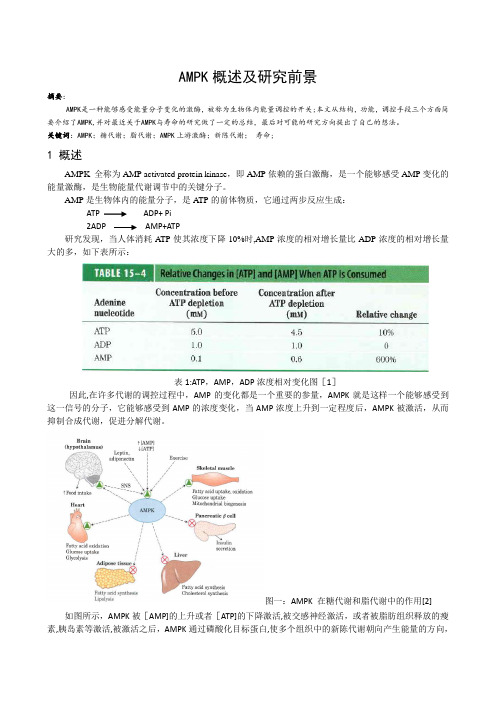
关键词:AMPK;糖代谢;脂代谢;AMPK上游激酶;新陈代谢;寿命;1 概述AMPK 全称为AMP activated protein kinase,即AMP依赖的蛋白激酶,是一个能够感受AMP变化的能量激酶,是生物能量代谢调节中的关键分子。
AMP是生物体内的能量分子,是A TP的前体物质,它通过两步反应生成:ATP ADP+ Pi2ADP AMP+ATP研究发现,当人体消耗A TP使其浓度下降10%时,AMP浓度的相对增长量比ADP浓度的相对增长量大的多,如下表所示:表1:ATP,AMP,ADP浓度相对变化图[1]因此,在许多代谢的调控过程中,AMP的变化都是一个重要的参量,AMPK就是这样一个能够感受到这一信号的分子,它能够感受到AMP的浓度变化,当AMP浓度上升到一定程度后,AMPK被激活,从而抑制合成代谢,促进分解代谢。
图一:AMPK 在糖代谢和脂代谢中的作用[2] 如图所示,AMPK被[AMP]的上升或者[ATP]的下降激活,被交感神经激活,或者被脂肪组织释放的瘦素,胰岛素等激活,被激活之后,AMPK通过磷酸化目标蛋白,使多个组织中的新陈代谢朝向产生能量的方向,削弱糖原,脂肪酸,胆固醇的合成,促进肝脏内的糖酵解,促进脂肪酸的氧化,向下丘脑传递信号促进摄食。
达到调控新陈代谢的目的。
因此AMPK也被称作新陈代谢的开关。
1AMPK的结构AMPK是一个异源三聚体,由三个不同的亚基构成,分别为催化亚基α,调节亚基β和γ。
如图二所示,α亚基为催化亚基,由550个氨基酸构成,在N末端含有一个丝氨酸/苏氨酸蛋白激酶的催化域,该催化域内172位上含有一个苏氨酸残基可被磷酸化.在C末端,有一段大约含有150氨基酸的部位用于与γ亚基和β亚基连接。

运动对棕色脂肪功能的影响及作用机制付鹏宇;龚丽景;胡扬【摘要】研究综述了运动对棕色脂肪组织(brown adipose tissue,BAT)功能的影响及其发挥作用的可能通路机制.随着肥胖发病率的日益增加,由此引发的多种慢性疾病正严重威胁着人类的健康.近年来,BAT以其耗能产热能力而备受关注.随着研究的深入,发现活化的BAT具有对抗肥胖所致慢性炎症状态和促进糖脂代谢的功能,以上特性都使BAT成为对抗肥胖及代谢相关疾病的新靶点.运动作为减脂降重和预防慢病的有效手段,其发挥促进健康作用的机制可能与激活BAT有关.具体作用机制如下:1)运动发挥促进BAT产热功能可能与VEGF信号通路、PI3K-Akt信号通路、PPAR信号通路有关;2)发挥抗炎作用可能与ErbB信号通路、Jak-STAT信号通路、TGF-β信号通路、胰岛素信号通路有关;3)发挥促进糖脂代谢的作用可能与PPAR信号通路、AMPK信号通路、胰岛素信号通路有关.综上,运动可通过调控多条信号通路而发挥促进BAT产热、提高抗炎特性及调控糖脂代谢变化的作用.【期刊名称】《体育科学》【年(卷),期】2018(038)011【总页数】6页(P92-97)【关键词】运动;棕色脂肪;产热;抗炎;糖脂代谢【作者】付鹏宇;龚丽景;胡扬【作者单位】北京体育大学运动人体科学学院,北京 100084;北京体育大学中国运动与健康研究院,北京 100084;北京体育大学中国运动与健康研究院,北京 100084【正文语种】中文【中图分类】G804.7棕色脂肪组织(brown adipose tissue,BAT)可通过非颤抖性产热调节体温,其多室的棕色脂肪细胞以含有大量线粒体和高表达解耦联蛋白1(uncoupling protein 1,UCP-1)等特异性基因为特征,在调节全身能量平衡中发挥关键作用,有助于控制肥胖及其相关疾病的发展[17,19,32]。
BAT在调控代谢中也发挥着重要作用,表现在BAT氧化葡萄糖和脂质,进而调节血糖平衡并降低血脂;BAT还有抑制巨噬细胞炎性特征的能力,可抵抗肥胖所致慢性炎症状态[17]。
台盼蓝染色细胞存活率检测试剂盒产品编号 产品名称包装 C0011台盼蓝染色细胞存活率检测试剂盒100次产品简介:碧云天生产的台盼蓝染色细胞存活率检测试剂盒(Trypan Blue Staining Cell Viability Assay Kit ),是利用正常的健康细胞能够排斥台盼蓝,而丧失细胞膜完整性的细胞可以被台盼蓝染色研制而成。
严格来说,台盼蓝染色检测的是细胞膜的完整性,通常认为细胞膜丧失完整性,即可认为细胞已经死亡。
台盼蓝染色后,通过显微镜下直接计数或显微镜下拍照后计数,就可以对细胞存活率进行比较精确的定量。
台盼蓝染色后的HeLa 细胞请参考图1。
图1. 细胞台盼蓝染色效果图。
胰酶消化下来的HeLa 细胞,1000g 离心1分钟,弃上清,用适量细胞重悬液重悬后,用等体积台盼蓝染色液(2X)染色。
箭头所示为台盼蓝染色的死亡细胞。
注:本HeLa 细胞经过一定的细胞坏死诱导处理。
台盼蓝染色只需3-5分钟即可完成,并且操作非常简单。
本试剂盒足够检测100个细胞样品。
包装清单:产品编号 产品名称 包装 C0011-1 台盼蓝染色液(2X) 10ml C0011-2细胞重悬液 100ml —说明书1份保存条件:4ºC 保存,一年有效。
注意事项:本试剂盒提供的两种溶液都是无菌的,使用时最好在超净台内进行,避免细菌污染。
本产品长时间存放可能会出现少量颗粒状沉淀,可在37ºC 水浴约10分钟以充分溶解沉淀。
沉淀完全溶解后即可正常使用。
台盼蓝对人体有毒,操作时请特别小心,并注意有效防护以避免直接接触人体或吸入体内。
本产品仅限于专业人员的科学研究用,不得用于临床诊断或治疗,不得用于食品或药品,不得存放于普通住宅内。
为了您的安全和健康,请穿实验服并戴一次性手套操作。
使用说明:1. 收集细胞:对于贴壁细胞先用胰酶和/或EDTA 消化下细胞。
对于悬浮细胞,则可以直接收集细胞。
把收集的细胞在1000-2000g 离心1分钟,弃上清,用1毫升或根据细胞的量用适当细胞重悬液重新悬起细胞。
腺苷酸活化蛋白激酶与肿瘤的研究进展孙启天(综述);高宇(审校)【期刊名称】《重庆医学》【年(卷),期】2014(000)017【总页数】3页(P2221-2223)【关键词】腺苷酸活化蛋白激酶;基因;肿瘤【作者】孙启天(综述);高宇(审校)【作者单位】承德医学院;承德医学院附属医院内分泌科,河北承德 067000【正文语种】中文腺苷酸活化蛋白激酶(AMP-activated protein kinase,AMPK)是细胞重要的能量感受器,在能量缺乏时被激活,能量充足时被抑制。
AMPK可以被多种激素、细胞因子及上游基因LKB1激活,并通过与哺乳动物雷帕霉素靶蛋白(mTOR)、p53的相互作用、对脂肪酸合酶及其他激酶的调节实现对细胞生长代谢的抑制,近期的研究发现AMPK在连接代谢综合征和肿瘤中起到重要作用,它可以通过缺氧诱导因子(HIF-1)和肿瘤抑制基因p53降低肿瘤细胞糖酵解水平,有可能会成为日后治疗肿瘤的新靶点。
现就近年来AMPK的研究新进展综述如下。
AMPK属于丝氨酸/苏氨酸蛋白质激酶家族,它包括3个亚单位,1个催化亚基(α)和2个调节亚基(β和γ)。
在哺乳动物中,每个亚基都包括不同的亚型(α1,α2;β1,β2; γ1,γ2和γ3),当细胞面临代谢压力时,细胞内AMP/ATP比例增高,AMP会与γ亚基发生连接,这种连接有两方面的作用,变构催化作用和防止α亚基活化环上的172位苏氨酸被磷酸酯酶去磷酸化。
AMP与γ亚基连接后,α亚基可以通过多种途径被磷酸化进而使ATP的生成增多,利用减少,以维持AMP/ATP的平衡,为细胞的生存提供足够的能量[1]。
1 AMPK的激活AMPK可以被一些激素及细胞因子激活,其中包括瘦素、脂联素、白细胞介素-6(IL-6)和睫状神经营养因子(CNTF)[2]。
AMPK还可以被多种药物激活,最典型的是5-氨基-4-氨甲酰咪唑核糖核苷酸(5-Aminoimidazole-4-carboxamide1-β-D-ribofuranoside AICAR),AICAR是一种细胞通透的磷酸化物质,可以在进入细胞后转化为AMP类似物(ZMP)。
AMPK信号通路在肝癌中的研究进展原发性肝癌发病率在全世界常见恶性肿瘤中位居第5位,死亡率位居第3位,其中90%以上的原发性肝癌属肝细胞肝癌。
AMPK信号通路是一种高度保守的丝氨酸/苏氨酸蛋白通路,是广泛存在于哺乳动物体内的一种能量调节器,通过促进分解代谢、抑制合成代谢来调控肿瘤细胞的生长代谢。
有研究证实当AMPK在肝脏中频繁发生异常表达及激活突变,其通过多种机制促进肝癌的发生发展,而且与肝癌的治疗及预后密切相关。
因此,AMPK信号通路可作为一个新的靶标,为肝癌的诊断和治疗提供新的策略与思路。
恶性肿瘤是全球的公共健康问题,而原发性肝癌发病率在全球常见恶性肿瘤中位居第5位,死亡率排第3位[1]。
从组织学划分上,原发性肝癌可分为三种类型,分别为肝细胞肝癌(hepatocellular carcinoma,HCC)、胆管细胞肝癌和混合型肝癌,而其中肝细胞肝癌占了90%以上。
流行病学调查显示,HCC在不同的地区发病率有所不同,其中高发病率集中在东南亚和撒哈拉沙漠以南的非洲这些发展中国家,而发达国家的发病率相对较低,如北美、南美以及北欧这些国家。
导致HCC的致病因素很多,其中病毒性肝炎引起HCC占绝大多数,此外脂肪肝、黄曲霉素、酗酒以及不洁水的长期饮用均是引起HCC的高危因素。
由于HCC 起病隐匿、发展速度快、恶性程度高且缺乏典型的临床表现,导致多数患者在确诊时已处于晚期,出现肿瘤的远处转移。
肝癌一旦发生其他脏器的转移则明显影响预后,术后生存率也随之降低,这是引起肝癌死亡的重要因素。
因此,對肝癌发生、发展的早认识及有效治疗成为新的挑战。
AMPK广泛存在于动植物体内,是一种高度保守的丝氨酸/苏氨酸结合蛋白,其作为一种能量平衡器,在能量代谢调节方面起到关键性作用,而且对肿瘤细胞的生长、增殖、自噬、新生血管形成、侵袭转移、应激反应和极性调控方面均具有重要意义。
有研究显示,AMPK的磷酸化和肝癌组织大小呈明显负相关[2]。
⼼⾎管领域⼗⼤进展2011年是⼼⾎管医学持续丰收和充满活⼒的⼀年。
全世界今年累计发表了约50000篇相关论⽂,约较2010年增长了6%。
这⼀年,在⼼⾎管研究领域虽然缺乏⼗分耀眼的明星分⼦和重⼤突破性的进展,但在⼼⾎管细胞⽣物学、分⼦⽣物学、⽣理病理学、转化医学、临床诊断和治疗药学等⽅⾯,都在持续地深⼊和扩展,并且有许多重要的研究成果和进展。
这⾥我们将着重介绍其中的⼗项主要进展和100项研究成果。
其研究进展主要有以下⼗个⽅⾯:⼼⾎管⼲细胞和再⽣医学的研究主要包括⼼脏内在⾃⾝⼲细胞研究(Cell Stem Cell 9:527);⼼⾎管残存的前体细胞(JMCC 50:269;50:304);脂肪、系膜细胞和⽪肤细胞可诱导的⼼脏多能⼲细胞(Nature 2⽉;Circ Res 109:923),并成功地应⽤⼲细胞⼈⼯培养出⼼肌细胞、⾎⼩板、红细胞、起搏细胞、⾎管和⼼脏(Nature Cell Biol 13:215;Science TM 2⽉;Acta Biomaterial 1⽉,PLoS One 3⽉、Blood 11⽉);证明损伤⼼肌可以⾃我修复(Nature,6⽉;PNAS 5⽉;Cir Res 109:1415);现已将⼲细胞成功应⽤于临床治疗⼼梗(Lancet 378:1847)等等。
⼼⾎管全基因组相关联研究(GWAS)包括动脉粥样硬化、⼼房纤颤、脑卒中、⾼⾎压、糖尿病、⾎管硬化、⾎脂异常等多数⼼脑⾎管病均⼴泛地开展了GWAS的研究,发现了⼀系列疾病相关基因和遗传变异,包括⼼肌⽼化基因(JBC 2⽉、6⽉);肥胖基因(Nature Genet 5⽉);衰⽼基因(PLoS Gent 4⽉);ACD相关基因(Nat.Gent 6⽉);⾼⾎压相关基因(Nat.Gent. 6⽉)等等,为⼼⾎管病发病的遗传背景、表型差异、诊断和个体治疗提供了研究基础(Nature Genetics 3⽉、9⽉;Nature Doi :10.1038, Nature 12⽉等等)。
Berberine Improves Kidney Function in Diabetic Mice via AMPK ActivationLong Zhao,Li-Na Sun,Hui-Bin Nie,Xue-Ling Wang,Guang-Ju Guan*Nephrology Research Institute,the Second Hospital of Shandong University,Jinan,Shandong,ChinaAbstractDiabetic nephropathy is a major cause of morbidity and mortality in diabetic patients.Effective therapies to prevent the development of this disease are required.Berberine(BBR)has several preventive effects on diabetes and its complications.However,the molecular mechanism of BBR on kidney function in diabetes is not well defined.Here,we reported that activation of AMP-activated protein kinase(AMPK)is required for BBR-induced improvement of kidney function in vivo.AMPK phosphorylation and activity,productions of reactive oxygen species(ROS),kidney function including serum blood urea nitrogen(BUN),creatinine clearance(Ccr),and urinary protein excretion,morphology of glomerulus were determined in vitro or in vivo.Exposure of cultured human glomerulus mesangial cells(HGMCs)to BBR time-or dose-dependently activates AMPK by increasing the thr172phosphorylation and its activities.Inhibition of LKB1by siRNA or mutant abolished BBR-induced AMPK activation.Incubation of cells with high glucose(HG,30mM)markedly induced the oxidative stress of HGMCs,which were abolished by5-aminoimidazole-4-carboxamide ribonucleoside,AMPK gene overexpression or BBR.Importantly,the effects induced by BBR were bypassed by AMPK siRNA transfection in HG-treated HGMCs.In animal studies, streptozotocin-induced hyperglycemia dramatically promoted glomerulosclerosis and impaired kidney function by increasing serum BUN,urinary protein excretion,and decreasing Ccr,as well as increased oxidative stress.Administration of BBR remarkably improved kidney function in wildtype mice but not in AMPK a2-deficient mice.We conclude that AMPK activation is required for BBR to improve kidney function in diabetic mice.Citation:Zhao L,Sun L-N,Nie H-B,Wang X-L,Guan G-J(2014)Berberine Improves Kidney Function in Diabetic Mice via AMPK Activation.PLoS ONE9(11): e113398.doi:10.1371/journal.pone.0113398Editor:Shang-Zhong Xu,University of Hull,United KingdomReceived June9,2014;Accepted October23,2014;Published November19,2014Copyright:ß2014Zhao et al.This is an open-access article distributed under the terms of the Creative Commons Attribution License,which permits unrestricted use,distribution,and reproduction in any medium,provided the original author and source are credited.Data Availability:The authors confirm that all data underlying the findings are fully available without restriction.All relevant data are within the paper.Funding:This work was supported by the National Key Technology Research and Development Program of the Ministry of Science and Technology of China 2011BA110B05.Competing Interests:The authors have declared that no competing interests exist.*Email:guangj@IntroductionDiabetes mellitus represents a global threat for premature morbidity and mortality[1].Diabetic nephropathy(DN)is a major chronic microvascular complication of diabetes and characterized by a progressive increase in albuminuria and a decline in glomerular filtration rate[2].Mechanisms underlying nephropathy in diabetes include a range of hemodynamic and metabolic factors[3].A growing number of studies support that the generation of reactive oxygen species(ROS)as a common downstream pathway of most of these mechanisms ultimately leads to inflammation and fibrosis[4].However,the exact pathogenesis of DN is complex and poorly understood.Berberine(BBR)is an isoquinolone alkaloid found in plants such as Phellodendron chinense and Coptis chinensis.In traditional Chinese medicine,BBR from Coptidis rhizoma is used as a constituent of the herbal medicine Huanglian[5].In this form it is reported to exert anti-fungal,anti-bacterial/viral,and anti-oncogenic effects,as well as a beneficial effect on diabetes,and anti-atherogenic properties[6,7].Several mechanisms are report-ed to be associated with the beneficial properties of BBR including improvement of sugar and lipid metabolism[8].BBR has also been reported to reduce the incidence of diabetes through suppression of oxidative stress.Whether BBR exerts its beneficialThe AMP-activated protein kinase(AMPK)is a heterotrimeric protein composed of a,b,and c subunits.The a subunit imparts catalytic activity,while the b subunit contains a glycogen-binding domain that also regulates the activity and the c subunit forms the broad base of the protein and is required for AMP binding.AMPK is well-conserved among eukaryotic cells and is expressed in endothelial cells of different origins.Activation of AMPK requires phosphorylation of Thr172in the activation loop of the a subunit and is mediated by at least two kinases,Peutz-Jeghers syndrome kinase LKB1(LKB1)and Ca2+/calmodulin-dependent protein kinase kinase.AMPK has been shown to mediate angiogenic and anti-inflammatory effects[9].Indeed,BBR has been reported to activate AMPK in several cell types,such as endothelial cells, smooth muscle cells[10],cardiomyocytes[11,12],cancer cells[13], b-cell[14],liver cells[15],macrophages[16,17],adipocytes[18]. Based on these reports,we hypothesized that the protective effects of BBR on kidney function in diabetes may be due,in part,to the ability of BBR to stimulate AMPK to suppress oxidative stress. Materials and MethodsMaterialsBBR,streptozotocin(STZ)and5-aminoimidazole-4-carboxa-Louis,MO,USA).BBR was dissolved in DMSO to make a 500mM stock solution(0.1%v/v final concentration)and stored at280u C.AMPK a1/2siRNA,LKB1siRNA,and all primary antibodies were purchased from Cell Signaling Company or Santa Cruz Biotechnology.The siRNA delivery agent Lipofectamine 2000was purchased from Invitrogen(Carlsbad,CA).Other chemicals were obtained from Sigma-Aldrich(St Louis,MO) unless otherwise indicated.AnimalsC57/BL6wildtype(WT)mice(6–8weeks of age,20–25g of bodyweight)were purchased from the animal department of Hunan Normal University.AMPK a2-/-mice were generated by Viollet B[19].They were maintained in a temperature-controlled (22u C)under a12h light/dark cycle,pathogen-free environment and fed a standard rodent chow diet and water ad libitum.Male WT mice,8–12weeks of age,20–25g,were obtained from the Jackson Laboratory(Bar Harbor,ME).A low-dose(50mg/kg/ day for5consecutive days)STZ induction regimen was used to induce pancreatic islet cell destruction and persistent hyperglyce-mia as described by the Animal Models of Diabetic Complications Consortium().Hyperglycemia was defined as a random blood glucose level of.300mg/dL for.2weeks after injection.The animal protocol was reviewed and approved by the University of Shandong,Animal Care and Use Committee. Primary cell cultureFor isolation of human glomerulus mesangial cells(HGMCs), collected kidney from surgery were washed twice with PBS at4u C and cut into small pieces.These pieces were purified by100-mesh sieve to get glomerular.Then the glomerular was incubated in a 0.2%collagenase solution at37u C for15–20minutes.Cell pellets were incubated in1640medium with full components.Purity of HGMCs was confirmed through positive staining for collagen and fibronectin.The participants have provided their written informed consent.We recorded the general information and got the permission from the patients to use their kidneys for studying only.The study protocol was approved by the Ethical Committee of the Second Hospital of Shandong University.Adenovirus infections in cellsAd-GFP,a replication-defective adenoviral vector expressing green fluorescence protein(GFP),served as control.An adenoviral vector expressing a constitutively active mutant of AMPK (AMPK-CA),was subcloned from a rat cDNA encoding residues 1-312of AMPK and bearing a Thr172-to-Asp mutation(T172D) into a shuttle vector(pShuttle CMV[cytomegalovirus]).Cells were infected with GFP or AMPK-CA in medium with2%feta calf serum overnight.The cells were then washed and incubated in fresh growth medium without feta calf serum for an additional 12h before ing these conditions,infection efficiency was typically.80%,as determined by GFP expression. Transfection of siRNA into cellsTransient transfection of siRNA was carried out according to Santa Cruz’s protocol.Briefly,the siRNAs were dissolved in siRNA buffer(20mM KCl;6mM HEPES,pH7.5;0.2mM MgCl2)to prepare a10m M stock solution.Cells grown in6-well plates were transfected with siRNA in transfection medium containing liposomal transfection reagent(Lipofectamine2000, Invitrogen).For each transfection,100m l transfection medium containing4m l siRNA stock solution was gently mixed with100m l 30-min incubation at room temperature,siRNA-lipid complexes were added to the cells in1.0ml transfection medium,and cells were incubated with this mixture for6h at37u C.The transfection medium was then replaced with normal medium,and cells were cultured for48h.Western blot analysisAs described previously[20],the protein content in total cell lysates or homogenized tissues was determined using the bicinchoninic acid protein assay reagent(Pierce,USA).Twenty micrograms of protein was separated by SDS-PAGE and then transferred to a membrane.The membrane was incubated with a 1:1,000dilution of primary antibody and a1:2,000dilution of horseradish peroxidase-conjugated secondary antibody.Protein bands were visualized by enhanced chemiluminescence(GE Healthcare).The intensity(area6density)of the individual bands on western blots was measured by densitometry(model GS-700, Imaging Densitometer;Bio-Rad).The background was subtracted from the calculated area.The control was set to100%.AMPK activity assayAMPK activity was assayed using the SAMS peptide.The activity was determined in the presence and absence of AMP (200m M).AMPK activity was calculated by determining the difference in activity between both conditions. Determination of kidney morphologyTo assess the glomerular sclerotic injury,renal tissues were embedded in paraffin and4m m-thick sections were prepared for periodic acid schiff(PAS)staining.Glomerulosclerosis(GS)was evaluated by determining the percentage of glomeruli exhibiting sclerotic lesions(%GS).200consecutive glomeruli were examined for each mouse.The data were averaged.Lesion scores related to the glomerular and mesangial area were graded according to the following scale:0=preservation of the architecture,1=5-15% glomerular expansion or mesangial expansion and PAS positivity, 2=15–30%glomerular expansion or mesangial expansion and PAS positivity,3=30–50%glomerular expansion or mesangial expansion and PAS positivity,and4=.50%glomerular expan-sion or mesangial expansion and PAS positivity,which was described previously[21].Renal function measurementBlood samples were centrifuged at2000g for10min,serum were collected for measuring BUN by using a commercially available kit(ABIN,Cat:577679).Urine samples were centrifuged in the same way to remove any suspended particles and the supernatant was used to detect24-hour urine albumin protein by using Albumin Mouse ELISA Kit(abcam,Cat:108792).The creatinine was measured with a Creatinine Companion kit (Exocell,Cat:1012).The renal function was assessed by measurement of Cr clearance(Ccr)as urinary Cr X urine volume/serum Cr and expressed as milliliters per minute. ImmunohistochemistryThe expressions of3-nitrotyrosine(3-NT)and malondialdehyde (MDA)in kidney were measured by immunohistochemistry in paraffin embedded sections following the process as previously described[22].All morphological analyses and cell counting were performed on blinded slides.Detection of ROSTissue or cell O 2-levels were measured using the DHE fluorescence/HPLC assay with minor modifications [23].Briefly,tissues or cells were incubated with DHE (10m M)for 30min,homogenized,and subjected to methanol extraction.HPLC was performed using a C-18column (mobile phase:gradient of acetonitrile and 0.1%trifluoroacetic acid)to separate and quantify oxyethidium (product of DHE and O 2-)and ethidium (a product of DHE auto-oxidation).O 2-production was determined by conver-sion of DHE into oxyethidine.Statistical AnalysisResults are expressed as the mean 6SD.Statistical significance for comparison between two groups was calculated using the two-tailed Student’s t test.To assess comparisons between multiple groups,analysis of variance (ANOVA)followed by the Bonferroni procedure was performed using the Graph-Pad Prism 4program (GraphPad Software,Inc,San Diego,CA).A P value of ,0.05was considered to be statistically significant.ResultsBBR activates AMPK via Thr172phosphorylation intime-and dose-dependent manner in cultured HGMCsEarlier studies had established that phosphorylation of AMPK at Thr172correlates with AMPK activity [24].BBR is well characterized as a drug to protect diabetes and the vascular complications.To investigate whether BBR activates AMPK in kidney,confluent HGMCs was treated with varying concentra-tions of BBR from 10to 200m M in culture medium for 2hours.AMPK activation was indirectly assessed by western blot analysis of AMPK phosphorylation at Thr172,which is essential for AMPK activity.As shown in Figure 1A,BBR of 100m M did not affect phosphorylation of AMPK at 1hour point.In contrast,BBR began to activate AMPK at 2hours.Increasing incubating time of BBR (100m M)further enhanced AMPK phosphorylation.In Figure 1B,the phosphorylation of AMPK gradually increased beginning from 25m M after incubation with BBR for 2hours and reached peak levels at 100m M in HGMCs.BBR treatmentdidFigure 1.BBR activates AMPK in cultured HGMCs.(A )Cultured primary HGMCs were incubated with BBR (100m M)for indicated time after starvation overnight.AMPK thr172phosphorylation in total cell lysate was detected by western blot.The blot is a representative of three blots from three independent experiments.*P ,0.05VS control.(B )Dose-dependent effects of BBR on AMPK-Thr172phosphorylation in HGMECs.N is 3in each group.*P ,0.05VS control.(C )Confluent HGMCs were treated with BBR (100m M)for 2hours.AMPK activity was assayed using the SAMS peptide as a substrate.N is 5in each group.*P ,0.05VS control.doi:10.1371/journal.pone.0113398.g001Figure 2.BBR-induced AMPK activation in cultured HGMCs is LKB1-dependent.(A )HGMCs were transfected with control siRNA or LKB1siRNA for 48h.Then cells were treated with BBR (100m M)for 2hours.AMPK thr172phosphorylation in total cell lysate was detected by western blot.The blot is a representative of three blots from three independent experiments.*P ,0.05VS control.NS indicates no significance.(B )HGMCs were infected with adenovirus containing LKB1-WT,LKB1-S428A or LKB1-S307A for 48h.The infected cells were then treated with BBR (100m M)for 2hours.AMPK-Thr172phosphorylation was detected by Western blot.The blot is a representative blot from three independent experiments.*P ,0.05VS ad-GFP.NS indicates no significance.Figure3.AMPK upregulations by AICAR or gene overexpression attenuate HG-induced oxidative stress in cultured HGMCs.(A) HGMCs were incubated with D-glucose(30mM)in presence or absence of AICAR(2mM)for12hours.ROS productions were detected by DHE fluorescence.N is5in each group.*P,0.05vs.control,#P,0.05vs HG alone.(B)HGMCs infected with Ad-GFP or Ad-AMPK-CA for48hours were incubated with HG for12hours.ROS productions were detected by DHE fluorescence.The picture is a representative from3independent experiments.*P,0.05vs.control GFP.NS indicates no significance.doi:10.1371/journal.pone.0113398.g003Figure4.BBR via AMPK activation reverses HG-induced oxidative stress in HGMCs.HGMCs were transfected with AMPK a1/2siRNA for 48hours.Then cells were pre-incubated with BBR(100m M)for30minutes followed by treatment with D-glucose(30mM)for12hours.(A)AMPK a protein expression in total cell lysates was assayed by Western blot.(B)ROS productions were detected by DHE fluorescence.(C)Quantitative data ofROS productions.N is5in each group.*P,0.05vs.control,#P,0.05vs HG plus control siRNA.NS indicates no significance.not alter total levels of AMPK(Figure1A and1B),suggesting that BBR-induced phosphorylation of AMPK was not due to altered expression of the total protein.In addition,increased AMPK phosphorylation was associated with elevated AMPK activity,as measured by the SAMS peptide assay[25](Figure1C).BBR-induced AMPK phosphorylation in HGMCs isLKB1-dependentTo study how BBR activates AMPK,we suppressed LKB1 expression,which is an AMPK upstream kinase by applying siRNA in cells[26].We found that LKB1siRNA,but not control siRNA,inhibited BBR-induced phosphorylation of AMPK at Thr172if LKB1protein expression was silenced by siRNA (Figure2A).These experiments offer solid support for the notion that LKB1is required for BBR-induced AMPK activation in cells. We next evaluated whether LKB1phosphorylation at Ser307or Ser428was required for BBR-induced AMPK activation[27]. Using site-directed mutagenesis,we developed an LKB1mutant in which an amino acid essential for LKB1activation,serine307or 428,was mutated to alanine.Since BBR treatment resulted in increased phosphorylation of AMPK in cells infected with adenovirus encoding WT-LKB1,we next investigated whether adenoviral overexpression of the LKB1mutant would prevent BBR-induced phosphorylation of AMPK.As expected,LKB1-S307A or LKB1-S428A mutant did abolish BBR-enhanced phosphorylation of AMPK-Thr172(Figure2B).These data suggest that LKB1is essential for BBR-induced AMPK activation Activation of AMPK attenuates HG-induced oxidative stressPermanent hyperglycemia is the most common single cause of kidney failure.Oxidative stress and inflammation are proved to be critical for the pathogenesis of diabetes mellitus.In order to induce the injury of kidney cells,we mimicked hyperglycemia by using high glucose(30mM D-glucose)in culture medium to treat HGMCs.As shown in Figure3A and3B,incubation of HGMCs with high glucose(HG)significantly increased the ROS produc-tions.These results are consistent with previous report[25]. Further,activation of AMPK by AICAR(Figure3A)or gene overexpression(Figure3B)abolished HG-induced oxidative stress in HGMCs,indicating that HG-induced oxidative stress is AMPK-dependent.BBR via AMPK activation suppresses HG-induced oxidative stressWe next determined whether BBR suppressed HG-induced oxidative stress through AMPK activation.To test this notion,we performed siRNA transfection to knockdown AMPK a1and a2 protein levels.As depicted in Figure4A,the level of AMPK protein was silenced significantly by specific siRNA,but not control siRNA.As expected,BBR treatment dramatically suppressed HG-induced oxidative stress in cultured HGMCs as assayed by DHE fluorescence(Figure4B and4C).Collectively, these results demonstrate that AMPK plays a key role in BBR-Figure5.BBR prevents hyperglycemia-induced renal dysfunction in WT but not in AMPK a2-/-mice.Permanent hyperglycemia in WT and AMPK a2-/-mice was induced by a low-dose STZ induction.All mice were received with or without BBR administration(200mg/kg body weight daily) for8weeks after the stable diabetic model was established.At the end of experiments,mice were sacrificed.(A)Blood glucose in all mice.5–10mice in each group.*P,0.05vs.WT alone,#P,0.05vs STZ plus WT.NS indicates no significance.(B)Morphological and quantitative analysis of glomerulus by HE staining.a,WT;b,WT+STZ;c,WT+STZ+BBR;d,AMPK a2-/-+STZ;e,AMPK a2-/-+STZ+BBR.(C)Serum BUN,(D)creatine clearance rate(Ccr), and(E)urinary protein excretion were assayed.5–10mice in each group.*P,0.05vs.control,#P,0.05vs DM in WT.NS indicates no significance. doi:10.1371/journal.pone.0113398.g005Hyperglycemia induces kidney dysfunction inSTZ-injected miceThen we investigated whether administration of BBR improved kidney function in diabetic mice.After16-week STZ-induced hyperglycemia duration,morphology of kidneys from all mice was assayed by PAS staining(Figure5A).Compared to control WT mice,hyperglycemia remarkably caused fibrosis,containing abnormal glomeruli.Similar to formation of glomerulosclerosis, diabetes also induced renal dysfunction as increased serum BUN (Figure5B),decreased Ccr(Figure5C),and enhanced urinary albumin excretion(Figure5D).Treatment of mice with BBR attenuates diabetes-induced kidney dysfunction in WT but not in AMPK a2-/-mice Then we determined the effects of BBR on diabetes-induced renal dysfunction.Injection of STZ dramatically increased blood glucose in WT and AMPK a2-/-mice.Interestingly,the increases of blood glucose induced by STZ in AMPK a2-/-mice were slightly higher that WT mice.As shown in Figure5B,BBR significantly inhibited the hyperglycemia-induced the formation of the glomerulosclerosis in WT but not in AMPK a2-/-diabetic mice. Quantitative analysis indicated that STZ significantly increased lesion score in WT mice.BBR remarkably reduced the lesion score in WT but not in AMPK a2-/-diabetic mice.Similarly,BBR reversed the increased serum BUN(Figure5C),decreased Ccr (Figure5D),and enhanced urinary albumin excretion(Figure5E) induced by hyperglycemia,suggesting that AMPK is a key mediator to perform the protective effects of BBR in diabetic Diabetes-induced oxidative stress in kidney is reversed by BBR in WT but not in AMPK a2-/-mice Pathophysiological processes of diabetes is extreme complex and controversial.A growing number of evidences showed that oxidative stress and inflammation might play important roles in the development of DN.We finally checked the oxidative stress in diabetic mice by assaying3-NT and MDA,which are makers of peroxidation of protein or lipid[28].As shown in Figure6A,6B, and6C,our data showed that hyperglycemia increased the levels of3-NT and MDA in kidney as compared to control group. Treatment of BBR significantly decreased levels of3-NT and MDA in WT diabetic mice but not in AMPK a2-/-diabetic mice, suggesting BBR via AMPK activation reduced oxidative stress in diabetic mice.DiscussionIn the present study,we have shown that LKB1-AMPK signaling mediates BBR-protected kidney function in diabetic mice.The underlying mechanism in this process is due to a novel pathway in which kidney dysfunction is inhibited by BBR as a result of AMPK activation.These findings indicate that AMPK pathway is an important mediator for kidney cell function and suggest that BBR therapy that modulates AMPK signaling may be beneficial in treating kidney dysfunction in diabetic nephropathy. BBR is a natural compound isolated from plants and with multiple pharmacological activities[8].The emerging role of BBR in modifying sugar and lipid metabolism has been verified in a large amount of experimental and clinical studies.Notably,due toFigure6.BBR via AMPK prevents hyperglycemia-induced oxidative stress in mice.Permanent hyperglycemia in WT and AMPK a2-/-mice was induced by a low-dose STZ induction as described in method section.All mice were received with or without BBR administration(200mg/kg body weight daily)for8weeks after the stable diabetic model was established.At the end of experiments,mice were sacrificed to detect MDA and3-NT by IHC.(A)Representative pictures of MDA and3-NT.(B)Quantitative analysis of3-NT.(C)Quantitative analysis of MDA.5–10mice in each group. *P,0.05vs.control,#P,0.05vs DM in WT.NS indicates no significance.doi:10.1371/journal.pone.0113398.g006chronic hepatitis.Beneficial effects of BBR were also observed in combating diabetic complications[29].Diabetes-related endothe-lial dysfunction,nephropathy,and neuropathy could be relieved after BBR administration.Recently,some groups reported that BBR has some protective effects in different stages of diabetes in rats,such as diabetic nephropathy[30–33].However,this present study not only demonstrated that BBR improves kidney function and inhibits glomerular fibrosis in diabetic mice,but further uncovered the molecular mechanisms,which BBR activates LKB1-AMPK signaling to suppress oxidative stress in glomerulus mesangial cells.Most importantly,the protective effects of BBR on kidney function in diabetic mice were mediated by AMPK because BBR was unable to protect liver functions in AMPK a2-/-mice, indicating the important role of AMPK in BBR-induced beneficial effects.In fact,BBR has been reported to activate AMPK in vascular endothelial cells and suppressed formation of atheroscle-rosis[34,35].Combined with these observations,BBR would be explored to treat kidney and cardiovascular disease,which are related to diabetic complications.Interestingly,we discovered that STZ-induced type1diabetes damaged renal function and induced glomerulosclerosis in mice. This is inconsistent with Qi’s report[36].We reasoned that the discrepancy should be due to the time difference of persistent hyperglycemia.Actually,Zhang M et al.also reported that the kidney function and the morphology of glomerular started to be abnormal at the6th week and were serve at the9th week during hyperglycemia[37],as well as the8th week which we reported.Another issue is that the relationship between AMPK and ROS remains controversial.Recently,Kumar Sharma et al.demon-strated that superoxide is increased by AMPK,which is renal-protective[38].In fact,AMPK functions as a redox sensor and modulator,in which slight increase of ROS can activate AMPK. However,activation of AMPK reduces ROS production.The disagreement would be due to the different level of ROS productions.Further studies are required to test these possibilities. In summary,we have demonstrated that BBR attenuates diabetic nephropathy in vivo via AMPK activation.In addition, the improvement of kidney function by BBR appears to be mediated by suppression of oxidative stress.The data presented here support the concept that AMPK may be an important therapeutic target for treating diabetic nephropathy and other kidney diseases related to oxidative stress.Therefore,till now, there is no report about the effects of BBR on diabetic nephropathy in clinical patients,indicating the potential applica-tion for BBR on treating diabetic kidney disease in patients in future.Author ContributionsConceived and designed the experiments:LZ GJG.Performed the experiments:LZ LNS HBN XLW.Analyzed the data:LZ GJG. Contributed to the writing of the manuscript:GJG.Revised the manuscript:LNS HBN XLW.References1.Roglic G,Unwin N,Bennett PH,Mathers C,Tuomilehto J,et al.(2005)Theburden of mortality attributable to diabetes:realistic estimates for the year2000.Diabetes Care28:2130–2135.2.Molitch ME,DeFronzo RA,Franz MJ,Keane WF,Mogensen CE,et al.(2004)Nephropathy in diabetes.Diabetes Care27Suppl1:S79–83.3.Jha JC,Jandeleit-Dahm KA,Cooper ME(2014)New Insights Into the Use ofBiomarkers of Diabetic Nephropathy.Adv Chronic Kidney Dis21:318–326.4.Forbes JM,Coughlan MT,Cooper ME(2008)Oxidative stress as a majorculprit in kidney disease in diabetes.Diabetes57:1446–1454.5.Yin J,Zhang H,Ye J(2008)Traditional chinese medicine in treatment ofmetabolic syndrome.Endocr Metab Immune Disord Drug Targets8:99–111.6.Liang KW,Ting CT,Yin SC,Chen YT,Lin SJ,et al.(2006)Berberinesuppresses MEK/ERK-dependent Egr-1signaling pathway and inhibits vascular smooth muscle cell regrowth after in vitro mechanical injury.Biochem Pharmacol71:806–817.7.Cho BJ,Im EK,Kwon JH,Lee KH,Shin HJ,et al.(2005)Berberine inhibits theproduction of lysophosphatidylcholine-induced reactive oxygen species and the ERK1/2pathway in vascular smooth muscle cells.Mol Cells20:429–434. 8.Li Z,Geng YN,Jiang JD,Kong WJ(2014)Antioxidant and Anti-InflammatoryActivities of Berberine in the Treatment of Diabetes Mellitus.Evid Based Complement Alternat Med2014:289264.9.Wang S,Xu J,Song P,Viollet B,Zou MH(2009)In vivo activation of AMP-activated protein kinase attenuates diabetes-enhanced degradation of GTP cyclohydrolase I.Diabetes58:1893–1901.10.Liang KW,Yin SC,Ting CT,Lin SJ,Hsueh CM,et al.(2008)Berberineinhibits platelet-derived growth factor-induced growth and migration partly through an AMPK-dependent pathway in vascular smooth muscle cells.Eur J Pharmacol590:343–354.11.Chang W,Zhang M,Li J,Meng Z,Wei S,et al.(2013)Berberine improvesinsulin resistance in cardiomyocytes via activation of59-adenosine monophos-phate-activated protein kinase.Metabolism62:1159–1167.12.Lv X,Yu X,Wang Y,Wang F,Li H,et al.(2012)Berberine inhibitsdoxorubicin-triggered cardiomyocyte apoptosis via attenuating mitochondrial dysfunction and increasing Bcl-2expression.PLoS One7:e47351.13.Fan LX,Liu CM,Gao AH,Zhou YB,Li J(2013)Berberine combined with2-deoxy-d-glucose synergistically enhances cancer cell proliferation inhibition via energy depletion and unfolded protein response disruption.Biochim Biophys Acta1830:5175–5183.14.Shen N,Huan Y,Shen ZF(2012)Berberine inhibits mouse insulin genepromoter through activation of AMP activated protein kinase and may exert beneficial effect on pancreatic beta-cell.Eur J Pharmacol694:120–126.15.Cao S,Zhou Y,Xu P,Wang Y,Yan J,et al.(2013)Berberine metabolitesexhibit triglyceride-lowering effects via activation of AMP-activated protein 16.Mo C,Wang L,Zhang J,Numazawa S,Tang H,et al.(2014)The crosstalkbetween Nrf2and AMPK signal pathways is important for the anti-inflammatory effect of berberine in LPS-stimulated macrophages and endotox-in-shocked mice.Antioxid Redox Signal20:574–588.17.Jeong HW,Hsu KC,Lee JW,Ham M,Huh JY,et al.(2009)Berberinesuppresses proinflammatory responses through AMPK activation in macro-phages.Am J Physiol Endocrinol Metab296:E955–964.18.Zhou L,Wang X,Yang Y,Wu L,Li F,et al.(2011)Berberine attenuates cAMP-induced lipolysis via reducing the inhibition of phosphodiesterase in3T3-L1 adipocytes.Biochim Biophys Acta1812:527–535.19.Viollet B,Andreelli F,Jorgensen SB,Perrin C,Flamez D,et al.(2003)Physiological role of AMP-activated protein kinase(AMPK):insights from knockout mouse models.Biochem Soc Trans31:216–219.20.Wang S,Peng Q,Zhang J,Liu L(2008)Na+/H+exchanger is required forhyperglycaemia-induced endothelial dysfunction via calcium-dependent calpain.Cardiovasc Res80:255–262.21.Liao J,Soltani Z,Ebenezer P,Isidro-Carrion AA,Zhang R,et al.(2010)Tesaglitazar,a dual peroxisome proliferator-activated receptor agonist(PPAR alpha/gamma),improves metabolic abnormalities and reduces renal injury in obese Zucker rats.Nephron Exp Nephrol114:e61–68.22.Wang S,Xu J,Song P,Wu Y,Zhang J,et al.(2008)Acute inhibition ofguanosine triphosphate cyclohydrolase1uncouples endothelial nitric oxide synthase and elevates blood pressure.Hypertension52:484–490.23.Fernandes DC,Wosniak J Jr.,Pescatore LA,Bertoline MA,Liberman M,et al.(2007)Analysis of DHE-derived oxidation products by HPLC in the assessment of superoxide production and NADPH oxidase activity in vascular systems.Am J Physiol Cell Physiol292:C413–422.24.Wang S,Liang B,Viollet B,Zou MH(2011)Inhibition of the AMP-activatedprotein kinase-alpha2accentuates agonist-induced vascular smooth muscle contraction and high blood pressure in mice.Hypertension57:1010–1017. 25.Wang S,Zhang M,Liang B,Xu J,Xie Z,et al.(2010)AMPKalpha2deletioncauses aberrant expression and activation of NAD(P)H oxidase and consequent endothelial dysfunction in vivo:role of26S proteasomes.Circ Res106:1117–1128.26.Shen Y,Honma N,Kobayashi K,Jia LN,Hosono T,et al.(2014)Cinnamonextract enhances glucose uptake in3T3-L1adipocytes and C2C12myocytes by inducing LKB1-AMP-activated protein kinase signaling.PLoS One9:e87894.27.Homolya L,Fu D,Sengupta P,Jarnik M,Gillet JP,et al.(2014)LKB1/AMPKand PKA control ABCB11trafficking and polarization in hepatocytes.PLoS One9:e91921.28.Wang S,Zhang C,Zhang M,Liang B,Zhu H,et al.(2012)Activation of AMP-activated protein kinase alpha2by nicotine instigates formation of abdominal aortic aneurysms in mice in vivo.Nat Med18:902–910.。