美国农业部风险评估案例分析. 沙门氏菌(陈伟生,姜艳彬,王海主编)思维导图
- 格式:xmin
- 大小:6.53 KB
- 文档页数:1


动物源性食品中沙门氏菌的风险评估 王 军 郑增忍 1) * 王晶钰 * ( 西北农林科技大学动物科技学院 陕西 杨凌 712100) 动物源性食品是人类食物的重 要组成部分。
在各种动物源性食品 中, 细菌性食物中毒最为常见, 而由 沙门氏菌引起的食物中毒病例在食 物中毒中屡居首位。
在我国, 细菌性 食物中毒中有 70%~80% 是由沙门 氏菌引起的, 而且大多数来源于动 物源性食品。
沙门氏菌菌型繁多, 分 布广泛, 是重要的人畜共患病的病 原体, 人们一旦摄入了含有大量沙 的理解和认识, 加强自我保护能力; 二是根据我国目前动物源性食品沙 门氏菌的污染情况, 提出了风险管 理的建议, 为今后开展其他食品、其 他微生物的风险评估提供依据。
起人的食物中毒。
沙门氏菌对外界 环境有一定的抵抗力, 如在水、牛奶、肉类和蛋类制品中可存活数周至数月, 在粪便中可存活 1~10 个 月, 冰雪中可存活 3~4 个月, 在 18~ 20℃、盐分为 5%~8% 时可存活 30 多天。
对热有一定的抵抗力, 60℃经 1 h , 70℃经 20 min , 75℃经 5 min 才 沙门氏菌的风险评估 危害识别 1 1.1 沙门氏菌的生物学特性 沙 门 氏 菌 为 肠 杆 菌 科 ( Enter- 1.1.1 能灭活, 在肉类产品中还需延长时沙 门 氏 菌 属 间才能将其杀死。
obacteriaceae ) 、 门氏菌的动物源性食品, 就会引起 的革兰氏阴性需氧或 流行病学研究沙门氏菌病是世界范围内报道 ( Salmonella ) 1.1.2 细菌性感染, 进而在毒素的作用下 兼性厌氧杆菌, 大小为( 1~3) μm ×( 0.4~0.6) μm , 无芽孢和荚膜, 绝大多数有鞭毛, 能运动。
一般沙门氏菌在普通培养基上生长、发育良好。
大 多数沙门氏菌在普通琼脂平板上, 发生食物中毒。
人和动物的沙门氏 菌病一直是一个世界性问题, 沙门 氏菌也一直作为食品中致病菌检测 的一项重要指标。


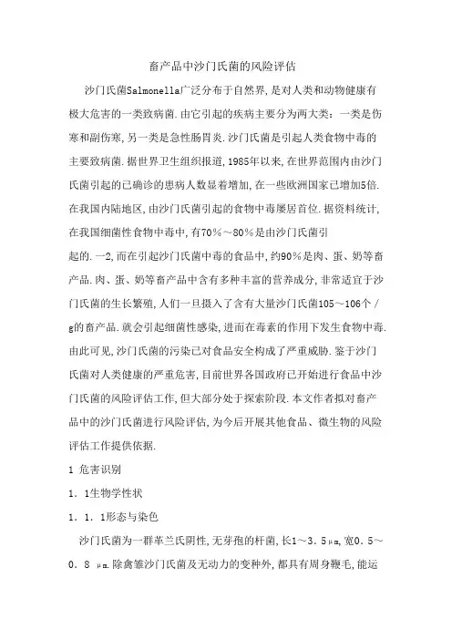
畜产品中沙门氏菌的风险评估沙门氏菌Salmonella广泛分布于自然界,是对人类和动物健康有极大危害的一类致病菌.由它引起的疾病主要分为两大类:一类是伤寒和副伤寒,另一类是急性肠胃炎.沙门氏菌是引起人类食物中毒的主要致病菌.据世界卫生组织报道,1985年以来,在世界范围内由沙门氏菌引起的已确诊的患病人数显着增加,在一些欧洲国家已增加5倍.在我国内陆地区,由沙门氏菌引起的食物中毒屡居首位.据资料统计,在我国细菌性食物中毒中,有70%~80%是由沙门氏菌引起的.一2,而在引起沙门氏菌中毒的食品中,约90%是肉、蛋、奶等畜产品.肉、蛋、奶等畜产品中含有多种丰富的营养成分,非常适宜于沙门氏菌的生长繁殖,人们一旦摄入了含有大量沙门氏菌105~106个/g的畜产品.就会引起细菌性感染,进而在毒素的作用下发生食物中毒.由此可见,沙门氏菌的污染已对食品安全构成了严重威胁.鉴于沙门氏菌对人类健康的严重危害,目前世界各国政府已开始进行食品中沙门氏菌的风险评估工作,但大部分处于探索阶段.本文作者拟对畜产品中的沙门氏菌进行风险评估,为今后开展其他食品、微生物的风险评估工作提供依据.1 危害识别1.1生物学性状1.1.1形态与染色沙门氏菌为一群革兰氏阴性,无芽孢的杆菌,长1~3.5μm,宽0.5~0.8 μm.除禽雏沙门氏菌及无动力的变种外,都具有周身鞭毛,能运动.1.1.2沙门氏菌的培养一般沙门氏菌易在普通培养基上生长,发育良好.但也有少数菌型,如甲型副伤寒、羊流产、猪伤寒、仙台、鸡雏沙门氏菌等,在普通琼脂上发育较差.大多数沙门氏菌在普通琼脂平板上,经18~24 h培养后,其菌落大小一般为2~3μm.光滑型菌落圆形,半透明,表面光滑,边缘整齐;粗糙型者,边缘不整齐,表面干燥,无光泽.在肉汤培养基内,光滑型呈均匀浑浊生长;粗糙型者可形成沉淀,上部澄清.1.1.3生化反应在肠杆菌科细菌分类鉴定中,生化特性检查有着重要的意义.绝大多数菌株能有规律地发酵葡萄糖并产生气体,但偶而亦有不产气者.该属细菌不能发酵侧金盏花醇、蔗糖,不产生吲哚,不分解尿素,不形成乙酰甲基甲醇.1.2 流行病学1.2.1 胃肠炎这是沙门氏菌感染中最常见的一型,约占病例的70%.潜伏期一般为4~24 h,发病大多急剧,有畏寒、发热,多伴有头痛、头晕、恶心、呕吐、腹痛,继以腹泻.亦有偶带脓血或呈血性便者.吐泻严重者,可出现脱水和电解质紊乱.偶有呈霍乱样的爆发性胃肠炎者,呕吐,腹泻剧烈,体温在病初上升后即下降,脉弱而速,尿少或尿闭等,如抢救不及时,可引起死亡.病例长短不一,一般为3~6 d,重者可延至1~3周才恢复.1.2.2菌血病或败血症沙门氏菌侵入血液并不少见,表现为畏寒、发热、出汗、面色苍白等中毒现象.细菌可随血液流到身体任何部位发生局部病灶.本型最常见的是猪霍乱沙门氏菌感染.1.2.3伤寒和其他肠热症型典型和严重的肠热症是伤寒,它是由伤寒沙门氏菌引起的.其他沙门氏菌,特别是甲型、乙型副伤寒沙门氏菌,也能引起本症.伤寒菌的唯一宿主是人.2 危害描述伤寒、甲型、乙型、丙型副伤寒沙门氏菌均为人类致病菌.在自然条件下,只能使人得病,而不能使动物自然感染.大部分其他血清型沙门氏菌,能使动物与家禽产生肠炎、败血症或伤寒样疾患家禽如鸡、鸭、鹅,家畜如猪、牛、马、羊,以及各种兽类、鱼类、鼠类均可带菌,甚至某些昆虫也可以分离出沙门氏菌.食用污染细菌的蛋类、肉类、奶制品常是引起人类沙门氏菌感染的重要原因.2.1 侵袭力有Vi抗原的沙门氏菌具有侵袭力,能穿过小肠上皮到达固有层.细菌在此部位常被吞噬细胞吞噬,但由于Vi抗原的保护作用,被吞噬后的细菌在细胞内不被破坏,反而在细胞内继续生长繁殖,并随游走的吞噬细胞将细菌带至机体的其他部位.2.2 内毒素沙门氏菌有较强的内毒素,可引起发热、白细胞改变、中毒性休克,并能激活补体系统产生多种生化效应,导致一系列病理生理变化. 2.3 肠毒素某些沙门氏菌如鼠伤寒沙门氏菌能产生类似大肠埃希菌的肠毒素.3 暴露评估3.1 沙门氏菌对禽肉的污染禽肉在生产加工线上连续被电击、屠宰、放血、烫洗和拔毛.烫毛和电流浸没式烫洗过程,已被证实是禽肉中沙门氏菌污染和交叉污染的主要来源.在禽肉运输过程中,由于其脚、毛、皮肤很容易沾上粪便,因此,沙门氏菌能存在于饲养场中并在加工操作开始时传染给禽类.3.2沙门氏菌对禽蛋的污染沙门氏菌对禽蛋的污染首先作用于蛋壳表面,或者通过其他途径进入禽蛋内部而造成污染.沙门氏菌既可以通过被感染的母鸡、母鸭水平传播,又可以通过产蛋进行垂直传播.3.2.1禽蛋表面的污染环境卫生状况差是造成禽蛋表面沙门氏菌污染的最重要因素.沙门氏菌首先对禽蛋表面造成污染.如果产蛋禽类体内携带有沙门氏菌,当其下蛋时,禽蛋表面已被感染了,因此孵化室会得到受感染的禽蛋.被污染的种蛋在孵化过程中,一部分中途死亡,一部分孵出病雏,而病雏通过与健雏接触,使沙门氏菌在整个禽类中传播.傅启勇等1991用常规方法对105枚市售禽蛋作了带染沙门氏菌的监测,从蛋壳分离到2株鸡伤寒沙门氏菌,说明通过市售蛋类可传播沙门氏菌.张彦明等1995研究了100枚样品鸡蛋蛋壳外表沙门氏菌污染情况,其阳性检出率为40%,检出鸡伤寒和鸡自痢沙门氏菌各2株.3.2.2禽蛋内部的污染近年来,蛋的内部受沙门氏菌污染的事件有上升趋势.由沙门氏菌引起的蛋污染主要是由于沙门氏菌对母禽繁殖器官侵袭力强有关.Oka—mura2001比较了6种不同血清型沙门氏菌对蛋污染和在机体组织器官中分布情况,分别采用6种沙门氏菌株对产蛋母禽接种,蛋黄中沙门氏菌的检出率为70%,这说明沙门氏菌能寄居在禽卵巢中并能转移到蛋黄中.如果禽蛋黄被沙门氏菌感染会导致:在孵化之前禽就死亡,孵出有病的禽,长成健康带菌的禽.禽类食囊中的食物会缓慢释放人胃中.Shackelford1988研究表明,从孵房中孵出的禽会被蛋壳上的沙门氏菌所污染,这些“健康”的禽可分泌高达10个沙门氏菌儋粪便,这是沙门氏菌污染的最大来源.另外没冰过的蛋类是最容易受污染的.4 风险描述畜产品中的沙门氏菌污染一般分为内源性污染和外源性污染两个方面.所谓内源性污染,是指活畜禽已经患有沙门氏菌病,如猪副伤寒、牛肠炎、鸡白痢等,这些患病畜禽不但其血液、内脏、肌肉中均可能含有大量的沙门氏菌,甚至在其卵中也可能会含有沙门氏菌.如禽蛋,健康的禽所产的蛋中是不含沙门氏菌的,但蛋禽一旦感染了沙门氏菌病,蛋壳形成前,经卵巢污染,产卵时污染,其体内的沙门氏菌就可能进入蛋内.外源性污染则是指畜产品在屠宰、加工、运输、储存和销售过程中,受到污水、粪便、加工工具等的污染而感染沙门氏菌.因此,要有效控制畜产品中沙门氏菌的污染,就必须针对其污染源,有区别地采取不同的监控措施.4.1禽体病禽或健康带菌禽的体内都存在大量沙门氏菌.病禽未彻底清除、带菌禽末被检出都可能造成再次污染.4.2饲料饲料中的主要污染源是含肉成分的原料,特别是鱼粉、血粉、骨粉等蛋白质饲料更易受沙门氏菌的污染.沙门氏菌被发现在鱼粉和肉骨粉中的含量为0.2%~4%I川.据英国国家兽医监察员抽样调查发现,80家蛋白质饲料加工厂中有21家的蛋白质成分“无沙门氏菌”熊谷进,1997.王玲扎2001通过对固体饲料酵母进行生化鉴定和血清学鉴定,结果发现检测样品中检出沙门氏菌.陈沁等2002通过常规分离培养鉴定技术,对上海口岸2001年l~6月份进口的动物性饲料498份进行了沙门氏菌的分离鉴定.结果共分离到沙门氏菌23株,分离率为4.62%.其中,鱼粉阳性率3.66%;肉骨粉阳性率为13.95%;明虾壳阳性率18.52%;乳清粉和饲料添加剂类阳性率0.4.3环境及其他因素禽舍地面、笼具、供饲设备、饮水器等环境条件都会成为沙门氏菌的传播源.带菌蛋、孵化器内环境中的胎绒,被沙门氏菌污染的空气,可引起同群雏禽的呼吸道感染.其他动物如犬、猫、鼠和野鸟等都可带菌,这些动物一旦进入禽舍也会带来传播的危险.5 风险管理5.1控制养殖场的污染应把对畜产品中沙门氏菌污染控制的焦点放在其首要环节一养殖场,从源头上确保畜产品不受沙门氏菌的污染.在这方面瑞典是一个很好的例子.过去10年来,瑞典设法将畜禽类中的沙门氏菌消除,从而有效地避免了畜产品中沙门氏菌的污染.他们的做法是建立良好的畜禽生活环境,对养殖场的环境卫生、畜体卫生、饮水和饲料卫生等所有环节进行严格控制,保证畜禽饲养的环境能有效防止沙门氏菌的传播.5.2控制加工及流通环节的污染应加强对畜产品加工、流通环节的监管.要求生产加工企业严格遵循畜产品安全生产技术规程,降低生产加工过程中沙门氏菌污染的危险,对上市前的畜产品进行强制性抽检,确保受污染的畜产品不能进入市场.5.3控制饲料的污染控制饲料的污染是对畜产品进行风险管理的关键,具体措施如下:5.3.I加酸处理沙门氏菌在温度高于10℃、pH6~7.5内繁殖最快.在商品饲料生产条件下,饲料不可能作冷藏处理,但添加各种有机酸甲酸、乙酸、丙酸和乳酸降低饲料的pH,就可以消灭或抑制饲料中沙门氏菌生长,并可改善动物肠道的微生物区系.5.3.2合理使用抗菌剂肉禽日粮甲酸钙添加量大于0.72%时,可使生长和饲料效率下降.而添加0.5%~1%的富马酸可以明显地P<0.05促进生长.饲料中添加抗菌剂已证实能有效地抑制沙门氏菌.Bailey等1998研究了各种抗菌剂包括球虫药对口服沙门氏鼠伤寒杆菌培养物的雏禽的影响,发现各种抗菌剂结合使用,能有效地减少沙门氏菌在盲肠中繁殖. 5.3.3加热处理制粒过程中饲料所受到的热足以杀死沙门氏菌.Liu等1969发现,当饲料含水量为15%,加热到88℃时可完全将沙门氏菌杀灭.调查表明,41%的肉禽开食料和58%的蛋用种禽日粮样品都有沙门氏菌存在,经蒸汽调质和压粒后,这2种日粮大约只有4%的样品尚有沙门氏菌存在.5.4严格执法严格执行有关畜产品安全的法律法规,以法律的手段来约束整个畜产品生产链所有参与者的行为,尽量避免因沙门氏菌污染的畜产品而导致的食物中毒.在畜产品生产的整个链环中,对养殖、加工、销售等各个环节应全面推行HACCP危害分析关键控制点系统管理,即以沙门氏菌的流行病学为开端,沿着畜产品生产链一直追溯到养殖场实行全面控制,并将控制的重点放在对人类健康的直接危害上.只有这样,才能确保畜产品在到达消费者手中时是安全的.参考文献:1余贺.医学微生物学M.北京:人民卫生出版社,1983.303—305.2俞树荣.微生物学和微生物学检验:第2版M.北京:人民卫生出版社,1988.175—177.3张河战.沙门氏菌分类、命名及中国沙门氏菌病分布J.微生物学免疫学进展,2002,302:74—76.4饶正华.沙门氏菌检测方法研究进展J.饲料研究, 2002,10:15—17.5王章云.肠炎沙门氏菌引起的食物中毒细菌学凋查J.中国人兽共患病杂志,1999,153:115—117.。
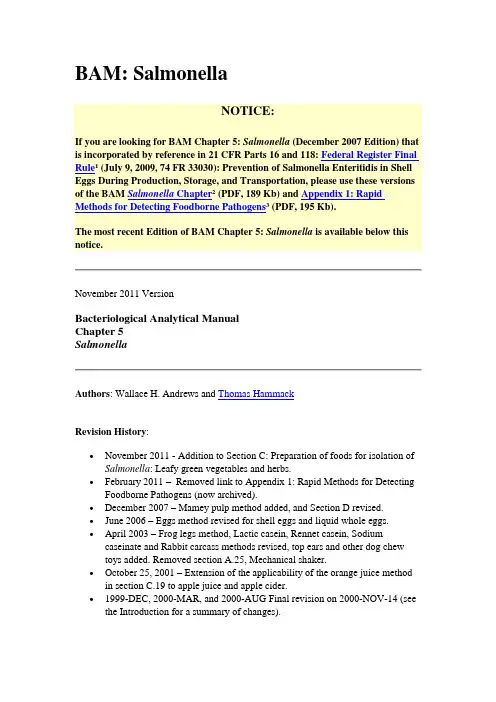
BAM: SalmonellaNOTICE:If you are looking for BAM Chapter 5: Salmonella (December 2007 Edition) that is incorporated by reference in 21 CFR Parts 16 and 118: Federal Register Final Rule1 (July 9, 2009, 74 FR 33030): Prevention of Salmonella Enteritidis in Shell Eggs During Production, Storage, and Transportation, please use these versions of the BAM Salmonella Chapter2 (PDF, 189 Kb) and Appendix 1: Rapid Methods for Detecting Foodborne Pathogens3 (PDF, 195 Kb).The most recent Edition of BAM Chapter 5: Salmonella is available below this notice.November 2011 VersionBacteriological Analytical ManualChapter 5SalmonellaAuthors: Wallace H. Andrews and Thomas HammackRevision History:∙November 2011 - Addition to Section C: Preparation of foods for isolation of Salmonella: Leafy green vegetables and herbs.∙February 2011 – Removed link to Appendix 1: Rapid Methods for Detecting Foodborne Pathogens (now archived).∙December 2007 – Mamey pulp method added, and Section D revised.∙June 2006 – Eggs method revised for shell eggs and liquid whole eggs.∙April 2003 – Frog legs method, Lactic casein, Rennet casein, Sodium caseinate and Rabbit carcass methods revised, top ears and other dog chewtoys added. Removed section A.25, Mechanical shaker.∙October 25, 2001 – Extension of the applicability of the orange juice method in section C.19 to apple juice and apple cider.∙1999-DEC, 2000-MAR, and 2000-AUG Final revision on 2000-NOV-14 (see the Introduction for a summary of changes).To obtain a copy of a prior version not currently posted, please contact Thomas HammackChapter Contents∙Introduction∙Equipment and Materials∙Media and Reagents∙Preparation of foods for isolation of Salmonella∙Isolation of Salmonella∙Identification of Salmonella∙ReferencesIntroductionSeveral changes are being introduced in this edition of BAM (8th Edition). The first change involves the expanded use of Rappaport-Vassiliadis (RV) medium4 for foods with both high and low levels of competitive microflora. In the previous edition, RV medium was recommended only for the analysis of shrimp. Based on the completion of AOAC precollaborative (5, 6) and collaborative (7, 8) studies, RV medium is now being recommended for the analysis of high microbial and low microbial load foods. RV medium replaces selenite cystine (SC) broth for the analysis of all foods, except guar gum. In addition, RV medium replaces lauryl tryptose broth for use with dry active yeast. Tetrathionate (TT)5 broth continues to be used as the second selective enrichment broth. However, TT broth is to be incubated at 43°C for the analysis of high microbial load foods and at 35°C for the analysis of low microbial load foods, including guar gum.The second change involves the option of refrigerating incubated preenrichments and selective enrichments of low-moisture foods for up to 72 h. With this option, sample analyses can be initiated as late as Wednesday or Thursday without weekend work being involved.The third change involves reducing the period of incubation of the lysine iron agar (LIA)6 slants. In the former edition (BAM-7), triple sugar iron agar (TSI)7 and LIA slants were incubated at 35°C for 24 ± 2 h and 48 ± 2 h, respectively. Unpublished data have demonstrated that the 48 h reading of LIA slants is without diagnostic value. Of 193 LIA slants examined, all gave definitive results within 24 ± 2 h of incubation.No significant changes altered the final test result when the slants were incubated an additional 24 h. Thus, both the TSI and LIA slants are now incubated for 24 ± 2 h.The fourth change involves the procedure for surface disinfection of shell eggs. In the previous edition (BAM-7), egg shells were surface-disinfected by soaking in 0.1% mercuric chloride solution for 1 h followed by soaking in 70% ethanol for 30 min. Mercuric chloride is classified as a hazardous waste, and is expensive to dispose of according to Environmental Protection Agency guidelines. In this edition (BAM-8) egg shells are now surface-disinfected by soaking for at least 10 sec in a 3:1 solution consisting of 3 parts of 70% alcohol (ethyl or isopropyl) to 1 part of iodine/potassium iodide solution.The fifth change involves the sample preparation of eggs. Egg contents (yolk and albumen) are thoroughly mixed before analysis. After mixing the egg contents, 25 g (ml) are added to 225 ml trypticase (tryptic) soy broth supplemented with ferrous sulfate.A method for the analysis of guar gum has been included. When guar gum is preenriched at a 1:9 sample/broth ratio, a highly viscous, nonpipettable mixture results. Addition of the enzyme cellulase to the preenrichment medium, however, results in a readily pipettable mixture.A method for orange juice (pasteurized and unpasteurized) has been included due to recent orange juice-related outbreaks.The directions for picking colonies from the selective plating agars have been made more explicit to reflect the intent of the method. In the absence of typical or suspect colonies on the selective plating agars, it is recommended that atypical colonies be picked to TSI and LIA slants. This recommendation is based on the fact that up to 4% of all Salmonella cultures isolated by FDA analysts from certain foods, especially seafoods, during the past several years have been atypical.Finally, since the publication of BAM-7, a 6-way comparison was conducted of the relative effectiveness of the three selective plating agars recommended in the BAM (bismuth sulfite8, Hektoen enteric9, and xylose lysine desoxycholate agars10) and three relatively new agars (EF-18, xylose lysine Tergitol 4, and Rambach agars). Our results (9) indicated no advantage in replacing any of the BAM-recommended agars with one or more of the newer agars. Thus, the combination of selective plating agars recommended in BAM-7 remains unchanged.A.Equipment and Materials1.Blender and sterile blender jars (see Chapter 111)2.Sterile, 16 oz (500 ml) wide-mouth, screw-cap jars, sterile 500 mlErlenmeyer flasks, sterile 250 ml beakers, sterile glass or paper funnelsof appropriate size, and, optionally, containers of appropriate capacityto accommodate composited samples3.Sterile, bent glass or plastic spreader rods4.Balance, with weights; 2000 g capacity, sensitivity of 0.1 g5.Balance, with weights; 120 g capacity, sensitivity of 5 mg6.Incubator, 35 ± 2 °C7.Refrigerated incubator or laboratory refrigerator, 4 ± 2°C8.Water bath, 49 ± 1°C9.Water bath, circulating, thermostatically-controlled, 43 ± 0.2°C10.Water bath, circulating, thermostatically-controlled,42 ± 0.2°C11.Sterile spoons or other appropriate instruments for transferring foodsamples12.Sterile culture dishes, 15 x 100 mm, glass or plastic13.Sterile pipets, 1 ml, with 0.01 ml graduations; 5 and 10 ml, with 0.1 mlgraduations14.Inoculating needle and inoculating loop (about 3 mm id or 10 5l),nichrome, platinum-iridium, chromel wire, or sterile plastic15.Sterile test or culture tubes, 16 x 150 mm and 20 x 150 mm;serological tubes, 10 x 75 mm or 13 x 100 mm16.Test or culture tube racks17.Vortex mixer18.Sterile shears, large scissors, scalpel, and forcepsmp (for observing serological reactions)20.Fisher or Bunsen burner21.pH test paper (pH range 6-8) with maximum graduations of 0.4 pHunits per color change22.pH meter23. Plastic bags, 28 x 37 cm, sterile, with resealable tape. (Items 23-24 areneeded in the analysis of frog legs and rabbit carcasses.)24.Plastic beakers, 4 liter, autoclavable, for holding plastic bag duringshaking and incubation.25.Sponges, non-bactericidal (Nasco cat # B01299WA), or equivalent.26.Swabs, non-bactericidal, cotton-tipped.B.Media12 and Reagents13For preparation of media and reagents, refer to Methods 967.25-967.28 inOfficial Methods of Analysis (1).ctose broth (M7414)2.Nonfat dry milk (reconstituted) (M11115)3.Selenite cystine (SC) broth (M13416)4.Tetrathionate (TT) broth (M14517)5.Rappaport-Vassiliadis (RV) medium (M13218). NOTE: RV mediummust be made from its individual ingredients. Commercialformulations are not acceptable.6.Xylose lysine desoxycholate (XLD) agar (M17919)7.Hektoen enteric (HE) agar (M6120)8.Bismuth sulfite (BS) agar (M1921)9.Triple sugar iron agar (TSI) (M14922)10.Tryptone (tryptophane) broth (M16423)11.Trypticase (tryptic) soy broth (M15424)12.Trypticase soy broth with ferrous sulfate (M18625)13.Trypticase soy-tryptose broth (M16026)14.MR-VP broth (M10427)15.Simmons citrate agar (M13828)16.Urea broth (M17129)17.Urea broth (rapid) (M17230)18.Malonate broth (M9231)19.Lysine iron agar (LIA) (Edwards and Fife) (M8932)20.Lysine decarboxylase broth (M87)3321.Motility test medium (semisolid) (M10334)22.Potassium cyanide (KCN) broth (M12635)23.Phenol red carbohydrate broth (M12136)24.Purple carbohydrate broth (M13037)25.MacConkey agar (M9138)26.Nutrient broth (M11439)27.Brain heart infusion (BHI) broth (M2440)28.Papain solution, 5% (M56a41)29.Cellulase solution, 1% (M18742)30.Tryptose blood agar base (M16643)31.Universal preenrichment broth (M18844)32.Universal preenrichment broth (without ferric ammonium citrate)(M188a45)33.Buffered peptone water (M19246)34.Dey-Engley broth (M19347)35.Potassium sulfite powder, anhydrous36.Chlorine solution, 200 ppm, containing 0.1% sodium dodecyl sulfate(R12a48)37.Ethanol, 70% (R2349)38.Kovacs' reagent (R3850)39.Voges-Proskauer (VP) test reagents (R8951)40.Creatine phosphate crystals41.Potassium hydroxide solution, 40% (R6552)42.1 N Sodium hydroxide solution (R7353)43.1 N Hydrochloric acid (R3654)44.Brilliant green dye solution, 1% (R855)45.Bromcresol purple dye solution, 0.2% (R956)46.Methyl red indicator (R4457)47.Sterile distilled water48.Tergitol Anionic 7 (R7858)49.Triton X-100 (R8659)50.Physiological saline solution, 0.85% (sterile) (R6360)51.Formalinized physiological saline solution (R2761)52.Salmonella polyvalent somatic (O) antiserum53.Salmonella polyvalent flagellar (H) antiserum54.Salmonella somatic group (O) antisera: A, B, C1, C2, C3, D1, D2, E1, E2,E3, E4, F, G, H, I, Vi, and other groups, as appropriate55.Salmonella Spicer-Edwards flagellar (H) antiseraC.Preparation of foods for isolation of SalmonellaThe following methods are based on the analysis of a 25 g analytical unit at a 1:9 sample/broth ratio. Depending on the extent of compositing, add enough broth to maintain this 1:9 ratio unless otherwise indicated. For samples not analyzed on an exact weight basis, e.g., frog legs, refer to the specific method for instructions.1.Dried egg yolk, dried egg whites, dried whole eggs, liquid milk(skim milk, 2% fat milk, whole, and buttermilk), and preparedpowdered mixes (cake, cookie, doughnut, biscuit, and bread),infant formula, and oral or tube feedings containing egg.Preferably, do not thaw frozen samples before analysis. If frozensample must be tempered to obtain analytical portion, thaw suitableportion as rapidly as possible to minimize increase in number ofcompeting organisms or to reduce potential of injuring Salmonellaorganisms. Thaw below 45°C for 15 min with continuous agitation inthermostatically controlled water bath or thaw within 18 h at 2-5°C.Aseptically weigh 25 g sample into sterile, wide-mouth, screw-cap jar(500 ml) or other appropriate container. For nonpowdered samples,add 225 ml sterile lactose broth65. If product is powdered, add about 15ml sterile lactose broth and stir with sterile glass rod, spoon, or tonguedepressor to smooth suspension. Add 3 additional portions of lactosebroth, 10, 10, and 190 ml, for total of 225 ml. Stir thoroughly untilsample is suspended without lumps. Cap jar securely and let stand 60 ±5 min at room temperature. Mix well by swirling and determine pHwith test paper. Adjust pH, if necessary, to 6.8 ± 0.2 with sterile 1 NNaOH or 1 N HCl. Cap jar securely and mix well before determining final pH. Loosen jar cap about 1/4 turn and incubate 24 ± 2 h at 35°C.Continue as in D, 1-11 , below.2.Eggsa.Shell eggs. Remove any adherent material from the shellsurface. Disinfect eggs with 3:1 solution consisting of 3 parts of70% alcohol (ethyl or isopropyl) to 1 part iodine/potassiumiodide solution. Prepare 70% alcohol solution either by diluting700 ml 100% alcohol with sterile distilled water for a finalvolume of 1,000 ml or by diluting 700 ml 95% alcohol withsterile distilled water for a final volume of 950 ml. Prepareiodine/potassium iodide solution by dissolving 100 g potassiumiodide in 200-300 ml sterile distilled water. Add 50 g iodineand heat gently with constant mixing until the iodine isdissolved. Dilute the iodine/potassium iodide solution to 1,000ml with sterile distilled water. Store iodine/potassium iodidesolution in amber glass-stoppered bottle in the dark. Prepare thedisinfection solution by adding 250 ml iodine/potassium iodidesolution to 750 ml 70% alcohol solution and mix well.Submerge eggs in disinfection solution for at least 10 seconds.Remove eggs and allow to air dry. Eggs with chipped, cracked,or broken shells are not included in the sample. Each sampleshall consist of twenty (20) eggs cracked aseptically into aWhirl-Pak bag, for a total of fifty (50) samples per poultryhouse. Eggs are cracked aseptically by gloved hands, with achange of gloves between samples. Mix samples thoroughly bygloved hands, with a change of gloves between samples. Mixsamples thoroughly by hand until yolks are completely mixedwith the albumen. Samples are held at room temperature(20-24°C) for 96 ± 2 h. After 96 ± 2 h, remove 25 ml portionfrom each sample of pooled eggs, and preenrich 25 ml testportion in 225 ml sterile trypticase soy broth (TSB)supplemented with ferrous sulfate66 (35 mg ferrous sulfateadded to 1000 ml TSB) and mix well by swirling. Let stand 60± 5 min at room temperature. Mix well by swirling anddetermine pH with test paper. Adjust pH, if necessary, to 6.8 ±0.2. Incubate 24 ± 2 h at 35°C. Continue as in D, 1-11, below.b.Liquid whole eggs (homogenized). Combine fifteen (15) 25ml test portions into a 375 ml composite contained in a 6-literErlenmeyer flask. Composites are held at room temperature(20-24°C) for 96 ± 2 h. After 96 ± 2 h, add 3,375 ml sterileTSB supplemented with ferrous sulfate67, as described above,and mix well by swirling. Let stand 60 ± 5 min at roomtemperature. Mix well by swirling and determine pH with testpaper. Adjust pH, if necessary, to 6.8 ± 0.2. Incubate 24 ± 2 hat 35°C. Continue as in D, 1-11, below.c.Hard-boiled eggs (chicken, duck, and others). If the eggshells are still intact, disinfect the shells as described above andaseptically separate the shells from the eggs. Pulverize the eggs(egg yolk solids and egg white solids) aseptically and weigh 25g into a sterile 500 ml Erlenmeyer flask or other appropriatecontainer. Add 225 ml TSB68 (without ferrous sulfate) and mixwell by swirling. Continue as described above.3.Nonfat dry milka.Instant. Aseptically weigh 25 g sample into sterile beaker (250ml) or other appropriate container. Using sterile glass or paperfunnel (made with tape to withstand autoclaving), pour 25 ganalytical unit gently and slowly over surface of 225 mlbrilliant green water contained in sterile 500 ml Erlenmeyerflask or other appropriate container. Alternatively, 25 ganalytical units may be composited and poured over the surfaceof proportionately larger volumes of brilliant green water.Prepare brilliant green water by adding 2 ml 1% brilliant greendye solution69 per 1000 ml sterile distilled water. Let containerstand undisturbed for 60 ± 5 min. Incubate loosely cappedcontainer, without mixing or pH adjustment, for 24 ± 2 h at35°C. Continue as in D, 1-11, below.b.Non-Instant. Examine as described for instant nonfat dry milk,except that the 25 g analytical units may not be composited.4.Dry whole milk. Examine as described for instant nonfat dry milk,except that the 25 g analytical units may not be composited.5.Caseinctic casein. Aseptically weigh 25 g sample into sterilebeaker (250 ml) or other appropriate container. Using sterileglass or paper funnel (made with tape to withstand autoclaving),pour 25 g analytical unit gently and slowly over the surface of225 ml Universal Preenrichment broth contained in sterile 500ml Erlenmeyer flask or other appropriate container. Analyticalunits (25 g) may be composited. Let container standundisturbed 60 ± 5 min. Incubate loosely capped container,without mixing or pH adjustment, for 24 ± 2 h at 35°C.Continue as in D, 1-11, below.b.Rennet casein. Aseptically weigh 25 g sample into sterilebeaker (250 ml) or other appropriate container. Using sterileglass or paper funnel (made with tape to withstand autoclaving),pour 25 g analytical unit gently and slowly over the surface of225 ml lactose broth contained in sterile 500 ml Erlenmeyerflask or other appropriate container. Analytical units (25 g) maybe composited. Let container stand undisturbed 60 ± 5 min.Incubate loosely capped container, without mixing or pHadjustment, for 24 ± 2 h at 35°C. Continue as in D, 1-11,below.c.Sodium caseinate. Aseptically weigh 25 g sample into sterile,wide-mouth, screw-cap jar (500 ml) or other appropriatecontainer. Add 225 ml sterile lactose broth and mix well.Analytical units may be composited. Let stand 60 min at roomtemperature with jar securely capped. Mix well by swirling anddetermine pH with test paper. Adjust pH, if necessary, to 6.8 ±0.2. Loosen jar about 1/4 turn and incubate 24 ± 2 h at 35°C.Continue as in D, 1-11, below.6.Soy flour. Examine as described for rennet casein, except 25 ganalytical units (25 g) may not be composited.7.Egg-containing products (noodles, egg rolls, macaroni, spaghetti),cheese, dough, prepared salads (ham, egg, chicken, tuna, turkey), fresh, frozen, or dried fruits and vegetables, nut meats,crustaceans (shrimp, crab, crayfish, langostinos, lobster), and fish.Preferably, do not thaw frozen samples before analysis. If frozensample must be tempered to obtain analytical portion, thaw below45°C for <15 min with continuous agitation in thermostaticallycontrolled water bath or thaw within 18 h at 2-5°C.Aseptically weigh 25 g sample into sterile blending container. Add 225 ml sterile lactose broth70 and blend 2 min. Aseptically transferhomogenized mixture to sterile, wide-mouth, screw-cap jar (500 ml) or other appropriate container and let stand 60 ± 5 min at roomtemperature with jar securely capped. Mix well by swirling anddetermine pH with test paper. Adjust pH, if necessary, to 6.8 ± 0.2.Mix well and loosen jar cap about 1/4 turn. Incubate 24 ± 2 h at 35°C.Continue as in D, 1-11, below.8.Dried yeast (active and inactive yeast). Aseptically weigh 25 gsample into sterile, wide-mouth, screw-cap jar (500 ml) or otherappropriate container. Add 225 ml sterile trypticase soy broth71. Mix well to form smooth suspension. Let stand 60 ± 5 min at roomtemperature with jar securely capped. Mix well by swirling anddetermine pH with test paper. Adjust pH, if necessary, to 6.8 ± 0.2,mixing well before determining final pH. Loosen jar cap 1/4 turn and incubate 24 ± 2 h at 35°C. Continue as in D, 1-11, below.9.Frosting and topping mixes. Aseptically weigh 25 g sample intosterile, wide-mouth, screw-cap jar (500 ml) or other appropriatecontainer. Add 225 ml nutrient broth72 and mix well. Cap jar securely and let stand 60 ± 5 min at room temperature. Mix well by swirlingand determine pH with test paper. Adjust pH, if necessary, to 6.8 ± 0.2.Loosen jar cap about 1/4 turn and incubate 24 ± 2 h at 35°C. Continue as in D, 1-11, below.10.Spicesa.Black pepper, white pepper, celery seed or flakes, chilipowder, cumin, paprika, parsley flakes, rosemary, sesameseed, thyme, and vegetable flakes.Aseptically weigh 25 g sample into sterile, wide-mouth,screw-cap jar (500 ml) or other appropriate container. Add 225ml sterile trypticase soy broth (TSB)73 and mix well. Cap jarsecurely and let stand 60 ± 5 min at room temperature. Mixwell by swirling and determine pH with test paper. Adjust pH,if necessary, to 6.8 ± 0.2. Loosen jar cap about l/4 turn andincubate 24 ± 2 h at 35°C. Continue as in D, 1-11, below.b.Onion flakes, onion powder, garlic flakes.Aseptically weigh 25 g sample into sterile, wide-mouth,screw-cap jar (500 ml) or other appropriate container. Preenrichsample in TSB74 with added K2SO3 (5 g K2SO3 per 1000 mlTSB, resulting in final 0.5% K2SO3 concentration). Add K2SO3to broth before autoclaving 225 ml volumes in 500 mlErlenmeyer flasks at 121°C for 15 min. After autoclaving,aseptically determine and, if necessary, adjust final volume to225 ml. Add 225 ml sterile TSB with added K2SO3 to sampleand mix well. Continue as in C-10a.c.Allspice, cinnamon, cloves, and oregano.At this time there are no known methods for neutralizing thetoxicity of these 4 spices. Dilute them beyond their toxic levelsto examine them. Examine allspice, cinnamon, and oregano at1:100 sample/broth ratio, and cloves at 1:1000 sample/brothratio. Examine leafy condiments at sample/broth ratio greaterthan 1:10 because of physical difficulties encountered byabsorption of broth by dehydrated product. Examine thesespices as described in C-10a, above, maintaining recommendedsample/broth ratios.11.Candy and candy coating (including chocolate). Aseptically weigh25 g sample into sterile blending container. Add 225 ml sterile,reconstituted nonfat dry milk75 and blend 2 min. Aseptically transfer homogenized mixture to sterile, wide-mouth, screw-cap jar (500 ml) or other appropriate container and let stand 60 ± 5 min at roomtemperature with jar securely capped. Mix well by swirling anddetermine pH with test paper. Adjust pH, if necessary, to 6.8 ± 0.2.Add 0.45 ml 1% aqueous brilliant green dye solution and mix well.Loosen jar caps 1/4 turn and incubate 24 ± 2 h at 35°C. Continue as in D, 1-11, below.12.Coconut. Aseptically weigh 25 g sample into sterile, wide-mouth,screw-cap jar (500 ml) or other appropriate container. Add 225 mlsterile lactose broth76, shake well, and let stand 60 ± 5 min at roomtemperature with jar securely capped. Mix well by swirling anddetermine pH with test paper. Adjust pH, if necessary, to 6.8 ± 0.2.Add up to 2.25 ml steamed (15 min) Tergitol Anionic 777 and mixwell. Alternatively, use steamed (15 min) Triton X-10078. Limit use of these surfactants to minimum quantity needed to initiate foaming. For Triton X-100 this quantity may be as little as 2 or 3 drops. Loosen jar cap about l/4 turn and incubate 24 ± 2 h at 35°C. Continue as in D,1-11, below.13. Food dyes and food coloring substances. For dyes with pH 6.0 orabove (10% aqueous suspension), use method described for driedwhole eggs (C-l, above). For laked dyes or dyes with pH below 6.0, aseptically weigh 25 g sample into sterile, wide-mouth, screw-cap jar (500 ml) or other appropriate container. Add 225 ml tetrathionatebroth79 without brilliant green dye. Mix well and let stand 60 ± 5 min at room temperature with jar securely capped. Using pH meter, adjust pH to 6.8 ± 0.2. Add 2.25 ml 0.1% brilliant green dye solution80 and mix thoroughly by swirling. Loosen jar cap about 1/4 turn and incubate24 ± 2 h at 35°C. Continue as in D, 3-11, below.14.Gelatin. Aseptically weigh 25 g sample into sterile, wide-mouth,screw-cap jar (500 ml) or other appropriate container. Add 225 mlsterile lactose broth81 and 5 ml 5% aqueous papain solution82 and mix well. Cap jar securely and incubate at 35°C for 60 ± 5 min. Mix well by swirling and determine pH with test paper. Adjust pH, if necessary, to 6.8 ± 0.2. Loosen jar cap about 1/4 turn and incubate 24 ± 2 h at35°C. Continue as in D, 1-11, below.15.Meats, meat substitutes, meat by-products, animal substances,glandular products, and meals (fish, meat, bone). Aseptically weigh25 g sample into sterile blending container. Add 225 ml sterile lactosebroth83 and blend 2 min. Aseptically transfer homogenized mixture to sterile wide-mouth, screw-cap jar (500 ml) or other appropriatecontainer and let stand 60 ± 5 min at room temperature with jarsecurely capped. If mixture is powder or is ground or comminuted,blending may be omitted. For samples that do not require blending,add lactose broth and mix thoroughly; let stand for 60 ± 5 min at room temperature with jar securely capped.Mix well by swirling and determine pH with test paper. Adjust pH, if necessary, to 6.8 ± 0.2. Add up to 2.25 ml steamed (15 min) Tergitol Anionic 7 and mix well. Alternatively, use steamed (15 min) Triton X-100. Limit use of these surfactants to minimum quantity needed to initiate foaming. Actual quantity will depend on composition of test material. Surfactants will not be needed in analysis of powderedglandular products. Loosen jar caps 1/4 turn and incubate samplemixtures 24 ± 2 h at 35°C. Continue as in D, 1-11, below.16.Frog legs. (This method is used for all domestic and imported froglegs.) Place 15 pairs of frog legs into sterile plastic bag and cover with sterile lactose broth at a 1:9 sample-to-broth (g/ml) ratio (see A, 23-24, above). If single legs are estimated to average 25 g or more, examine only one leg of each of 15 pairs. Place bag in large plastic beaker or other suitable container. Mix well and let stand 60 ± 5 min at roomtemperature. Mix well by swirling and determine pH with test paper.Adjust pH, if necessary, to 6.8 ± 0.2. Place plastic bag containing the frog legs and lactose broth into plastic beaker or other suitablecontainer. Incubate 24 ± 2 h at 35°C. Continue examination as in D, 1-11, below.17.Rabbit carcasses. (This method is used for all domestic and importedrabbit carcasses.) Place rabbit carcass into sterile plastic bag . Placebag in beaker or other suitable container. Add sterile lactose broth at a 1:9 sample-to-broth (g/ml) ratio to cover carcass (see A, 23-24, above).Mix well by swirling and let stand 60 ± 5 min at room temperature.Mix well by swirling and determine pH with test paper. Adjust pH, if necessary, to 6.8 ± 0.2. Incubate 24 ± 2 h at 35° C . Continueexamination as in D, 1-11, below.18.Guar gum. Aseptically weigh 25 g sample into sterile beaker (250 ml)or other appropriate container. Prepare a 1.0% cellulase solution (add 1g cellulase to 99 ml sterile distilled water). Dispense into 150 mlbottles. (Cellulase solution may be stored at 2-5°C for up to 2 weeks).Add 225 ml sterile lactose broth84 and 2.25 ml sterile 1% cellulasesolution to sterile, wide-mouth, screw-cap jar (500 ml) or otherappropriate container. While vigorously stirring the cellulase/lactose broth with magnetic stirrer, pour 25 g analytical unit quickly through sterile glass funnel into the cellulase/lactose broth. Cap jar securelyand let stand 60 ± 5 min at room temperature. Incubate loosely capped container without pH adjustment, for 24 ± 2 h at 35°C. Continue as in D, 1-11, below.19.Orange juice (pasteurized and unpasteurized), apple cider(pasteurized and unpasteurized), and apple juice (pasteurized)Aseptically add 25 ml sample to 225 ml Universal preenrichmentbroth85 in a sterile, wide mouth, screw-capped jar (500 ml) or other。

沙门氏菌实验活动风险评估报告一、生物学特性(一)种类和细菌分型沙门菌属(Salmonella lignieres)是一群寄生于人和动物肠道内的生化特性和抗原结构相似的革兰氏阴性杆菌,沙门菌属细菌的血清型已达2500多种。
根据DNA同源性,沙门菌属可分为两个种,即肠道沙门菌和邦戈沙门菌。
肠道沙门菌又分为6个亚种,而能感染人类的沙门菌血清型约有1400多种,都在第一亚种,即肠道沙门菌肠道亚种中。
沙门菌属中的少数血清型如伤寒沙门菌、甲型副伤寒沙门菌、肖氏沙门菌(原称乙型副伤寒沙门菌)和希氏沙门菌(原称丙型副伤寒沙门菌)是人的病原菌,对人类有直接的致病作用,引起肠热症。
沙门菌具有复杂的抗原结构,主要有菌体(O)抗原、鞭毛(H)抗原,少数菌中尚有一种表面(Vi)抗原,即英膜抗原。
1.O抗原O抗原存在于菌体细胞壁的最外层,化学成分为类脂一多糖一多肽复合物,由多糖决定其特异性,以阿拉伯数字顺序排列。
每个沙门菌血清型含一种或多种O抗原。
把含有相同抗原组分的归为一个群(组),这样可将本菌属中许多血清型细菌分为若干个菌群。
菌群分别以A、B、C……Z群,再以O51~O65等表示,作为定群分类的依据。
引起人类疾病的沙门菌血清型大多数在A~E群。
O抗原耐热、醇和酸,经100~121℃加热2.5h、用乙醇或盐酸处理而不失去抗原性,因此,菌体抗原已经被公认为沙门菌血清型分型的基础。
2.H抗原H抗原存在于鞭毛之中,为蛋白质,由肽链中氨基酸的排列顺序及空间构型决定其特异性。
不耐热,经加热和用乙醇及碱处理后其易变性。
H抗原分为第I相和第Ⅱ相两种,第I相为特异相,用小写英文字母表示,如:a、b、e、h等;第Ⅱ相为非特异相,用阿拉伯数字表示,如:1、2、5等,但也有少数菌含有第I相中的抗原e、n、x等成分。
一个菌株同时有第I相和第Ⅱ相H抗原的称为双相菌。
H抗原常有两相的变异。
每一组沙门菌根据H抗原不同,可进一步将组内沙门菌分成不同菌型。