丙球蛋白制作过程
- 格式:docx
- 大小:3.51 KB
- 文档页数:2
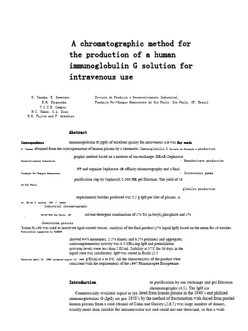
A chromatographic method forthe production of a humanimmunoglobulin G solution forintravenous useK. Tanaka, E. Sawatani, Divisão de Produção e Desenvolvimento Industrial,E.M. Shigueoka, Fundação Pró-Sangue Hemocentro de São Paulo, São Paulo, SP, BrasilT.C.X.B. Campos,H.C. Nakao, G.A. Dias,R.K. Fujita and F. ArashiroAbstractCorrespondence Immunoglobulin G (IgG) of excellent quality for intravenous use was Key wordsK. Tanaka obtained from the cryosupernatant of human plasma by a chromato-•Immunoglobulin G Divisão de Produção e productiongraphic method based on a mixture of ion-exchange, DEAE-SepharoseDesenvolvimento Industrial •Hemoderivate productionFF and arginine Sepharose 4B affinity chromatography and a finalFundação Pró-Sangue Hemocentro •Intravenous gammapurification step by Sephacryl S-300 HR gel filtration. The yield of 10de São Pauloglobulin productionexperimental batches produced was 3.5 g IgG per liter of plasma. AAv. Enéas C. Aguiar, 155, 1º andar•Industrial chromatography05403-000 São Paulo, SP solvent/detergent combination of 1% Tri (n-butyl) phosphate and 1%•Downstream processTriton X-100 was used to inactivate lipid-coated viruses. Analysis of the final product (5% liquid IgG) based on the mean for 10 batches Publication supported by FAPESP.showed 94% monomers, 5.5% dimers and 0.5% polymers and aggregates.Anticomplementary activity was 0.3 CH50/mg IgG and prekallikreinactivator levels were less than 5 IU/ml. Stability at 37o C for 30 days in theliquid state was satisfactory. IgG was stored in flasks (2.5Received April 28, 1998 Accepted August 18, 1998 g/flask) at 4 to 8o C. All the characteristics of the product wereconsistent with the requirements of the 1997 Pharmacopée Européenne.Introduction or purification by ion-exchange and gel filtrationchromatography (4,5). The IgG iso-Commercially available liquid or lyo-lated from human plasma in the 1940’s and philizedimmunoglobulins G (IgG) are pro-1950’s by the method of fractionation with duced from pooledhuman plasma from a cold ethanol of Cohn and Oncley (2,6,7) was large number of donors,usually more than suitable for intramuscular use and could not one thousand, so that a widevariety of anti-be administered intravenously because of bodies will be present in the product (1). undesirable effects due to the modifications Several production processes are employed, in the IgG molecule, such as aggregates re-most of them based on the method of Cohn sulting from the fractionation process with (2), i.e., fractionation with cold ethanol, with ethanol and other agents which increased its polyethyleneglycol (PEG) precipitation (3) anticomplement activity (8,9).Thus, the major goal of IgG producers was to develop methods that would guarantee intravenous tolerance, eliminating or preventing the aggregation of molecules without affecting the activity of the antibodies present in the IgG preparation. The first process for the production of intravenous(iv) IgG was based on treatment of IgG with a quantity of pepsin (10) or plasmin (11) that caused enzymatic cleavage of IgG molecules. This process, corresponding to the preparation of first-generation iv IgG, is today considered to be obsolete (9).Specific structural changes of IgG were introduced in the enzymatic methods to re-duce anticomplementary activity. The prod-ucts thus obtained usually have reduced in vivo survival times and their continuous use may cause an antigen response depending on the enzyme used (12). The second-gen-eration iv IgG consisted of chemically modi-fied preparations with more or less impaired Fc-related effector functions (9). From 1975 to 1989, the chemical modification of the protein was probably the most successful approach to the preparation of iv IgG (12). The reagents used for this modification range from ß-propiolactone (13) to reducing/alkylating agents (12,14), reducing/amidating agents (15) and reducing/sulfonating agents (16).Today, all of these preparations are being replaced with third-generation products con-taining intact IgG molecules which retain effector functions. The latest generation in-cludes preparations that are free of comple-ment-activating aggregates thanks to the use of small or trace amounts of pepsin, pH 4.0, PEG and bulk adsorption with ion-exchange gel (9). This development was encouraged by the fact that the chromatographic method provides a safe and effective iv IgG product meeting the 1982 requirements of the World Health Organization Committee (17). The choice of a third-generation preparation which exhibits all the functions of the IgG molecule will be determined on the basis of safety in terms of viral transmission and absence of contaminants. Furthermore, the preparation should contain a normal distri-bution of IgG subclass molecules and have a half-life after infusion (9) which is in the physiological range of 21 to 36 days. Material and MethodsProduction equipmentPharmacia liquid chromatography equip-ment was employed using a Bio Process controller (Uppsala, Sweden). The following columns were used: step 1, desalting, gel filtration on Sephadex G-25, 1 column, mo-del BPSS 400/600 (60 cm in height by 40 cm in diameter). Step 2, anion-exchange DEAE-Sepharose FF, 1 column, model PS-370/15 (15 cm in height and 37 cm in diameter). Step 3, affinity gel Arginine Sepharose 4B (40%) + anion-exchange DEAE-Sepharose FF (60%), 1 column, model PS-370/15 (15 cm in height by 37 cm in diameter) and cation-exchange CM-Sepharose FF, 1 column, model Index 200/500 (15 cm in height and 20 cm in diameter). Step 4, cation-exchange CM-Sepharose FF, 1 column, model Index 200/500. Step 5, gel filtration on Sephacryl S-300, 1 column, model BPG 200/ 950 (95 cm in height and 20 cm in diameter).Other instruments used were a tangential flow ultrafiltration Pellicon cassette system and a filtration system with a stainless steel sanitary filter holder, 293 mm in diameter (Millipore, Bedford, MA, USA), and a con-tinuous flow centrifuge model AG BKA-6 (Westphalia Separator, Oeld, Germany). Details about the buffer solutions are given in Table 1.Preparation of immunoglobulin G Approximately 1200 human plasma bagsstored at -30o C were thawed at a temperature of 2 to 4o C in order to form a 200-l pool. Thawed plasma was centrifuged at 2o C toBraz J Med Biol Res 31(11) 1998obtain a cryoprecipitate to be used for the production of factor VIII. The supernatant of the cryoprecipitate was cleared by filtration through a 30-S depth filter (Zeta Plus, Cuno, Meriden, CT, USA) and desalted with coarse Sephadex G-25 filtration gel on a BPSS 400/ 600 column using 5 mM sodium acetate as elution solution. The pH was adjusted to 5.2 with 1 M CH 3CO 2H and the protein solution (410 l) was allowed to stand overnight in a cold chamber at 4oC for euglobulin precipi tation. On the following day the preparation was centrifuged at 4oC to remove the pre cipitated euglobulins and the supernatant was cleared by filtration. The pH was readjusted to 5.2 with 1 M CH 3CO 2H and conductivity was adjusted to 1.4 mS/cm with NaCl. The sample was applied to a PS-370/ 15 column containing DEAE-Sepharose FF in order to separate gamma-globulin (unadsorbed) from other plasma proteins such as albumin, etc.The gamma-globulin fraction was submitted to chromatography for the preparation of IgG as described below. The albumin fraction was further purified by a chromatographic method (18). The pH of the gamma-globulin fraction (480 l) was adjusted to pH6.0 with 1 M NaOH and conductivity was adjusted to 1.4 mS/cm. The fraction was then applied to two columns, PS-370/15 and Index 200/500, coupled in series. The first column was packed with a mixture of two gels, 40% arginine Sepharose 4B and 60% DEAE-Sepharose FF, and the second with 8 l of CM-Sepharose FF gel.Arginine + DEAE-Sepharose and CM-Sepharose FF Equilibration 20 mM sodium acetate, pH 6.0, and conductivity 1.4 mS/cm Elution of arginine + DEAE-Sepharose FF 0.5 M sodium acetate, pH 7.0, and conductivity >20 mS/cm Elution of CM-Sepharose FF 1.0 M NaCl, pH 7.0 Cleaning and storage 0.5 M NaOH and 10 mM NaOHCM-Sepharose FF (after virusinactivation) Equilibration 10 mMglycine, pH 7.0 Elution 0.1 M glycine + 0.15 M NaCl, pH 9.0 Cleaning and storage 0.5 M NaOH and 10 mM NaOHSephacryl S-300 HR 1st Equilibration 0.1 M sodium acetate + 50 mM NaCl, pH 6.0 2nd Equilibration 0.15 M NaCl, pH 6.0 Elution with2nd equilibration buffer Cleaning and storage 0.5 M NaOH and 10 mM NaOHTable 1 - Buffer solutions used. Sephadex G-25 C Equilibration 5 mM sodium acetate, pH 7.0, and conductivity 0.37 mS/cmElutionwith equilibration buffer Cleaning and storage 0.5 M NaOH and 10 mM NaOHDEAE-Sepharose FFEquilibration 20 mM sodium acetate, pH 5.2, and conductivity 1.4 mS/cm1st Elution with equilibration buffer2nd Elution25 mM sodium acetate, pH 4.5, and conductivity 1.75 mS/cmThe proteins not of interest for the presentstudy, IgM, IgA, transferrin, etc, wereadsorbed to the first column. IgG wasadsorbed to the second column and eluted andconcentrated to 5% (w/v) using the-Thawing 4o CCryoprecipitate-CentrifugationCryosupernatantDesalting on Sephadex G-25 CSephadex fraction -Euglobulin precipitation at pH 5.2 -Kept overnight at 4o C -Centrifugation -Filtration SupernatantAlbumin fraction -Purification Unadsorbed + Gamma-globulin 60% DEAE-Sepharose FF 40% Arginine-Sepharose 4 BUnadsorbed protein fractionCM-Sepharose FFIgG -Concentration to 5% (w/ v) -S/D Virus inactivationCM-Sepharose FFIgG without S/D-Enzyme treatment (0.1 mg pepsin/g of IgG, pH 4) -Temperature 37o C/12 h -pH adjustment to 6.0 with 1.0 M NaOHSephacryl S-300IgG without enzyme or polymers-Concentration to 7% (w/v) -Formulation-Sterilization by filtrationFigure 1 - Flow diagram for the production of liquid intravenous IgG.Pellicon Cassette System 30,000 NMWL. The pH of the IgG solution (16 l) was adjusted to 5.5 and the material was submitted to viral inactivation with a solvent/detergent combination (19), i.e., 1% Tri (n-butyl) phosphate and 1% Triton X-100 at 35o C for 10 h. After viral inactivation, the solvent/deter-gent combination was removed by ion-ex-change chromatography on CM-Sepharose FF in an Index 200/500 column containing 8 l of gel. The IgG solution was concentrated to 7%, pepsin was added (0.1 mg/g protein), pH 4.0, and the solution was heated at 37o C for 10 h. The preparation was cooled to 20o C and then applied to a BPG 200/950 column containing 20 l of Sephacryl S-300 HR fil tration gel to remove the aggregated IgG molecules and pepsin. The eluted IgG solution was concentrated to 6.5% and then formulated as follows: the pH was adjusted to5.0 with 1 N HCl, the conductivity to 9 to 10 mS/cm with solid NaCl, and 7.5% (w/v) maltose and 0.1 M glycine were added as stabilizers (12,20). The material was steril-ized by filtration through a 0.22-µm mem-brane (Millipore) and the final product (14 l), containing 5% protein (w/v), was bottled in 50-ml type I (neutral) flasks, with 2.5 g IgG per flask and was stored at 4 to 8o C (see Figure 1).Analytical methodsThe characteristics of normal liquid 5% IgG for intravenous use were evaluated by the methods described in the Pharmacopée Européenne (1) and in Regulation No. 2.419 of 12/17/1996 of the Brazilian Ministry of Health.The immunochemical tests were carried out by cellulose acetate electrophoresis, by the micro-Ouchterlony method and by im-munoelectrophoresis using anti-total human protein and anti-animal protein antisera of domestic species such as horses, cows and sheep. Protein concentration was determined by the biuret method, pH was measured by Braz J Med Biol Res 31(11) 1998diluting to 1% in a 0.9% sodium chloride solution, and anti-A and anti-B hemaggluti-nins were determined by the indirect Coombs method. The presence of other plasma proteins such as IgA and IgM and of the IgG subclasses IgG1, IgG2, IgG3 and IgG4 was determined by radial immunodiffusion on Bindarid plates (The Binding Site Inc., San Diego, CA, USA).Anticomplementary activity was deter-mined by the method of Mayer using guinea pig complement and sheep red cells. The prekallikrein activator was determined using the S2302 chromogenic substrate of Chro-mogenix (Mölndal, Sweden).The biological safety of the product was evaluated by tests for the detection of anti-HIV, anti-HTLV-1 and 2, anti-HCV, and HB antigens at the Serology Laboratory of Fundação Pró-Sangue Hemocentro de São Paulo. Sterility was evaluated by the Steritest membrane method (Sterility Testing System, Millipore). Pyrogenicity and toxicity were tested at Medlab, São Paulo, Brazil.The distribution of monomers, dimers and polymers was evaluated by HPLC, and anti-polio I, II and III, anti-measles and anti-herpes activities were determined at Instituto Adolfo Lutz, São Paulo, Brazil. Anti-rubeola, anti-CMV and anti-streptolysin O activities were determined at the Immunology Laboratory of IAMSPE, São Paulo, Brazil. Stability was evaluated by incubating the preparation at 57o C for 4 h and observing the presence of jelling.Results and DiscussionThe size distribution of the product, evaluated by HPLC, indicated 94% monomers, 5.5% dimers and 0.5% polymers, corresponding to standard values. The profile of the IgG molecule did not show any alterations when the IgG preparation was incubated at 37o C for one month; the anticomplementary activ ity was 0.3 CH50/mg IgG, in agreement with the specifications that determine a value of less than 1 CH50/mg IgG, and the functional activity of Fc was unchanged, presenting an index of 125 to 128% of Fc, meeting the recommendationof Pharmacopée Européenne, whichrequires >60%. On the basis of these evaluations,we concluded that the product contained intactmolecules. The subclasses determined were IgG1= 65.6%, IgG2 = 28.7%, IgG3 = 4.2%, and IgG4= 1.4%, with no significant variations comparedto normal plasma. For prekallikrein activator(PKA) determination we used a reference PKAfrom the FDA and the S2302 chromogenicsubstrate of Chromogenix. Values of less than 5IU/ml 5% (w/v) IgG were obtained, well belowthe specification limit of 35 IU/ml of PKA. Theremaining data which characterize the productare presented in Tables 2 and 3, and celluloseacetateTable 2 - Characteristics of liquid intravenous IgG.N = 10. ND, Not detected. *Pharmacopée Européenne(1).Table 1 - Buffer solutions used.Sephadex G-25 CEquilibration 5 mM sodium acetate, pH 7.0, and conductivity 0.37mS/cmElution with equilibration bufferCleaning and storage 0.5 M NaOH and 10 mM NaOHDEAE-Sepharose FFEquilibration 20 mM sodium acetate, pH 5.2, and conductivity 1.4mS/cm1st Elution with equilibration buffer2nd Elution 25 mM sodium acetate, pH 4.5, and conductivity 1.75mS/cm3rd Elution 0.15 M sodium acetate, pH 4.0, and conductivity 8.0mS/cmCleaning and storage 0.5 M NaOH and 10 mM NaOHAnalysis Results Specifications*Protein concentration 5.0 ±0.2% >90 to <110%pH determination 4.5-5.0 4.0-7.4PKA determination <5.0 IU/ml <35 IU/mlPurity (electrophoresis) >99% >95%IgA ND -IgM ND -Anti-A hemagglutinin ≤1:8 <1:64Anti-B hemagglutinin ≤1:8 <1:64 Anticomplementaryactivity<0.3 CH50/mg IgG <1 CH50/mg IgGAluminum <20 µg/ml -Fc function 125-128% >60%electrophoresis and immunoelectrophoresis data are shown in Figures 2 and 3, respectively.We describe the preparation of intravenous 5% immunoglobulins in the liquid state from a pool of human plasma from 1200 donors by the chromatographic method. The analysesperformed for quality control showed that the IgG met international specifications. In thestability test involving heat-Table 3 - Antibodies detected in liquid intravenous IgG.N = 10.Antibody Method Titer and/or uniting to 57o C for 4 h there was no jelling, and in the quarantine test involving incubation in an oven at 37o C for 4 weeks, again there were no alterations in the IgG molecule.Viral inactivation was performed with a solvent/detergent system (19), i.e., 1% Tri (n-butyl) phosphate and 1% Triton X-100, pH 5.5, at 35o C for 10 h, which was then removed by the ion-exchange gel, CM-Sepharose FF. The protein aggregates gener ated during the process were removed by gel filtration chromatography through Sephacryl S-300 HR. Thus, the product obtained consisted mainly of the monomer, presenting only trace amounts of polymers, consistent with low anticomplementary activity, i.e.,less than 1 CH50/mg IgG.Anti-polio I Neutralization test 1:256 Anti-polio II Neutralization test 1:128 Of the several stabilizing agents tested,Anti-polio III Neutralization test 1:64 glucose, sucrose and maltose, the last one at Anti-measles Hemagglutination inhibition 1:64a 7.5% concentration, and 0.1 M glycine, pH Anti-rubeola ELISA 756 IU/ml5.0 (12,20), were found to be the most ap Anti-herpes Immunofluorescence 1:128 Anti-cytomegalovirus ELISA 898 IU/ml propriate. The IgG solution was limpid andAnti-streptolysin O Nephelometry 716 IU/mltransparent with no detectable molecular al-terations even when stored for more than 2years at 5 to 8o C.On the basis of in vitro and in vivo laboratorytests, we conclude that the product fullysatisfies all the requirements of thePharmacopée Européenne (1), as well as thenorms of the Brazilian Ministry of Health(Regulation No. 2419 of December 17, 1996).We are awaiting the results of clinical testscurrently underway to liberate the productfor use.Acknowledgments009 009Figure 2 - Cellulose acetate electrophoresis. a, Plasma pool (25 µl of 10 g/l); b, immunoglo-The authors wish to thank Dr. Hiroyoshi bulin G (25 µl of 30 g/l). Protein was detected with Ponceau S and the data are reported asIto from the Japanese Red Cross, Chitose, percent of total densitometer units.Hokkaido, Dr. Komei Ohashi fromKaketsuken, Kumamoto, and Dr. KentaroNakamura from Green Cross, Osaka, forFigure 3 - Immunoelectrophoretheir helpful suggestions and the opportunitysis. a , Plasma pool (2 µl of 10 g/ l); b , immunoglobulin G (2 µl ofof training at theirPlasmaFractionation a30 g/l). Protein was detected Plants inJapan, and Dr. Erica Kitahara, withlight green stain.bPharmacia Biotech, forsupplying the reagents.Braz J Med Biol Res 31(11) 199814. Pappenhagen A, Lundblad J & Schroeder D (1975). Pharmaceutical compositionscomprising intravenously injectable modified serum globulin, its production and use. US Patent No. 3,903,262.15. Schmidtberger R (1978). Amidated immune globulins and process for preparing them. US Patent No. 4,118,379.16. Masuho Y, Tomibes S, Matsuzawa K & Ohtdu A (1977). Development of an intravenous gamma-globulin with Fc activities.I. Preparation and characterization of S-sulfonated human gamma-globulin. VoxSanguinis , 32: 175-181.17. WHO Expert Committee on Biological Standardization (1982). Report of an informal meeting on intravenous immunoglobulins (human), Geneva.18. Tanaka K, Sawatani E, Nakao HC, Dias GA & Arashiro F (1996). An alternative column chromatographic process for the productionof human albumin. Brazilian Journal of Medical and Biological Research , 29: 185-191.19. Horowitz B, Wieb ME, Lippin A & Stryker H (1985). Inactivation of viruses in labile blood derivatives. Transfusion , 25: 516522. 20. McCue JP, Hein RH & Tendold R (1986). Three generations of immunoglobulin G preparations for clinical use. Reviews of InfectiousDiseases , 8 (Suppl 4): 374-381.Table 1 - Buffer solutions used. Sephadex G-25 C Equilibration 5 mM sodium acetate, pH 7.0, and conductivity 0.37 mS/cmElutionwith equilibration buffer Cleaning and storage 0.5 M NaOH and 10 mM NaOHDEAE-Sepharose FFEquilibration 20 mM sodium acetate, pH 5.2, and conductivity 1.4 mS/cm1st Elution with equilibration buffer2nd Elution 25 mM sodium acetate, pH 4.5, and conductivity 1.75 mS/cm3rd Elution0.15 M sodium acetate, pH 4.0, and conductivity 8.0 mS/cmCleaning and storage 0.5 M NaOH and 10 mM NaOH AnalysisResults Specifications* Protein concentration 5.0 ± 0.2% >90 to <110% pH determination 4.5-5.0 4.0-7.4 PKA determination <5.0 IU/ml <35 IU/ml Purity (electrophoresis) >99% >95% IgA ND - IgMND - Anti-A hemagglutinin ≤1:8 <1:64 Anti-B hemagglutinin ≤1:8<1:64Anticomplementary activity <0.3 CH 50/mg IgG <1 CH 50/mg IgG Aluminum <20 µg/ml - Fc function125-128%>60%Molecular distributionMonomers 94.0% mono + dimers = 90%Dimers5.5%A PRIVATE BRAZILIAN BIOTECHNOLOGY COMPANYLEADER IN THE BRAZILIAN INSULINMARKETDO YOU KNOW OUR COMPANY?If you don't know us or, if you already know us, did you realize we do all that? Call us! It will be a pleasure toserve you. BIOBRÁS S.A. Fax: 55-31-335-79-88。


人血丙种球蛋白制造及检定规程本品系由乙型肝炎疫苗免疫健康人的血浆或血清经低温乙醇法提取的免疫球蛋白制剂。
含两种球蛋白90%以上。
用于常见病毒性感染的被动免疫,主要用于预防麻疹和传染性肝炎。
1 制造1.1制造要求与《人血白蛋白(低温乙醇法)制造及检定规程》中1.1项规定相同。
1.2制造工艺1.2.1采用低温乙醇法。
可含适宜稳定剂(如含甘氨酸,应为0.3或0.4mol/L)。
1.2.2分批每批制品最少应由1000名以上健康献血员的血浆(清)混合制成。
同一制造工艺同一空中溶解稀释的制品作为一批,不同滤器除菌过滤或不同机柜冻干的制品应分为亚批。
1.2.3半成品检定液体制剂于除菌过滤后应做理化检查(残余乙醇含量≤0.03%)及热原质试验,并按亚批抽样做无菌试验。
直接分装时应留样做上述实验。
1.2.4 冻干半成品经检定合格后及时分装,并按《人血白蛋白(低温乙醇法)制造及检定规程》1。
2。
5项规定进行冻干,但制品温度不得超过35℃。
1.3剂型与规格1.3.1剂型分为液体及冻干两种。
1.3.2规格液体制剂蛋白质浓度为10%。
液体和冻干制剂每瓶(支)蛋白质装量均为150mg、300mg。
1.4制品重滤和再制与《人血白蛋白(低温乙醇法)制造及检定规程》中1.4项相同。
2 成品检定2.1抽样每批成品应抽样做全面检定,不同机柜冻干的制品应分别抽样做无菌试验及水分测定。
2.2物理检查2.2.1外观冻干制剂应为白色或灰白色的疏松体,无融化迹象。
液体制剂和冻干制剂溶解后溶液应为接近无色,可带乳光,或淡黄色澄明液体,不应含有异物、混浊或摇不散的沉淀。
2.2.2冻干制剂溶解时间冻干制剂加入20~25℃标示量的灭菌注射用水后,应在15分钟内完全溶解。
2.2.3热稳定性试验液体制剂置57±0.5℃水浴保温4小时后,应无凝胶化或絮状物。
2.3化学检定按《生物制品化学检定规程》进行。
2.3.1水分冻干制剂的水分含量应≤3%(g/g)。
凝胶层析分离丙种球蛋白与核黄素实验流程下载温馨提示:该文档是我店铺精心编制而成,希望大家下载以后,能够帮助大家解决实际的问题。
文档下载后可定制随意修改,请根据实际需要进行相应的调整和使用,谢谢!并且,本店铺为大家提供各种各样类型的实用资料,如教育随笔、日记赏析、句子摘抄、古诗大全、经典美文、话题作文、工作总结、词语解析、文案摘录、其他资料等等,如想了解不同资料格式和写法,敬请关注!Download tips: This document is carefully compiled by theeditor.I hope that after you download them,they can help yousolve practical problems. The document can be customized andmodified after downloading,please adjust and use it according toactual needs, thank you!In addition, our shop provides you with various types ofpractical materials,such as educational essays, diaryappreciation,sentence excerpts,ancient poems,classic articles,topic composition,work summary,word parsing,copy excerpts,other materials and so on,want to know different data formats andwriting methods,please pay attention!丙种球蛋白与核黄素的凝胶层析分离实验流程详解一、实验目的:本实验旨在通过凝胶层析法,有效分离并纯化血液中的丙种球蛋白(γ-球蛋白)和核黄素(维生素B2),以了解该方法在生物大分子分离中的应用。
凝胶层析分离丙种球蛋白与核黄素实验流程下载温馨提示:该文档是我店铺精心编制而成,希望大家下载以后,能够帮助大家解决实际的问题。
文档下载后可定制随意修改,请根据实际需要进行相应的调整和使用,谢谢!并且,本店铺为大家提供各种各样类型的实用资料,如教育随笔、日记赏析、句子摘抄、古诗大全、经典美文、话题作文、工作总结、词语解析、文案摘录、其他资料等等,如想了解不同资料格式和写法,敬请关注!Download tips: This document is carefully compiled by theeditor.I hope that after you download them,they can help yousolve practical problems. The document can be customized andmodified after downloading,please adjust and use it according toactual needs, thank you!In addition, our shop provides you with various types ofpractical materials,such as educational essays, diaryappreciation,sentence excerpts,ancient poems,classic articles,topic composition,work summary,word parsing,copy excerpts,other materials and so on,want to know different data formats andwriting methods,please pay attention!丙种球蛋白与核黄素的凝胶层析分离实验流程详解凝胶层析,又称为分子筛色谱,是一种广泛应用的生化分离技术。
丙球蛋白制作过程
丙球蛋白是一种重要的免疫球蛋白,它在人体中具有重要的免疫调节功能。
丙球蛋白的制备过程主要包括采集血浆、分离、纯化和灭菌等步骤。
下面将详细介绍丙球蛋白的制作过程。
丙球蛋白的制备需要采集血浆。
血浆是血液中没有凝固的部分,它含有丰富的蛋白质和其他营养物质。
采集血浆的过程一般通过静脉采血实现,采血后将血液置于离心机中离心分离,得到血浆。
接下来,需要对血浆进行分离。
分离的目的是将血浆中的各种成分分离出来,以得到丙球蛋白。
分离的方法有多种,其中最常用的是冷沉淀法。
该方法通过在低温下将血浆进行冷冻,然后将冷冻的血浆进行离心,将沉淀物与上清分离开来。
分离后的上清中含有丙球蛋白,但其中还夹杂着其他蛋白质和杂质。
因此,需要对上清进行纯化。
纯化的目的是去除杂质,提高丙球蛋白的纯度。
纯化的方法有多种,常用的是离子交换层析法和凝胶过滤法。
离子交换层析法是利用离子交换树脂的特性,将血浆中的蛋白质分离开来。
这种方法可以根据蛋白质的电荷大小来进行分离,从而得到纯净的丙球蛋白。
凝胶过滤法则是利用凝胶的孔隙大小来分离蛋白质,将丙球蛋白与其他蛋白质分离开来。
经过纯化后,得到的丙球蛋白需要进行灭菌处理,以确保其无菌。
灭菌的方法有多种,常用的是高温灭菌法和紫外线灭菌法。
高温灭菌法是将丙球蛋白置于高温环境中,使其中的微生物被杀灭。
紫外线灭菌法则是利用紫外线的杀菌作用,对丙球蛋白进行灭菌处理。
经过灭菌处理的丙球蛋白就可以进行包装和贮存了。
包装的目的是保护丙球蛋白不受外界环境的污染,贮存的目的是延长丙球蛋白的保质期。
常见的包装方式有玻璃瓶和注射器等,贮存一般在低温下进行,以确保丙球蛋白的质量。
丙球蛋白的制备过程包括采集血浆、分离、纯化、灭菌和贮存等步骤。
这个过程需要严格的操作和控制,确保丙球蛋白的质量和安全性。
丙球蛋白的制备是一项复杂的工艺,但通过科学的方法和技术,可以获得高纯度和高活性的丙球蛋白,为临床应用提供重要的免疫治疗药物。