Notch-1 activation-dependent p53 restoration contributes
- 格式:pdf
- 大小:508.75 KB
- 文档页数:6

p53通路相关基因p53通路与机体防御机制中起到重要作用的基因引言:在维持机体正常生理功能中,p53通路相关基因扮演着至关重要的角色。
p53是一种转录因子,它能够调控多个信号途径,参与细胞周期调控、DNA损伤修复以及细胞凋亡等关键过程。
本文将介绍几个与p53通路相关的基因,并探讨它们在维持机体健康中的作用。
I. BRCA1基因BRCA1 (Breast Cancer 1 Gene)是乳腺癌相关基因之一,也是与p53通路密切相关的基因。
BRCA1是一种抑癌基因,它参与了DNA修复途径中的核心机制。
具体而言,BRCA1与p53共同作用,通过参与细胞周期调控,维持基因组稳定性。
此外,一些研究还表明,BRCA1还能够调控p53的翻译水平,进一步增强了p53通路的功能。
II. MDM2基因MDM2 (Mouse Double Minute 2 Homolog)是p53通路中一个关键的负调控因子。
在正常情况下,MDM2通过与p53结合,促进p53的泛素化降解,从而调节p53的稳定性。
然而,在DNA损伤或应激情况下,MDM2的功能被抑制,从而导致p53的激活。
因此,MDM2在维持p53稳态的平衡中起到重要作用。
近年来,研究发现通过抑制MDM2-p53相互作用,可以提高p53的活性,从而对抗某些恶性肿瘤。
III. p21基因p21 (Cyclin Dependent Kinase Inhibitor 1A)是p53通路中的一个重要效应基因。
当细胞遭受DNA损伤时,p53通过与p21结合,抑制细胞周期的进行,从而给予细胞足够的时间进行DNA修复。
此外,p21还具有抑制细胞增殖的功能,能够抑制肿瘤的形成。
研究发现,p21的异常表达与多种肿瘤的发生发展密切相关,进一步证实了p53-p21途径的重要性。
IV. PUMA基因PUMA (p53 Upregulated Modulator of Apoptosis)是p53通路中一个重要的促凋亡基因。
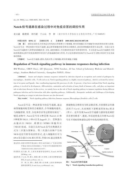
Notch信号通路在感染过程中对免疫应答的调控作用麦文豪陈楚溪刘巧媛宁云山李妍(南方医科大学检验与生物技术学院,广州 510515)中图分类号R392.12 文献标志码 A 文章编号1000-484X(2024)04-0872-08[摘要]感染启动的先天性和适应性免疫应答依赖于巨噬细胞、树突状细胞以及T细胞等对病原体的识别与控制。
Notch信号是一种高度保守的信号通路,通过相邻细胞间配受体结合被激活,进而协调细胞的重要生命过程。
目前已证实Notch信号通路参与多种免疫细胞发育、分化、成熟和激活,并在感染性疾病中发挥重要作用。
本文综述Notch信号通路在不同病原体感染过程中的免疫调控作用及其与其他通路的相互作用,并讨论在感染性疾病中以Notch信号为靶点的治疗方法及面临的挑战。
[关键词]Notch信号通路;感染;免疫应答;巨噬细胞;树突状细胞;T细胞Regulation of Notch signaling pathway in immune responses during infectionMAI Wenhao, CHEN Chuxi, LIU Qiaoyuan, NING Yunshan, LI Yan. School of Laboratory Medicine and Biotech⁃nology, Southern Medical University, Guangzhou 510515, China[Abstract]Innate and adaptive immune responses initiated by infection depend on recognition and control of pathogens by macrophages, dendritic cells, T cells and so on. Notch signaling pathway is a highly conserved pathway, which is activated by interac‑tion of receptors and ligands, thus coordinating important life processes of cells. At present, it has been confirmed that Notch signaling pathway is involved in development, differentiation, maturation and activation of many kinds of immune cells, and plays an important role in infectious diseases. In this review, we mainly focus on the role of Notch signaling pathway in immune regulation during different pathogens infection and its interaction with other signaling pathways. Additionally, therapeutic methods and challenges of developing Notch signaling as a target in infectious diseases are also discussed.[Key words]Notch signaling pathway;Infection;Immune response;Macrophages;Dendritic cells;T cellsNotch信号是一种高度保守的信号通路,通过相邻细胞间配受体结合被激活,介导细胞间通讯,与细胞增殖、发育、分化和存活等过程密切相关[1]。

Notch信号通路在乳腺癌干细胞中的研究进展郭瑢;张瑾【摘要】Breast cancer stem cells (BCSC) are group of cells exhibiting self-renewal and multi-directional differentiation potentials. These cells have an important role in the occurrence, development, metastasis, and recurrence of breast cancer. In normal circumstances, the ability of mammary stem cells to differentiate and undergo self-renewal is governed by related signaling pathways. After this mechanism is destroyed, breast stem cells undergo abnormal differentiation, forming breast cancer stem cells that unlimitedly proliferate to develop into breast cancer. As research on BCSC increasingly deepens, regulation of BCSC by notch signaling and its crosstalk with several signaling pathways have drawn a great deal of attention in this field. This paper reports the signaling pathways of breast cancer stem cells and latest studies on this field to better understand the essential role of notch signaling pathway in the occurrence and development of breast cancer and corresponding clinical targeted therapy.%乳腺癌干细胞是一群具有自我更新及多向分化潜能的细胞,在乳腺癌的发生、发展以及转移、复发中起着极其重要的作用。

沉默Notch1基因促进人乳腺癌MCF-7细胞JNK1和p53磷酸化袁磊;陈旭东;范文娟;杨旭光;王建国【摘要】目的:探究沉默Notch1基因对人乳腺癌MCF-7细胞JNK1和p53磷酸化的影响.方法:选取人乳腺癌MCF-7细胞作为研究对象,构建shRNA-Notch1真核表达质粒用于转染MCF-7细胞使Notch1基因沉默.采用Western blotting方法检测MCF-7细胞Notch1、Hes-1、PUMA和NOXA蛋白的表达,JNK1和p53蛋白磷酸化水平以及caspase-3活化水平的改变.应用流式细胞术检测细胞凋亡和线粒体膜电位的变化.结果:人乳腺癌MCF-7细胞Notch1基因被沉默后,Notch1和Hes-1蛋白表达量明显减少(P<0.01),细胞凋亡率显著升高(P<0.01),JNK1和p53的磷酸化水平明显高于对照组(P<0.01),PUMA和NOXA表达量显著升高(P<0.05),cleaved caspase-3蛋白明显多于对照组(P<0.01),线粒体膜电位明显下降(P<0.05).结论:沉默Notch1基因可能通过激活JNK1信号通路活化p53,促进PUMA和NOXA蛋白表达,进而通过线粒体途径导致人乳腺癌MCF-7细胞凋亡.%AIM:To investigate the effect of Notch1 gene silencing on phosphorylations of JNK1 and p53 in human breast cancer MCF-7 cells.METHODS:shRNA-Notch1 eukaryotic expression plasmid was constructed and transfected into MCF-7 cells.The expression of Notch1 and Hes-1 was observed by Western blotting after transfction.Apoptosis and mitochondrial membrane potential were detected by flow cytometry.Western blotting was also used to determine the protein levels of p-JNK1,p-p53,PUMA,NOXA and cleaved caspase-3 after Notch1 silencing was performed in MCF-7 cells.RESULTS:Silencing of Notch1significantly reduced the expression of Notch1 and Hes-1 in MCF-7 cells (P <0.01).In shNotch1 group,the number of apoptotic cells was much higher (P < 0.01) and mitochondrial membrane potential was much lower (P <0.05) than those in shControl group.The protein levels of p-JNK1,p-p53,PUMA,NOXA and cleaved caspase-3 increased obviously after silencing of Notch1 was performed in MCF-7 cells (P <0.05).CONCLUSION:Notch1 silencing induces apoptosis of human breast cancer MCF-7 cells through promoting phosphorylations of JNK1 andp53,and increasing the production of PUMA,NOXA and cleaved caspase-3.【期刊名称】《中国病理生理杂志》【年(卷),期】2013(029)006【总页数】6页(P1014-1019)【关键词】Notch1蛋白;短发夹RNA;JNKI蛋白;p53蛋白;MCF-7细胞【作者】袁磊;陈旭东;范文娟;杨旭光;王建国【作者单位】漯河医学高等专科学校,河南漯河462002;漯河医学高等专科学校,河南漯河462002;漯河医学高等专科学校,河南漯河462002;漯河医学高等专科学校,河南漯河462002;漯河医学高等专科学校,河南漯河462002【正文语种】中文【中图分类】R737.9近年来,在果蝇发育的研究中,发现了一条在细胞间传导相互作用的信号途径,称为Notch信号途径。

Notch信号转导通路在肝纤维化形成中的作用与分子机制陈方园1, 涂传涛2(1.复旦大学附属中山医院内科,上海 200032;2.复旦大学附属上海市公共卫生临床中心消化内科,上海 201508)摘要:慢性肝病及其并发症严重危害人类健康。
各种原因导致的慢性肝病可经过肝纤维化阶段最终进展至肝硬化,而肝纤维化甚至早期肝硬化是可逆转的。
因此,除强调针对肝病的基础病因防治外,临床中还迫切需要抗肝纤维化治疗。
然而,目前尚缺乏公认的行之有效的治疗药物。
因此,需要更深入地理解肝纤维化形成、进展和逆转的细胞分子机制,从而开发针对特异性靶点的药物,最终实现临床转化。
Notch信号转导通路在肝纤维化形成中的作用近年来倍受关注,其可能是抗肝纤维化治疗的新的潜在靶点,值得进一步探索。
本文就Notch信号转导通路在肝纤维化形成中的作用与分子机制进行综述。
关键词:肝纤维化;Notch信号转导通路;肝星状细胞;巨噬细胞;肝细胞;肝祖细胞The role and molecular mechanism of Notch signaling transduction pathway on liver fibrogenesisChen Fangyuan1, Tu Chuantao2(1.Department of Internal Medicine, Zhongshan Hospital, Fudan University,Shanghai 200032, China; 2.Department of Gastroenterology, Shanghai Public Health Clinical Center, FudanUniversity, Shanghai 201508, China)Abstract: Chronic liver disease and its complications endanger human health seriously. It is now clear thatchronic liver diseases of various causes eventually progress to cirrhosis through the stage of liver fibrosis.However, liver fibrosis and even early liver cirrhosis can be reversed. Therefore, beside the prevention andtreatment of the underlying causes of liver diseases, antifibrotic therapy is urgently needed. However, thereare currently no effective agents for liver fibrosis available. A greater understanding of molecular mechanismsregulating the liver fibrogenesis is needed for identification of novel targets for successful antifibrotic therapies.Recently, the role of Notch signaling transduction pathway in pathologic development of liver fibrosis hasreceived much attention, and current evidence suggests that the modulation of the Notch signaling transductionpathway may represent a new therapeutic target in liver fibrosis. This review highlights the recent advances inthe field indicating that Notch signaling transduction pathway is involved in the development of liver fibrosis.Key words: Liver fibrosis; Notch signaling transduction pathway; Hepatic stellate cell; Macrophage;Hepatocyte; Hepatic progenitor cells目前,慢性肝病仍是严重危害人类健康的重大疾病[1,2]。
小细胞肺癌中Notch1信号通路、ASCL1与EMT关系研究进展周怡然;辛宇;陈骏【摘要】小细胞肺癌(SCLC)是人类常见恶性肿瘤之一,其具有转移率和复发率高、对初始化疗敏感、易获得化学耐药等特征.上皮-间质转化(epithelial-mesenchymal transition,EMT)是肿瘤细胞发生侵袭、转移前重要的细胞表象,EMT的发生与多种细胞因子、信号传导通路及转录因子有关.尽管Notch途径激活在非小细胞肺癌(NSCLC)中起着致癌的作用,但在神经内分泌肿瘤(neuroendocrine tumors,NET)中,Notch通路激活抑制肿瘤生长.在SCLC中,Notch1通路激活可以诱导产生E-钙黏蛋白(E-Cadherin),增强细胞的粘附,诱导间质-上皮转化,从而抑制EMT过程.ASCL1(achaete-scute complex homologue 1)是SCLC一种系特异性基因,其编码的转录因子同样具有促进神经内分泌分化、EMT的功能.Notch1信号通路可以抑制ASCL1的转录,同时也可对ASCL1进行转录后调节.因此,可通过激活Notch1信号通路、抑制ASCL1表达从而抑制SCLC的发生、侵袭和转移.【期刊名称】《大连医科大学学报》【年(卷),期】2018(040)003【总页数】5页(P270-273,277)【关键词】SCLC;Notch1信号通路;ASCL1;EMT【作者】周怡然;辛宇;陈骏【作者单位】大连医科大学附属第二医院肿瘤科,辽宁大连116027;大连医科大学附属第二医院肿瘤科,辽宁大连116027;大连医科大学附属第二医院肿瘤科,辽宁大连116027【正文语种】中文【中图分类】R734小细胞肺癌(SCLC)是各种类型肺癌中恶性程度最高的肿瘤,约占所有肺癌的15%~20%,其5年生存率<6%[1-2]。
过去几十年中,SCLC的一线化疗方案仍以顺铂或卡铂加依托泊苷为主,尽管其暂时对放化疗敏感,但很快出现复发和转移[3-5]。
Notch1 的异常激活与T细胞型急性淋巴细胞白血病闫慧;刘兰波;莫茜【摘要】Notch1受体的异常激活在T细胞型急性淋巴细胞白血病(T-ALL)的发病中起主导作用.近期研究表明,超过60%的T-ALL患者中存在Notch1的激活性突变.文章简要介绍Notch1异常激活在T-ALL发病中的作用,以及针对Notch1异常激活的现有方法及其面临的问题,为T-ALL的研究及靶向治疗提供参考.%Abnormal activation of Notch1 plays pivotal roles in the molecular pathogenesis of human T-cell acutelymphoblastic leukemia (T-ALL). Activating Notch1 mutations present in over 60% of the T-ALL patients. However, so far,there is no therapy with little side effects that specifically targets the abnormally activated Notch1 pathway-induced T-ALL. Thepresent study briefly reviewed the effects of abnormal activation of Notch1 in the pathogenesis of T-ALL, as well as the currentapproaches targeting Notch1 and its limitations, thus providing some guidance for the research and development of clinicaltherapies targeting T-ALL.【期刊名称】《临床儿科杂志》【年(卷),期】2015(033)005【总页数】3页(P483-485)【关键词】Notch1;T细胞型急性淋巴细胞白血病;靶向治疗【作者】闫慧;刘兰波;莫茜【作者单位】上海交通大学医学院附属上海儿童医学中心上海 200127;上海交通大学医学院附属上海儿童医学中心上海 200127;上海交通大学医学院附属上海儿童医学中心上海 200127【正文语种】中文急性淋巴细胞白血病(acute lymphoblastic leukemia,ALL)是儿童和成人中最常见的恶性肿瘤之一,根据免疫分型可将其划分为T细胞型和B细胞型[1]。
ONCOLOGY REPORTS 26: 925-930, 2011Abstract. Glioblastoma is the most malignant form of adult brain tumor and is associated with a dismal prognosis. Emerging data suggest that Notch signaling participates principally in the formation and malignant progression of glioblastoma. Resveratrol is a terpenoid that exhibits broad pro-apoptotic activity in various types of cancers, including glioblastoma. However, the effects of resveratrol on Notch signaling in glioblastomas have not yet been fully elucidated. We demonstrated that resveratrol strongly suppressed cell growth and induced apoptosis in A172 and T98G glioblas-toma cells, which have low active Notch-1 expression and a heterozygous p53 mutation. Our results suggest that resveratrol significantly activates intracellular Notch-1 and restores wild-type p53 expression in a time-dependent manner. Significant de-phosphorylation of Akt, increased Bax expression, decreased Bcl-2 expression and cleavage of caspase-3 were also observed in resveratrol-induced apoptosis in glioblastoma cells. Moreover, simultaneous treatment with resveratrol and a Notch-1 inhibitor (MRK-003) partially attenuated the apoptosis and completely blocked the activation of Notch-1 and the increase in wild-type p53. This suggests that restoration of wild-type p53 expression depends on Notch-1 activation. In addition, the de-phosphorylation of Akt, increased expression of Bax and cleavage of caspase-3 were not fully reversed by MRK-003 treatment, suggesting that p53 restora-tion is not the only mechanism underlying resveratrol-induced apoptosis. Taken together, we confirmed the anti-proliferative and pro-apoptotic effects of resveratrol on glioblastoma cells and revealed Notch-1 activation-dependent restoration of p53 as an important causative mechanism.IntroductionGlioblastoma multiforme (GBM, WHO IV) is the most common adult intracranial malignancy which is characterized by rapid tumor proliferation and a strong tendency to diffuse and invade surrounding normal brain tissue (1). Despite ongoing improvements in conventional therapeutic regimens, including selective surgical resection and concurrent radiation, the prognosis for glioblastoma patients remains extremely dismal (2). The median survival and median progression-free survival for glioblastoma patients who received radiotherapy alone are 12 and 5 months, respectively (3). Concomitant and adjuvant chemotherapies have been shown to improve the progression and quality of life of glioblastoma patients; however, many glioblastoma patients do not benefit from these clinical interventions due to their minimal responses to anti-cancer agents that promote apoptosis (type I-programmed cell death). Therefore, identification of the genetic character-istics of glioblastomas that contribute to this low response is critical, as is the development of novel therapeutic agents and adjuvants that can effectively induce substantial apoptosis in glioblastoma cells (4).Emerging evidence suggests that tumor development and progression result from aberrant regulation of oncogenes and cancer-suppressor genes. With respect to genetic alterations, glioblastoma cells are known to exhibit aberrant expression of Notch-1 (5). Since it has been documented that overexpression of Notch-1 is fundamental for promoting malignant progres-sion and maintaining the self-renewal potential in various types of cancers, its potential role in the development and oncobiology of glioblastoma is intriguing. Previous studies have revealed that the Notch-1 protein gradually increases in gliomas according to pathological grade as compared to non-neoplastic brain tissue (6). Notch-1 expression in glioblas-tomas is lower than that in low-grade gliomas (7). However, its activated fragment, Notch-1 receptor intracellular domain (Notch-1ICD) and ligands, Jagged 1 (JAG1) and Delta-like 1 (DLL1), are endogenously expressed in glioblastomas (8). Given that Notch-1 activation depends on cell type andNotch-1 activation-dependent p53 restoration contributesto resveratrol-induced apoptosis in glioblastoma cellsHONG LIN*, WEI XIONG*, XIANG ZHANG, BOLIN LIU, WEI ZHANG,YONGQIANG ZHANG, JINXIANG CHENG and HUIYONG HUANGDepartment of Neurosurgery, Xijing Institute of Clinical Neuroscience,Xijing Hospital, Fourth Military Medical University, 710032 Xi'an, P.R. ChinaReceived April 27, 2011; Accepted June 11, 2011DOI: 10.3892/or.2011.1380Correspondence to: Dr Xiang Zhang, Department of Neurosurgery,Xijing Institute of Clinical Neuroscience, Xijing Hospital, FourthMilitary Medical University, 710032 Xi'an, P.R. ChinaE-mail: xiangzhang_fmmu@*Contributed equallyAbbreviations: IRS, immunoreactivity scores; Notch-1ICD, Notch-1receptor intracellular domainKey words: glioblastoma, resveratrol, Notch-1, p53, apoptosisLIN et al: RESVERATROL INDUCES APOPTOSIS BY RESTORING THE NOTCH-1-p53 SIGNALING PATHWAY 926context, the exact role of Notch-1 signals in the response of glioblastoma cells to pro-apoptotic agents cannot be easily predicted.Tumor protein 53 (p53) plays a primary role in control-ling carcinogenesis and helps to determine the response of cancer cells. Its tumor-suppressor function has been well documented in a number of epidemiological investigations and experimental studies in past years. The intracellular function of p53 is decreased by mutations or post-translational inactiva-tion. Attenuation of functional p53 enables the translation and activation of many pro-survival genes, such as protein kinase B (PKB/Akt), which promote the development of a malignant phenotype and apoptotic resistance in cancer cells (9). Low levels of functional p53 are characteristic of genetic alterations in several cancer types including glioblastoma (10). Immunohistochemical studies suggest that nearly half of the p53-positive cells in glioblastomas show high levels of p53 in the cytoplasm, where it is inactive. Cytoplasmic retention and inactivation predominantly account for the low levels of functional p53 in glioblastoma cells (11). Resveratrol (trans-3,4',5-trihydroxystilbene) is a phenolic compound extracted from Polygonum cuspidatum and red grape skin. Resveratrol exhibits anti-oxidant and anti-cancer activities and has been used to experimentally treat several types of cancers (12,13). Previous studies have revealed that resveratrol inhibits tumor cell growth, arrests the cell cycle and induces apoptosis in glioblastoma U251MG and C6 cells (14). However, more studies are required to investigate the underlying mechanisms before resveratrol can be used in clinical practice.The present study confirms resveratrol-induced apoptosis in human glioblastoma A172 and T98G cells. The expression and activation of Notch-1, wild-type p53, p-Akt, Bax and Bcl-2 were analyzed to determine the genetic features of glioblas-toma cells and the molecular mechanisms by which resveratrol exerts its anti-cancer activity in this cell type. Materials and methodsAgents. Resveratrol (Santa Cruz Biotechnology, Santa Cruz, CA) was dissolved in dimethyl sulfoxide (DMSO) (Gibco/ Invitrogen, NY, USA) to produce a 100-mM stock solution. Before each experiment, a resveratrol stock solution was dissolved in fresh Dulbecco's modified Eagle's medium (DMEM) (Sigma Chemical Co., St. Louis, USA) to obtain 50 and 100 µM working solutions. The Notch-1 inhibitor MRK-003 (BioSun Sci & Tech Co., Ltd., Shanghai, China) and a γ-secretase inhibitor were dissolved in DMSO and then added to the culture medium to obtain working solutions (5 µM). The final DMSO concentration used in the working solutions did not exceed 1‰ (v/v), so cell growth was not affected.Cell lines and cell culture. Human glioblastoma cell lines (A172 and T98G; cell bank at the Fourth Military Medical University, China) were cultured in DMEM supplemented with 10% fetal bovine serum (FBS) (BioSun Sci & Tech Co., Ltd.) in a 37˚C incubator with a humidified atmosphere of 5% CO2-95% O2. Primary cultured astrocytes were obtained from a brain tissue fragment from an informed and consenting volunteer with cerebral trauma under the approval of the local medical research ethics committee. Twenty-four hours before each experiment, cells were transferred to serum-free medium. Working solutions of resveratrol were then added in place of the culture medium.MTT assay. Cell viability was determined by the methyl thiazolyl tetrazolium (MTT) assay. Briefly, A172 and T98G glioblastoma cells and primary-cultured astrocytes were cultured in different 96-well plates (Greiner Bio-One GmbH, Frickenhausen, Germany) at a density of 5x10³ cells/ well. For the treatment groups, resveratrol was added to the culture medium at final concentrations of 50 and 100 µM. In the Notch-1 inhibition experiments, MRK-003 (5 µM) was added to the culture medium in combination with resvera-trol (50 µM). After incubating the cells for 24 or 72 h, MTT (Sigma), dissolved in PBS (0.01 M, pH 7.4), was added to each well at a final concentration of 5 mg/ml. All experiments were performed in triplicate.Hoechst 33342 nuclear staining. Briefly, cells (2x10⁴) were plated in 6-well plates and treated with resveratrol (50 µM) for 24 h. Cells were then washed in PBS (0.01 M, pH 7.4) and fixed in 70% ethanol for 2 h at 4˚C. Cell nuclei were stained with Hoechst 33342 (5 µg/ml; Sigma). After a final wash in PBS, changes in nuclear morphology were visualized by fluores-cence microscopy (Leica Microsystems, Wetzlar, Germany) using excitation wavelengths between 330 and 380 nm.Annexin V/propidium iodide. To estimate the apoptosis induced by resveratrol, we performed flow cytometric analyses as previously described (15). Briefly, cells (1x10⁵) were plated in a cell culture flask and were treated with resveratrol (50 µM) for 24 h. For the Notch-1 inhibition experiments, MRK-003 (5 µM) was added to the culture medium in combination with resveratrol (50 µM).Western blotting. Western blot analyses were performed as previously described (15). Equivalent amounts (25 µg) of protein lysates were separated in each cell line. The following primary antibodies were used: anti-Notch-1 (diluted 1:300, mouse monoclonal), anti-Akt-1 (diluted 1:1000, mouse monoclonal), anti-p-Akt-1 (Ser 473, diluted 1:300, rabbit polyclonal), anti-p53 (C-11, diluted 1:300, mouse monoclonal), anti-Bax (diluted 1:600, rabbit polyclonal), anti-Bcl-2 (diluted 1:600, rabbit polyclonal), anti-cleaved caspase-3 p11 (diluted 1:200, goat polyclonal) and anti-β-actin (diluted 1:1000, mouse monoclonal). The following secondary antibodies were used: HRP-conjugated anti-goat IgG (diluted 1:2000), HRP-conjugated anti-rabbit IgG (diluted 1:2000) and HRP- conjugated anti-mouse IgG (diluted 1:2000). All antibodies were purchased from Santa Cruz Biotechnology.Immunocytochemistry. Immunocytochemistry and evaluation of immunoreactivity scores (IRS) for nuclear p53 were performed as previously described (16). A172 and T98G cells were treated with or without resveratrol (50 µM) for 24 h and cultured on glass slides. The primary antibody was anti-p53 mouse monoclonal antibody (1:50), and the secondary antibody was biotinylated goat anti-mouse IgG (1:50). Both antibodies were purchased from Santa Cruz Biotechnology.ONCOLOGY REPORTS 26: 925-930, 2011927Statistical analysis. Data are expressed as the mean ± standard error of the mean (SEM) of separate experiments. All data were tested for significance by one-way analysis of variance (ANOVA) followed by Fisher's post hoc test using SPSS 13.0 software (IBM SPSS, Chicago, IL, USA). p<0.05 was considered statistically significant.ResultsResveratrol suppresses the cell viability of glioblastoma cells but does not affect primary cultured astrocytes. The cell viabi-lity and proliferative capacity of glioblastoma A172 and T98G cells were significantly decreased by resveratrol treatment in a concentration-dependent manner. The inhibitory effect was more pronounced in A172 cells than in T98G cells. Exposure to resveratrol (100 µM) for 24 h decreased A172 and T98G cell viabilities to 42.3±0.4 and 65.8±0.6% compared to the vehicle-treated controls, respectively (Fig. 1A). Resveratrol treatment did not affect cell viability in primary cultured astrocytes. Simultaneous inhibition of Notch-1 activation via MRK-003 treatment partially rescued the resveratrol-induced inhibition of glioblastoma cell growth. While exposure to resveratrol (50 µM, 24 h) treatment alone reduced A172 and T98G cell viabilities to 54.7±0.8 and 78.4±0.6%, respectively, treatment with MRK-003 (5 µM) significantly attenuated the resveratrol (50 µM)-induced inhibitory effect to just 69±0.5 and 90±0.4%, respectively (Fig. 1B). Simultaneous MRK-003 (5 µM) and resveratrol (50 µM) treatment for 24 h did not affect astrocyte cell growth.Resveratrol induces apoptosis in glioblastoma cells. After treatment with resveratrol (50 µM) for 24 h, glioblastoma cells experienced apoptotic morphological alterations. Many glioblastoma A172 cells had disrupted cytoskeletons, which were round and contracted, and the residual viable cells displayed shortened and narrowed synapses (Fig. 2A). Morphological alterations in the T98G cells were similar to those of the A172 cells; however, the affected population was smaller and the degree was not as severe. Hoechst 33342 nuclear staining revealed that resveratrol exposure significantly condensed the nucleus in both glioblastoma cell lines (Fig. 2B).Flow cytometric analyses were performed using an Annexin V-FITC/PI staining kit to analyze the apoptosisinduced by resveratrol. Treatment with resveratrol (50 µM) for 24 h induced apoptosis in 34±3.4 and 21±2.8% of the A172 and T98G cells, respectively. In the Notch-1 inhibition experiments, treatment with both resveratrol (50 µM) and MRK-003 (5 µM) induced apoptosis in only 17±1.6 and12±2.4% of the A172 and T98G cells, respectively (Fig. 3A).Figure 1. Effects of resveratrol on glioblastoma and astrocyte cell viabilities. (A) Resveratrol inhibited cell viability in glioblastoma A172 and T98G cells in a concentration-dependent manner but rarely affected cell viability in astrocytes. (B) The Notch-1 inhibitor MRK-003 (5 µM) significantly attenuated the resveratrol (50 µM)-induced inhibitory effect on glioblastomaA172 and T98G cell viabilities.Figure 2. (A and B) Effects of resveratrol (50 µM, 24 h) on cellular and nuclear structures in glioblastoma cells. (A) Resveratrol shortened and narrowed neurites and induced contraction and condensation in glioblastoma A172 and T98G cells. Co-treatment with the Notch-1 inhibitor MRK-003 (5 µM) decreased the number of apoptotic glioblastoma cells and ameliorated their morphological alterations. (B) Resveratrol treatment induced condensed and bud-forming apoptotic nuclei and increased the diopter in glioblastoma cells. MRK-003 (5 µM) lessened the morphologic alterations of the apoptotic nuclei. (C and D) Effects of resveratrol (50 µM, 24 h) on p53 expression and nuclear location. (C) Resveratrol significantly increased p53 expression and nuclear localization in glioblastoma A172 and T98G cells. The Notch-1 inhibitor MRK-003 (5 µM) partially inhibited these effects. (D) Immunoreactivity scores for nuclear p53 in glioblastoma cells treated with or without resveratrol.LIN et al : RESVERATROL INDUCES APOPTOSIS BY RESTORING THE NOTCH-1-p53 SIGNALING PATHWAY928This suggests that inhibition of Notch-1 activation significantly attenuates resveratrol-induced apoptosis (Fig. 3B).Resveratrol promotes nuclear translocation of p53 in A172 and T98G cells. To determine whether p53 plays a functional role in the response to resveratrol exposure, the subcellular distribution of the p53 protein in glioblastoma cells was investigated by immunocytochemistry (Fig. 2C and D). Weak p53 expression was observed mainly in the cytoplasm of the vehicle-treated glioblastoma A172 and T98G cells. After resveratrol treatment (50 µM) for 24 h, p53 expression was significantly increased in both glioblastoma cell lines. Data indicated that p53 accumulated in the nucleus of apoptotic glioblastoma cells (IRS = 10±1 in A172 cells; IRS = 6.33±1.45 in T98G cells). These results suggested that p53 function is restored in resveratrol-induced apoptosis. In addition, simulta-neous MRK-003 treatment significantly rescued the nuclear accumulation of p53 in both cell types (IRS = 7±1 in A172 cells; IRS = 5.33±0.67 in T98G cells).Resveratrol increases active Notch-1 and p53 expression in glioblastoma cells. To further explore the molecular mechanisms underlying the effect of resveratrol on glioblas-toma cells, we quantified changes in the expression levels of active Notch-1 and p53 during treatment. After treatment with resveratrol (50 µM) for 24 h, expression of both active Notch-1 and p53 was significantly increased (Fig. 4A). Western blot analysis revealed that the expression level of the Notch-1 intracellular domain (Notch-1ICD , functional form of Notch-1) was 3.2-fold (p=0.001) and 2.9-fold (p=0.001) higher in the resveratrol-treated A172 and T98G cells, respectively, compared to the vehicle-treated controls (Fig. 4B). Expression of p53 was 1.2-fold (p=0.04) and 1.1-fold (p=0.03) higher in the resveratrol-treated A172 and T98G cells, respectively, compared to the vehicle-treated controls. In addition, exposure to MRK-003 completely blocked the expression of Notch-1ICD and significantly reversed the increase in p53 expression. These results suggest that activation of the Notch-1 pathway and enhanced expression of p53 occur during resveratrol-induced apoptosis in glioblastoma cells.Resveratrol decreases Akt-1 phosphorylation and Bcl-2 expression and increases Bax expression in glioblastoma cells. Akt-1 was significantly de-phosphorylated in the resveratrol-treated glioblastoma cells compared to the vehicle-treated controls (Fig. 4A). As early as 12 h after treatment with resveratrol (50 µM), phosphorylated Akt-1 (Ser 473) protein expression began to decrease; it remained low for the rest of the experiment. Moreover, pro-apoptotic Bax protein expression was up-regulated, and the pro-survival Bcl-2 protein expression was down-regulated in resveratrol-induced apoptosis. A colorimetric assay was then performed using BandScan 4.3 software (Glyko, Inc., USA), with protein levels standardized to the β-actin loading control. The p-Akt-1/β-actin ratio was reduced to 17.2 and 35.8% in the A172 and T98G cells, respectively, after treatment with resveratrol (50 µM) for 24 h. In addition, resveratrol exposure reduced the Bcl-2/ β-actin ratio to 18.7 and 26.4% in the A172 and T98G cells, respectively. Resveratrol increased the Bax/β-actin ratio to 523 and 636% in the A172 and T98G cells, respectively. The p-Akt-1/β-actin, Bcl-2/β-actin and Bax/β-actin ratios of the vehicle-treated control cells were set to 100%.Resveratrol induces cleavage and activation of caspase-3 in glioblastoma cells. Cleavage and activation of caspase-3 is one of the most important mechanisms of apoptosis. An increase in the active 11-kDa caspase-3 fragment was noted in the resveratrol-treated glioblastoma cells (Fig. 4A). Western blot analysis indicated that in response to resveratrol treatment, pro-caspase-3 became cleaved and produced active fragments within 24 h. The colorimetric assay indicated that the cleaved caspase-3/β-actin ratio increased by 460 and 338% in the A172 and T98G cells, respectively. The cleaved caspase-3/β-actin ratio of the vehicle-treated control cells was set to 100%.DiscussionOur results confirmed the therapeutic potential of resvera -trol in glioblastoma A172 and T98G cells. Treatment with resveratrol (50-100 µM) significantly inhibited cell viabilityand induced apoptosis in both glioblastoma cell lines within 24 h. Resveratrol treatment also resulted in an increased expression of active Notch-1 in both cell lines. Furthermore, increased expression and nuclear translocation of wild-type p53, de-phosphorylation of Akt-1, up-regulated expression of the pro-apoptotic Bax protein and down-regulated expressionof the pro-survival Bcl-2 protein were also demonstrated. InFigure 3. Flow cytometric analysis. (A) Resveratrol (50 µM, 24 h) induced pronounced apoptosis in glioblastoma A172 and T98G cells. The Notch-1 inhibitor MRK-003 (5 µM) partially rescued this apoptosis. (B) Resveratrol significantly induced early and late apoptosis in both glioblastoma cell lines. Simultaneous MRK-003 (5 µM) treatment reduced the number of cells exhibiting early and late apoptosis.ONCOLOGY REPORTS 26: 925-930, 2011929addition, MRK-003 significantly blocked Notch-1 activation and partially reversed resveratrol-induced apoptosis. Taken together, these data suggest that Notch-1 activation and restoration of functional p53 play critical roles in the effect of resveratrol on glioblastoma.The polyphenolic resveratrol is a natural product which has attracted much interest for its use as a therapy for multiple cancer types as it has promising growth-inhibitory potency in cancer cells and rarely harms the surrounding normal parenchyma. Previous studies have suggested that, at therapeutic doses, resveratrol has minimal cytotoxic effects and is protective in neurons, human lens epithelial cells, human endothelium and mouse fibroblast 3T3 cells (14). Our study also found that resveratrol did not affect the cell viability of primary cultured astrocytes, which are substantially distributed in normal brain parenchyma. However, despite the anti-cancer properties and biologic safety of resveratrol, its functional mechanisms are still not fully understood.Research in the past two decades has revealed that resvera-trol induces apoptosis in cancer cells through dually regulating pro-survival oncogenes and pro-apoptotic cancer-suppressor genes. On the one hand, resveratrol causes loss of mitochondrial membrane potential, release of cytochrome C, and activation of pro-apoptotic proteins such as Bax and caspases; however, it also antagonizes pro-survival proteins such as Bcl-2. Our results are concordant with this research and demonstrate that resveratrol treatment down-regulates Bcl-2 expression and up-regulates Bax expression. In addition, previous studies have also suggested that modulation of Notch signaling is an important mechanism in the anti-cancer effect of resveratrol. Resveratrol affects Notch signaling at the transcriptional and post-transcriptional levels (affecting the proteolytic cleavage-dependent nuclear translocation and activation). It has been shown that resveratrol induces Notch-2 mRNA expression and promotes profound growth inhibition and apoptosis in medullary thyroid cancer cells (17) and human GI carcinoid BON cells (8). It has also been shown that resveratrol represses Notch-1 activation in human T-cell acute lymphoblastic leukemia MOLT-4 cells (19).Dysregulated Notch signaling has been shown to partici-pate in the apoptosis-survival switch in certain cancer cells. In glioblastoma, activation of Notch-1 has been suggested to predict poor prognosis in glioblastoma patients (20). Inhibition of Notch-1 signaling by RNA interference (RNAi) or γ-secretase inhibitors (GSIs) experimentally blocks cell growth and induces apoptosis in glioblastoma cells and inhibits repopulation and tumorigenesis in glioblastoma-derived neurospheres. Furthermore, Notch-1 inhibition also leads to enhanced chemosensitivity of glioblastoma cells to temozolo-mide treatment (21). However, strengthened activation of Notch-1 in glioblastoma A172 and T98G cells was noted during resveratrol-induced apoptosis, evidenced by increased expression of Notch-1ICD . To investigate whether this increase in Notch-1 signaling acts as an initiating factor in resveratrol-induced apoptosis or as a self-protective event to antagonize resveratrol stimulation, A172 and T98G cells were treated with resveratrol and the GSI MRK-003 simultaneously. This treatment completely blocked the activation of Notch-1 and partially rescued the apoptosis induced by resveratrol in both A172 and T98G cells, suggesting a critical role for Notch-1 activation in resveratrol-induced apoptosis. Prior studies have demonstrated a close association between Notch-2 activation and apoptosis and found that Notch-1 signaling minimally contributes to apoptosis in glioblastoma cells (6). The present results suggest an alternative mechanism for resveratrol-induced apoptosis in glioblastoma cells. In our preliminaryFigure 4. Effects of resveratrol (50 µM, 24 h) on protein expression profiles in glioblastoma A172 and T98G cells. (A) Western blot analysis. (B) Relative expression of the Notch-1 intracellular domain (Notch-1ICD ) in glioblastoma cells treated with or without resveratrol.LIN et al: RESVERATROL INDUCES APOPTOSIS BY RESTORING THE NOTCH-1-p53 SIGNALING PATHWAY 930study, we investigated the activation of Notch-1 signaling in five untreated glioblastoma cell lines. A172 and T98G cells have less active Notch-1 than SHG44, U87MG and U251MG cells (data not shown). Glioblastomas are genetically heteroge-neous, and the expression of Notch receptors and ligands differs between individual glioblastoma cell lines. Therefore, it is possible that the contributions of Notch-1 and -2 signaling to cell growth and apoptosis primarily depend on their expression status. Given that A172 cells are more sensitive to resveratrol treatment than T98G cells, we concluded that glioblastoma cells with less active Notch-1 are more susceptive to resveratrol. However, since we measured changes in Notch-1 activation in only two glioblastoma cell lines, the association between resveratrol-induced apoptosis and Notch-1 activation should be confirmed in more glioblastoma cell lines and primary glioblastoma cultures from clinical specimens with less active Notch-1.Since p53 is one of the major cancer-suppressor genes and its restoration is an important mechanism for various anti-cancer agents, p53 expression and activation were investi-gated in resveratrol-treated glioblastoma cells. This study suggests that resveratrol significantly increases intracellular p53 content and promotes its nuclear translocation. Our results indicate that the restoration and activation of p53 are important events in resveratrol-induced apoptosis. To investigate the possible association between Notch-1 activation and p53 restoration, p53 expression and activation were compared in glioblastoma cells treated with or without the Notch-1 inhibitor MRK-003 in combination with resveratrol. Simultaneous MRK-003 treatment slightly decreased p53 restoration in both A172 and T98G cells and significantly inhibited the nuclear translocation of p53, especially in A172 cells. These data suggest that Notch-1 activation can augment p53 expression and restore p53 function in glioblastoma A172 and T98G cells. Activation of the Notch-1-p53 signaling pathway appears to be an initiating factor in resveratrol-induced apoptosis, with augmented Bax expression and decreased Bcl-2 expression promoting the activation of the caspase cascade and eventual apoptosis.The present study suggests that resveratrol induces apoptosis in a sub-population of glioblastoma cells that have mutated p53 and express less active p53 by activating Notch-1. This novel mechanism differs from prior notions that i) induction of Notch-2, not Notch-1, plays a dominant role in the induction of apoptosis in glioblastoma cells by anti-cancer agents; and ii) inhibition but not induction of Notch-1 signaling represents a unique mechanism for inhibiting glioblastoma cell growth and promoting apoptosis. Our results suggest that the activated p53 protein, which was reinstated by Notch-1 activation, plays an important mediator in resveratrol-induced apoptosis in glioblastoma cells by up-regulating Bax expression, down-regulating Bcl-2 expression, de-phosphor-ylating Akt-1 and activating caspase-3. Taken together, we demonstrated the chemotherapeutic potential of resveratrol in glioblastomas and highlighted the importance of evaluating gene expression profiles prior to selecting a chemotherapeutic strategy. Further investigations are needed to improve our understanding of Notch signaling in glioblastomas and to determine the mechanisms underlying the anti-cancer activity of resveratrol.AcknowledgementsThe authors would like to thank Dr Xuewu Liu of the Department of Biochemistry, Fourth Military Medical University, China for helpful discussions.References1. Khasraw M and Lassman AB: Advances in the treatment of malignant gliomas. Curr Oncol Rep 12: 26-33, 2010.2. Jahraus CD and Friedman AH: Chemopotentiation by ultra-fractionated radiotherapy in glioblastoma resistant to conventional therapy. Tumori 96: 771-775, 2010.3. Stupp R, Mason WP, van den Bent MJ, et al: Radiotherapy plus concomitant and adjuvant temozolomide for glioblastoma. N Engl J Med 352: 987-996, 2005.4. Quant EC and Wen PY: Novel medical therapeutics in glio-blastomas, including targeted molecular therapies, current and future clinical trials. Neuroimaging Clin N Am 20: 425-448, 2010.5. Kanamori M, Kawaguchi T, Nigro JM, et al: Contribution of Notch signaling activation to human glioblastoma multiforme. J Neurosurg 106: 417-427, 2007.6. Lino MM, Merlo A and Boulay JL: Notch signaling in glio-blastoma: a developmental drug target? BMC Med 8: 72, 2010.7. Margareto J, Leis O, Larrarte E, Idoate MA, Carrasco A and Lafuente JV: Gene expression profiling of human gliomas reveals differences between GBM and LGA related to energy metabolism and Notch signaling pathways. J Mol Neurosci 32: 53-63, 2007.8. Li JL, Sainson RC, Shi W, et al: Delta-like 4 Notch ligand regulates tumor angiogenesis, improves tumor vascular function, and promotes tumor growth in vivo. Cancer Res 67: 11244-11253, 2007.9. Soussi T and Wiman KG: Shaping genetic alterations in human cancer: the p53 mutation paradigm. Cancer Cell 12: 303-312, 2007.10. Zhang WB, Wang Z, Shu F, et al: Activation of AMP-activated protein kinase by temozolomide contributes to apoptosis in glioblastoma cells via p53 activation and mTORC1 inhibition. J Biol Chem 285: 40461-40471, 2010.11. Berger B, Capper D, Lemke D, et al: Defective p53 antiangiogenic signaling in glioblastoma. Neuro Oncol 12: 894-907, 2010. 12. Schneider JG, Alosi JA, McDonald DE and McFadden DW: Pterostilbene inhibits lung cancer through induction of apoptosis. J Surg Res 161: 18-22, 2010.13. He X, Wang Y, Zhu J, Orloff M and Eng C: Resveratrol enhances the anti-tumor activity of the mTOR inhibitor rapamycin in multiple breast cancer cell lines mainly by suppressing rapamycin-induced AKT signaling. Cancer Lett 301: 168-176, 2011.14. Zhang W, Fei Z, Zhen HN, Zhang JN and Zhang X: Resveratrol inhibits cell growth and induces apoptosis of rat C6 glioma cells. J Neurooncol 81: 231-240, 2007.15. Lin H, Zhang X, Cheng G, et al: Apoptosis induced by ardipusilloside III through BAD dephosphorylation and cleavage in human glioblastoma U251MG cells. Apoptosis 13: 247-257, 2008.16. Lin H, Wang YG, Zhang X, Liu BL, Zhang W and Cheng JX: Prognostic significance of kappaB-Ras1 in gliomas. Med Oncol: Feb 8, 2011 (Epub ahead of print).17. Truong M, Cook MR, Pinchot SN, Kunnimalaiyaan M and Chen H: Resveratrol induces Notch2-mediated apoptosis and suppression of neuroendocrine markers in medullary thyroid cancer. Ann Surg Oncol 18: 1506-1511, 2011.18. Pinchot SN, Jaskula-Sztul R, Ning L, et al: Identification and validation of Notch pathway activating compounds through a novel high-throughput screening method. Cancer 117: 1386-1398, 2011.19. Cecchinato V, Chiaramonte R, Nizzardo M, et al: Resveratrol-induced apoptosis in human T-cell acute lymphoblastic leukaemia MOLT-4 cells. Biochem Pharmacol 74: 1568-1574, 2007. 20. Li J, Cui Y, Gao G, Zhao Z, Zhang H and Wang X: Notch1 is an independent prognostic factor for patients with glioma. J Surg Oncol: doi:10.1002/jso.21851, 2011.21. Gilbert CA, Daou MC, Moser RP and Ross AH: Gamma-secretase inhibitors enhance temozolomide treatment of human gliomas by inhibiting neurosphere repopulation and xenograft recurrence. Cancer Res 70: 6870-6879, 2010.。