卵巢癌组织中的表达状况及临床意义
- 格式:pdf
- 大小:499.11 KB
- 文档页数:5

AGR2在卵巢癌组织中表达及临床意义万敏;杨胜晗【摘要】目的:通过分析人前梯度蛋白2(AGR2)在不同卵巢组织中的表达及其与卵巢癌各临床病理参数间的关系,探讨AGR2在卵巢癌发生、发展中的作用.方法:采用免疫组化SP法检测AGR2在15例正常卵巢组织、20例卵巢良性肿瘤组织及50例卵巢癌组织中的表达情况,并结合卵巢癌各临床病理参数进行分析.结果:AGR2在卵巢癌组织中的表达水平明显高于卵巢正常组织和卵巢良性肿瘤组织(P<0.05);AGR2在卵巢癌组织中表达水平与患者的年龄、文化程度及病理类型无关(P>0.05),而与患者的临床分期、淋巴结转移密切相关(P<0.05).结论:AGR2有望成为卵巢癌早期诊断的一种新的肿瘤标记物.【期刊名称】《淮海医药》【年(卷),期】2017(035)006【总页数】3页(P639-641)【关键词】卵巢肿瘤;人前梯度蛋白2;免疫组织化学;肿瘤标志物【作者】万敏;杨胜晗【作者单位】安徽省蚌埠市第三人民医院妇产科,233000;安徽省蚌埠市第三人民医院妇产科,233000【正文语种】中文【中图分类】R737.31卵巢癌是女性生殖道常见的恶性肿瘤之一,其发病率仅次于子宫颈癌和子宫内膜癌而列居妇科肿瘤第3位,但死亡率却居第一位[1]。
由于卵巢位于盆腔深部,发病隐匿,目前又缺乏有效的检测手段,因此很难早发现和早诊断。
大部分卵巢癌患者确诊时已是晚期,仅有25%的患者早期被发现,而晚期卵巢癌患者的5年生存率较早期的95%降至20%~30%[2]。
近年来随着越来越多的肿瘤标志物被发现,使得早期卵巢癌的诊断率逐渐升高。
研究发现人前梯度蛋白2(anterior gradient 2,AGR2)与多种恶性肿瘤发生发展相关[3],被称谓“一个新的癌症诊断标记”[4],是目前研究的热点之一。
本资料旨在探讨AGR2在卵巢癌组织中的表达情况及其临床意义,以便为卵巢癌的早期诊断寻找新的分子生物学依据。
Nestin和Ki67在卵巢上皮性癌组织中的表达及临床意义段爱红;孙志强;杨丽萍;胡志强【摘要】目的探讨Nestin及Ki67在卵巢上皮性癌组织中的表达及其与临床病理特征的关系.方法对2007年1月~2009年1月邯郸市中心医院妇科60例卵巢癌患者及20例卵巢正常患者的病理资料进行回顾性分析,采用免疫组化SP法检测卵巢上皮性癌标本及正常组织中Nestin及Ki67的表达,分析并比较正常卵巢组织及不同病理类型、FIGO分期、残余肿瘤直径的卵巢组织中Nestin及Ki67的表达情况.结果①卵巢上皮性癌标本中Nestin和Ki67阳性表达率(38.5%、76.7%)明显高于正常卵巢组织(5.0%、15.0%),两者相比,差异均有统计学意义(均P<0.05).②组织分化程度高级别的卵巢癌组织中Nestin及Ki67阳性表达率明显高于低级别者(P< 0.05);FIGO分期越高、残余肿瘤直径越大,Nestin、Ki67阳性表达率越高(均P<0.05).③Nestin与Ki67在卵巢癌中的表达无明显相关性(r=0.176,P=0.13).结论 Nestin与Ki67的表达与卵巢癌的发生、发展有密切关系,对Nestin与Ki67的检测可用于判断卵巢上皮性肿瘤的恶性程度及其预后.【期刊名称】《中国医药导报》【年(卷),期】2014(011)005【总页数】4页(P24-27)【关键词】卵巢癌;Nestin;Ki67;免疫组化【作者】段爱红;孙志强;杨丽萍;胡志强【作者单位】河北省邯郸市中心医院妇科,河北邯郸 056001;河北省邯郸市中心医院神经外科,河北邯郸 056001;河北省邯郸市中心医院妇科,河北邯郸 056001;河北省邯郸市中心医院放射科,河北邯郸 056001【正文语种】中文【中图分类】R737.31卵巢上皮性癌(epithelial ovaian cance,EOC)简称卵巢癌,占卵巢恶性肿瘤的90%,是病死率最高的生殖道恶性肿瘤,卵巢癌属于对化疗中-高度敏感肿瘤,目前的标准治疗是肿瘤细胞减灭术辅以铂类为基础的6~8个疗程的联合化疗。
doi: 10.13241/ki.pmb.2020.19.034上皮性卵巢癌患者组织P53、K i67表达及其临床意义*青措吉艾小庆崔英杨晓燕董艳(青海红十字医院妇科青海西宁810000)摘要目的:探讨上皮性卵巢癌患者组织P53、K i67表达及其临床意义。
方法:选择卵巢肿瘤102例,其中病理诊断为良性卵巢肿 瘤40例(良性组)和上皮性卵巢癌62例(恶性组),采用免疫组化法检测组织P53、K i67表达水平,调查患者的临床病理特征并进行 相关性分析。
结果:恶性组的P53、K i67表达阳性率为80.6 %和72.6%,显著高于良性组的10.0%和12.5 %(P<0.05)。
在恶性组中,不同浸润转移、分化程度、病理分期患者的P53、K i67表达阳性率对比差异有统计学意义(P<0.05)。
直线相关分析显示上皮性卵巢 癌患者P53表达阳性率与K i67表达阳性率呈现显著正相关性(r=0.872,P=0.000)。
多因素logistic回归分析显示浸润转移、分化程 度、病理分期都为影响P53、K i67表达阳性率的主要因素(P<0.05)。
结论:上皮性卵巢癌患者组织P53、K i67都呈现高表达状况,与患者的临床病理特征显著相关,两者也可互相影响,共同参与上皮性卵巢癌的发生与发展。
关键词:上皮性卵巢癌;P53;K i67;病理特征;相关性中图分类号:R737.31文献标识码:A文章编号:1673-6273(2020)19-3757-04Expression of P53 and Ki67 in Patients with Epithelial Ovarian Cancer andIts Clinical Significance*QING Cuo-ji, AlXiao-qing, CUI Ying, Y A N G Xiao-yan, D O N G Yan(Department o f G ynecology, Qinghai R ed Cross Hospital, Xining, Qinghai, 810000, China)A B S T R A C T Objective: T o investigate the expression of P53 a nd K i67 in patients with epithelial ovarian cancer a nd i t s clinical significance. Methods: 102 patients with ovarian tumors wer e selected a nd treated, including 40 cases of benign ovarian tumors (benign group) and 62 cases of epithelial ovarian cancer (malignant group). T h e expression levels of P53 a nd K i67 wer e detected b y chemical method, and the clinicopathological features of the patients w ere investigated a nd correlation analysis were performed Results: T h e positive rates of P53 a nd K i67expression in the malignant group wer e 80.6 % a nd 72.6 %, w h i c h wer e significantly higher than those in the benign group (10.0 % an d 12.5 %)(P<0.05). In the malignant group, the positive rates of P53 an d K i67 expression in patients with different invasive metastasis, differentiation a nd pathological stage w ere statistically significant (P<0.05). Linear correlation analysis s h o w e d that the positive rate of P53 expression in patients with epithelial ovarian cancer w ere significantly positively correlated with the positive rate of K i67expression (r=0.872, P=0.000). Multivariate logistic regression analysis s h o w e d that infiltration, differentiation and pathological stage wer e the m a i n factors affecting the positive rate of P53 and K i67 expression (P<0.05). Conclusion: P53 and K i67 in patients with epithelial ovarian cancer have high expression status, w h i c h is significantly correlated with the clinicopathological features of the patients. T h e y can also affect each other a nd participate in the occurrence and development of epithelial ovarian cancer.K e y w o r d s: Epithelial ovarian cancer; P53; Ki67; Pathological characteristics; CorrelationChinese Library Classification(CLC): R737.31 D o c u m e n t code: AArticle ID: 1673-6273(2020)19-3757-04刖目上皮性卵巢癌是女性常见恶性肿瘤之一,由于卵巢位于盆 腔深部,起病隐匿,早期病变难以发现,约70%的患者在首次 就诊时已属晚期,使得生存率在30%以下M。
Nestin和Ki67在卵巢上皮性癌组织中的表达及临床意义作者:段爱红孙志强杨丽萍胡志强来源:《中国医药导报》2014年第05期[摘要] 目的探讨Nestin及Ki67在卵巢上皮性癌组织中的表达及其与临床病理特征的关系。
方法对2007年1月~2009年1月邯郸市中心医院妇科60例卵巢癌患者及20例卵巢正常患者的病理资料进行回顾性分析,采用免疫组化SP法检测卵巢上皮性癌标本及正常组织中Nestin及Ki67的表达,分析并比较正常卵巢组织及不同病理类型、FIGO分期、残余肿瘤直径的卵巢组织中Nestin及Ki67的表达情况。
结果①卵巢上皮性癌标本中Nestin和Ki67阳性表达率(38.5%、76.7%)明显高于正常卵巢组织(5.0%、15.0%),两者相比,差异均有统计学意义(均P < 0.05)。
②组织分化程度高级别的卵巢癌组织中Nestin及Ki67阳性表达率明显高于低级别者(P < 0.05);FIGO分期越高、残余肿瘤直径越大,Nestin、Ki67阳性表达率越高(均P < 0.05)。
③Nestin与Ki67在卵巢癌中的表达无明显相关性(r = 0.176,P = 0.13)。
结论 Nestin与Ki67的表达与卵巢癌的发生、发展有密切关系,对Nestin与Ki67的检测可用于判断卵巢上皮性肿瘤的恶性程度及其预后。
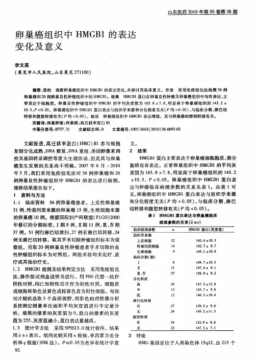
[关键词] 卵巢癌;Nestin;Ki67;免疫组化[中图分类号] R737.31 [文献标识码] A [文章编号] 1673-7210(2014)02(b)-0024-04Expression and clinical significance of Nestin and Ki67 in epithelial ovarian cancerDUAN Aihong1 SUN Zhiqiang2 YANG Liping1 HU Zhiqiang31.Department of Gynecology, Central Hospital of Handan City, Hebei Province, Handan 056001, China;2.Department of Neurosurgery, Central Hospital of Handan City, Hebei Province, Handan 056001, China;3.Department of Radiology, Central Hospital of Handan City, Hebei Province, Handan 056001, China[Abstract] Objective To investigate the expression of Nestin and Ki67 in epithelial ovarian cancer and the relationship between their expression and clinicopathologic characteristics. Methods pathological files of 60 patients with epithelial ovarian carcinoma and 20 patients with nomal ovarian tissues from January 2007 to January 2009 in Gynecology Department of Central Hospital of Handan City were retrospective analyzed, the Nestin and Ki67 were detected by immunohistochemistry (SP method), the expression of Nestin and Ki67 in nomal ovarian and epithelial ovarian cancer tissues with different pathological pattern, FIGO stages and residual tumor size were analyzed and compared. Results ①The positive expression rate of Nestin Ki67 in ovarian epithelial carcinomas (38.5%, 76.7%) were higher than those in normal ovarian tissues(5.0%, 15.0%), thedifferences were statistically significant(all P < 0.05). ②The positive expression of Nestin andKi67 in epithelial ovarian cancer tissues with high differentiation degree was higher than those in with low differentiation degree (all P < 0.05). The higher FIGO stage and larger residual tumor size,the higher positive expression of Nestin and Ki67 in epithelial ovarian cancer tissues. ③There was no significant correlation between Nestin and Ki67 expression (r = 0.176, P = 0.13). Conclusion The expressions of Nestin and Ki67 are associated with the development and progression of the ovarian epithelial neoplasm, the detection of Nestin and Ki67 may be helpful to estimate the malignant degree and prognosis of the epithelial ovarian carcinoma.[Key words] Ovarian cancer; Nestin; Ki67; Immunohistochemistry卵巢上皮性癌(epithelial ovaian cance,EOC)简称卵巢癌,占卵巢恶性肿瘤的90%,是病死率最高的生殖道恶性肿瘤,卵巢癌属于对化疗中-高度敏感肿瘤,目前的标准治疗是肿瘤细胞减灭术辅以铂类为基础的6~8个疗程的联合化疗。
mir-100在上皮性卵巢癌患者组织中的表达及临床意
义的探讨的开题报告
1. 研究背景:上皮性卵巢癌是最常见的卵巢恶性肿瘤之一,其发病
率和死亡率均居女性恶性肿瘤之首。
miRNA(microRNA)是一类重要的
非编码RNA,已被证实在癌症的发生和发展中发挥关键作用。
其中,mir-100被认为是具有抑制肿瘤作用的miRNA,在多种癌症中已有研究报道。
然而,关于mir-100在上皮性卵巢癌患者中是否具有潜在的临床意义仍缺乏深入研究。
2. 研究目的:探究mir-100在上皮性卵巢癌患者组织中的表达及其
与临床病理特征的关系,为进一步研究上皮性卵巢癌的病理生理机制提
供新思路和方法。
3. 研究方法:采集上皮性卵巢癌患者组织样本和对照组正常卵巢组
织样本,通过实时定量PCR技术检测mir-100的表达水平,并与患者的
临床资料(如年龄、病理分期、淋巴结转移等)进行比较分析。
同时,
建立上皮性卵巢癌细胞株,通过过表达或靶向抑制方法进一步探究mir-100对癌细胞增殖、转移和凋亡的影响。
4. 研究意义:mir-100可能成为上皮性卵巢癌的新的分子标志物和
治疗靶点,有望提高上皮性卵巢癌的诊断和治疗水平,为患者的生命质
量和生存期提供保障。
血管内皮生长因子在卵巢癌组织中的表达及临床意义作者:杨春秋来源:《中国实用医药》2012年第29期【摘要】目的研究血管内皮生长因子(VEGF)在卵巢癌组织中的表达以及分析其与年龄、病理分型、病理分级、临床分期及淋巴结转移的关系。
方法应用免疫组化S—P法对卵巢癌、卵巢良性肿瘤及正常卵巢组织中血管内皮生长因子的表达进行检测。
结果血管内皮生长因子在正常卵巢组织中的表达为阴性,在卵巢良性肿瘤中的阳性表达率为17.5%,在卵巢癌中阳性表达率为85%,卵巢良性肿瘤与正常卵巢组织比较,差异无统计学意义(P>0.05);卵巢癌与正常卵巢组织和卵巢良性肿瘤比较,差异具有统计学意义(P0.05),病理分级、临床分期及淋巴结转移与血管内皮生长因子在卵巢癌组织中的表达相关(P【关键词】卵巢癌;血管内皮生长因子;免疫组织化学卵巢癌是目前死亡率最高的妇科恶性肿瘤,临床就诊时约70%的患者已经出现转移[1]。
因此,及早的发现并诊断卵巢癌是降低其死亡率的关键。
相关的研究表明[2],肿瘤的生长和转移与血管的生长密不可分,血管内皮生长因子是目前所知道的促血管生成因子中最强的一种,与多种肿瘤的发生、发展和预后相关。
本研究通过探讨卵巢癌患者肿瘤组织中血管内皮生长因子的表达方式和特点,进一步明确血管内皮生长因子与卵巢癌的相关性。
1 资料与方法1.1 一般资料选取2010年2月至2011年2月我院手术切除卵巢癌组织标本40例,手术切除卵巢良性肿瘤组织标本40例,正常卵巢组织标本10例。
年龄29~68岁,平均年龄(47.5±13.4)岁。
病理分型:浆液性囊腺癌28例,黏液性囊腺癌12例;病理分级:G1级6例,G2级9例,G3级25例;FIGO分期:Ⅰ~Ⅱ期16例,Ⅲ~Ⅳ期24例;淋巴结转移19例。
所有患者术前均未服用非甾体类抗炎药,未进行过放疗及化疗。
均经病理明确诊断。
1.2 方法鼠抗人单克隆血管内皮生长因子抗体、链霉菌抗生物素蛋白—过氧化酶免疫组化染色(S—P)试剂盒、DAB显色试剂盒等均购自深圳晶美生物工程有限公司。
趋化因子在卵巢癌组织中的表达及其意义目的:探讨卵巢癌组织中趋化因子(RANTES)蛋白与mRNA的表达水平及临床意义。
方法:采用免疫组化法和原位杂交技术检测卵巢癌组织中RANTES 蛋白和mRNA表达,分析其与卵巢癌生物学特征之间的关系。
结果:RANTES 蛋白在卵巢癌及转移灶中的阳性率明显高于卵巢良性肿瘤,两组差异有统计学意义(P<0.05)。
RANTES mRNA在卵巢癌及卵巢良性肿瘤中的阳性率分别为75.55%和11.76%,两组差异有统计学意义(P<0.05)。
RANTES蛋白与mRNA 表达与癌的组织学分级及TNM分期有关(P<0.05)。
结论:RANTES蛋白在卵巢癌中有阳性表达,其表达水平与卵巢癌侵袭发展和淋巴结转移有关。
标签:卵巢癌RANTES 原位杂交免疫组织化学Expression and their biological significances of RANTES proteins and mRNA in human ovarian carcinomaZHENG Lihong, CHEN Ping, MEI Qingbu, LIU Feng, WU Nan(Qiqihar Medical College, Heilongjiang Province, Qiqihar 161006, China)[Absrtact] Objective: To study the expression of regulated on activation, normal T cell expressed and secreted (RANTES) protein and mRNA in human ovarian carcinoma (OC), evaluate the relationship between the expression of RANTES protein and the clinic pathology factors, and lymph node metastasis of OC and to analyze the clinical significance. Methods: Expression of RANTES was examined by immunohistochemistry in benign ovarian tumors and m alignant ovarian carcinoma. The expression of RANTES and mRNA in these specimens were detected by in-situ hybridization. The relationship between expression and biologic characteristic of ovarian carcinoma was analyzed. Results: The positive expression of RANTES protein in ovarian carcinoma was higher than that in benign ovarian tumors, there was a significant statistic difference between two groups (P<0.05). The expression rate of RANTES protein was closely correlated with the histological grade, lymphatic metastasis and TNM grade in ovarian carcinoma (P<0.05). The expression rates of RANTES mRNA in ovarian carcinoma and benign ovarian tumors were 75.55%, 11.76% respectively, there was a significant statistic difference between two groups (P <0.05). The expression rate of RANTES mRNA was closely correlated with the histological grade and TNM grade in ovarian carcinoma (P<0.05). Conclusion: Higher expression of RANTES may be related to development, invasion and metastasis of ovarian carcinoma.[Key words] Ovarian carcinoma; RANTES; In-situ hybridization; Immunohistochemistry卵巢癌(ovarian carcinoma,OC)是女性生殖系统常见恶性肿瘤。
四川生理科学杂志 2021, 43(4) 541·临床论著·卵巢癌患者癌组织中nm23-H1蛋白和N-cad 的表达及意义#戎凤敏* 秦海霞△张景航 张娇(新乡医学院第一附属医院妇科,河南 新乡 453100)摘要 目的:探讨卵巢癌患者癌组织中nm23-H1、神经型钙粘附蛋白(N-cadherin ,N-cad )表达及其临床意义。
方法:选取我院2011年1月至2013年12月间收治的118例卵巢癌患者,根据卵巢癌病理分期分为Ⅰ级(n=49)、Ⅱ级(n=40)、Ⅲ级(n=29),采用免疫组化法检测癌组织及癌旁组织中nm23-H1蛋白和N-cad 的表达情况,分析癌组织nm23-H1蛋白、N-cad 表达阳性率与卵巢癌病理分级、淋巴结转移间的关系。
结果:癌组织中nm23-H1蛋白表达阳性率明显低于癌旁组织(P<0.05),而N-cad 蛋白表达阳性率明显高于癌旁组织(P<0.05),其中病理分期Ⅰ级的患者卵巢癌组织中nm23-H1表达总阳性率最高(P<0.05),N-cad 表达总阳性率最低(P<0.05);转移者nm23-H1蛋白表达阳性率更低(P<0.05),N-cad 表达阳性率更高(P<0.05);nm23-H1蛋白低表达组浆液型腺癌比例高(P<0.05),nm23-H1蛋白低表达组和N-cad 高表达组病理分级Ⅲ级比例高(P<0.05)、FIGO 分期中Ⅲ~Ⅳ比例高(P<0.05)、3年生存率更低(P<0.05)。
结论:卵巢癌组织中nm23-H1及N-cad 表达与肿瘤细胞分化程度、癌症类型、癌症病情进展及转移均密切相关,可影响卵巢癌患者的生存期。
关键词:卵巢癌;nm23-H1蛋白;神经型钙粘附蛋白Expressions and significance of nm23-H1 and N-cad in ovarian cancer patients #Rong Feng-min*, Qin Hai-xia △, Zhang Jing-hang, Zhang Jiao(Department of Gynecology, The First Affiliated Hospital of Xinxiang Medical College, Xinxiang 453100, Henan, China)Abstract Objective: To explore the expressions and clinical significance of nm23-H1 and N-cadherin (N-cad) in ovarian cancer patients. Methods: Immunohistochemistry was used to detect the expressions of nm23-H1 protein and N-cadherin (N-cad) inovarian cancer tissues of 118 cases from January 2011 to December 2013. According to pathological stage of ovarian cancer, the patients were divided into grade I (n=49), grade II (n=40) and grade III (n=29). The positive expression rates of nm23-H1 protein and N-cad were compared between cancer tissues and adjacent tissues, and the relationship between positive expression rates of nm23-H1protein and N-cad and pathological grade and lymph node metastasis of ovarian cancer was analyzed. Results:The positive expression rate of nm23-H1 protein in cancer tissues was lower than that in adjacent tissues (P<0.05) while the positive expression rate of N-cad protein was higher than that in adjacent tissues (P<0.05). The total positive rate of nm23-H1 expression in grade Iovarian cancer tissues was the highest (P<0.05), and the total positive rate of N-cad expression was the lowest (P<0.05). The positive rate of nm23-H1 protein expression was lower (P<0.05) while the positive rate of N-cad expression was higher (P<0.05) among patients with metastasis. The proportion of serous adenocarcinoma was higher innm23-H1 low expression group(P<0.05), and the proportion of grade III in pathological grade was higher (P<0.05)and the proportion of III~IV in FIGO stage was higher(P<0.05) and the 3-year survival rate was lower (P<0.05) in nm23-H1 low expression groupand N-cad high expression group. Conclusion: The expressions of nm23-H1protein and N-cad in ovarian cancer tissues are related to the tumor cell differentiation degree, cancer types, cancer progression and metastasis, which may affect the survival time of patients with ovarian cancer. Key words: Ovarian cancer; nm23-H1 protein; N-cadherin卵巢癌是临床常见恶性肿瘤之一,发病率位居女性生殖系统恶性肿瘤第3位,死亡率位居妇科恶性肿瘤第1位[1]。
3本课题受国家自然科学基金资助(No 130560157);广西壮族自治区年“研究生科研创新资助项目”资助(N 15M 3)△通信作者收稿日期22Apoa Ⅱ基因在卵巢恶性肿瘤组织中的表达及其临床价值3许莉莉 王 琪 黎丹戎 张 玮 阳志军 潘忠勉 李 力△(广西医科大学附属肿瘤医院妇瘤科 南宁 530021)摘要 目的:探讨载脂蛋白A Ⅱ(Apoa Ⅱ)基因在卵巢恶性肿瘤组织中的表达及其临床意义。
方法:采用R T 2PCR 技术检测39例卵巢恶性肿瘤、16例良性肿瘤及18例正常卵巢组织Apoa ⅡmRNA 的表达情况,分析其与临床诊断、治疗及预后的关系。
结果:①卵巢恶性肿瘤表达Apoa Ⅱ基因,正常卵巢及良性肿瘤不表达Apoa Ⅱ基因,即Apoa Ⅱ基因与临床诊断有关(P <0105)。
②Apoa Ⅱ基因与患者年龄有关(P <0105),但Apoa Ⅱ基因与组织学类型、病理分期及预后无关(P >0105)。
结论:Apoa Ⅱ基因表达异常可能不是卵巢肿瘤细胞基因的改变,而是化疗引起机体代谢变化的结果或是先期化疗不足以引起卵巢肿瘤细胞分子水平的改变。
关键词 卵巢恶性肿瘤;载脂蛋白A Ⅱ;基因表达;RT 2PCR 中图分类号:R73714 文献标志码:A 文章编号:10052930X (2008)022*******THE EXP RESSIO N OF APO A ⅡGENE IN O VARIAN CARCINOMA AND ITS CL INICAL SIG 2NIFICANCEXu Lili ,Wang Qi ,Li Da nrong ,et al.(Depart ment of G ynecology a nd Obstet rics ,Affiliat ed Tumor Hospit al of Guangxi Medical U niversty ,Nanni ng 530021Chi na )Abstract Object ive :To i nvest igat e t he exp ression of Apoa Ⅱgene in ovaria n carci no ma cell s and different ovaria n t i ssue ,a nd explore it s cl inical si gnificance i n human ovarian cancer.Met hods :R T 2PCR wa s used to det ect t he expression of Apoa Ⅱm RNA i n 73pat ient s (18pati ent s wit h nor mal ovaria n ,16wi t h be ni gn o 2varian t ume r and 39pa tient s wi t h ovarian t umer ).The relation bet ween t he expression a nd t he clinical 2pat hological changes was analyzed stat ist ically.Resul t :Apoa Ⅱm RNA level s showed si gnificant l y higher i n t he pat ient s wit h ovarian t umor compa red to t he normal and benign t umor (P <0105),and t here were diff ere nces bet ween different ages (P <0105).On t he ot her ha nd ,no significant a ssociation was found be 2t ween Apoa Ⅱe xp re ssion and hi stological t ypi ng ,st ages and prognosis of ovarian carci noma.Conclusion :The cause of abnor mal expression of Apoa Ⅱin ovarian mali gna nt i s t hat met abolize change by chemot hera 2py ,but not i t s gene ,or neoadj uva nt chemot herapy doesn ’t make t he change of ovarian mali gna nt cell mole 2cule level s.K ey w or ds ovarian carci noma ;Apoa Ⅱ;gence expression ;R T 2PCR 卵巢癌死亡率占女性生殖系统恶性肿瘤的首位,至今发病机制尚未阐明,亦缺乏有效的早期诊断和治疗方法[1]。
miR—185在卵巢癌组织中的表达及其临床意义作者:王淑芬来源:《医学信息》2014年第01期摘要:目的探讨miR-185(Homo sapiens miR-185,miR-185)在卵巢癌组织中的表达及其临床意义。
方法运用qRT-PCR检测miR-185在卵巢癌及癌旁组织中的表达情况,分析其表达与卵巢癌临床分期及淋巴结转移间的关系。
结果 qRT-PCR检测显示,在37例卵巢癌组织中miR-185的表达值为(0.57±0.06),与癌旁对照组(1.02±0.11)相比,差异有统计学意义(P关键词:miR-185;卵巢癌;生长与侵袭miRNAs是一组长度约为22个核苷酸的非编码单链RNA,其作为基因表达的一个重要调控因子,通过对靶基因的翻译抑制和mRNA降解从而在转录后水平调控基因的表达。
miRNA 参与多种细胞生物学过程,包括细胞的分化、增殖、迁移、代谢、以及调亡。
以前的研究已经证实miRNA在肿瘤中的潜在作用,并指出miRNA的表达异常与肿瘤的发生发展相明显相关。
miRNA通过调控肿瘤相关基因以及不同的通路在癌症的发病机理中起重要作用[1-2]。
miR-185位于染色体22q11.21,是一个具有抑癌特性的miRNAs。
研究报道miR-185在很多癌症中表达下调,包括前列腺癌、胶质瘤、卵巢癌以及肺癌等[3-5]。
这说明miR-185表达下调可能在肿瘤的发生发展中具有重要作用。
本研究通过采用qRT-PCR研究检测miR-185在卵巢癌组织中的表达,分析其表达与临床分期及淋巴结转移间的关系,探讨卵巢癌发病的分子机制和寻找更有效的治疗方法提供实验依据。
1资料与方法1.1标本收集标本均取自于本院2009年7月~2012年10月卵巢癌和癌旁正常组织标本46例,组织立即放于液氮保存。
所有组织标本活检后均经病理学诊断确认。
1.2逆转录常规Trizol试剂(Invitrogen公司)抽提组织总RNA。