P53 信号通路译文
- 格式:docx
- 大小:23.44 KB
- 文档页数:4
p53通路相关基因p53通路与机体防御机制中起到重要作用的基因引言:在维持机体正常生理功能中,p53通路相关基因扮演着至关重要的角色。
p53是一种转录因子,它能够调控多个信号途径,参与细胞周期调控、DNA损伤修复以及细胞凋亡等关键过程。
本文将介绍几个与p53通路相关的基因,并探讨它们在维持机体健康中的作用。
I. BRCA1基因BRCA1 (Breast Cancer 1 Gene)是乳腺癌相关基因之一,也是与p53通路密切相关的基因。
BRCA1是一种抑癌基因,它参与了DNA修复途径中的核心机制。
具体而言,BRCA1与p53共同作用,通过参与细胞周期调控,维持基因组稳定性。
此外,一些研究还表明,BRCA1还能够调控p53的翻译水平,进一步增强了p53通路的功能。
II. MDM2基因MDM2 (Mouse Double Minute 2 Homolog)是p53通路中一个关键的负调控因子。
在正常情况下,MDM2通过与p53结合,促进p53的泛素化降解,从而调节p53的稳定性。
然而,在DNA损伤或应激情况下,MDM2的功能被抑制,从而导致p53的激活。
因此,MDM2在维持p53稳态的平衡中起到重要作用。
近年来,研究发现通过抑制MDM2-p53相互作用,可以提高p53的活性,从而对抗某些恶性肿瘤。
III. p21基因p21 (Cyclin Dependent Kinase Inhibitor 1A)是p53通路中的一个重要效应基因。
当细胞遭受DNA损伤时,p53通过与p21结合,抑制细胞周期的进行,从而给予细胞足够的时间进行DNA修复。
此外,p21还具有抑制细胞增殖的功能,能够抑制肿瘤的形成。
研究发现,p21的异常表达与多种肿瘤的发生发展密切相关,进一步证实了p53-p21途径的重要性。
IV. PUMA基因PUMA (p53 Upregulated Modulator of Apoptosis)是p53通路中一个重要的促凋亡基因。
P53 信号通路P53是一个肿瘤抑制蛋白,调节各种各样基因的表达,包括细胞凋亡,生长抑制,抑制细胞周期进程,分化和加速DNA修复,基因毒性和细胞应激后的衰老。
作为一个转录因子,p53是N端激活域、DNA中央特定结合域和C-端四聚体化域的组成部分,而且其调控域富含碱性氨基酸。
p53半衰期很短,在26S蛋白酶体作用下,通过持续的泛素化和后期降解,p53在无刺激的哺乳类动物细胞中维持较低的含量。
去磷酸化的p53在MDM2(鼠双微体基因-2)泛素连接酶作用下被泛素化。
MDM2结合p53使其无活性是通过两种途径:第一,MDM2结合p53的转录激活域,阻止转录元件的相互作用。
第二,介导p53共价结合泛素蛋白,泛素化的p53被蛋白水解酶降解。
通过使p53的失活,MDM2扮演着p53抑癌基因的主要监视者。
当细胞面临着DNA损伤、缺氧、细胞因子、代谢改变、病毒感染或者致癌基因等刺激时,导致p53泛素化被抑制和p53在细胞核内积累,通过多个共价修饰包括磷酸化和乙酰化,p53被激活并稳定存在。
p53的磷酸化大多数出现在N-末端激活域的Ser6,Ser9,Ser15,Thr18,Ser20,Ser33,Ser37,Ser46,Thr55和Thr81残基上,另外还有一些p53磷酸化出现在C-末端连接处和碱性区域的Ser315, Ser371, Ser376, Ser378, 和 Ser392残基上。
大多数位点上的磷酸化是由DNA损伤诱发的,还有一些例如Thr55 and Ser376在基因毒性应激下被压抑。
P53磷酸化是由几个细胞激酶所介导,包括Chks,CSNK1-Delta,CSNK2,PKA,CDK7,DNA-PK,HIPK2,CAK, p38和JNK。
显然,由ATM/ATR作用在Ser15上磷酸化,直接作用或者通过Chk1/Chk2,在Chk1/Chk2作用下的Ser20磷酸化已经证实能够减缓抑制或减慢降解p53,导致p53稳定并活化。
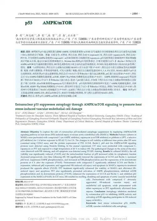
Extranuclear p53suppresses autophagy through AMPK/mTOR signaling to promote heat stress-induced vascular endothelial cell damageLI Li 1,2,ZOU Zhimin 1,2,LI Qin 1,2,ZHANG Kun 1,2,SU Lei 3,GU Zhengtao 1,21Treatment Center for Traumatic Injuries,Third Affiliated Hospital of Southern Medical University,Guangzhou 510630,China;2Academy of Orthopedics of Guangdong Province//Orthopedic Hospital of Guangdong Province//Guangdong Provincial Key Laboratory of Bone and Joint Degenerative Diseases,Guangzhou 510630,China;3Department of Critical Medicine,General Hospital of Southern Theater Command of PLA,Guangzhou 510010,China摘要:目的阐明核外p53通过调控蛋白激酶(AMPK )/雷帕霉素靶体(mTOR )信号通路介导自噬抑制在热打击后血管内皮细胞(VECs )损伤的分子机制。
方法实验分为:对照组、热打击组、热打击组+Compound C 组、热打击组+rapamycin 组、热打击组+PFT 组,并分别使用AMPK 抑制剂Compound C 、mTOR 抑制剂(自噬激活剂)rapamycin 、p53线粒体转位抑制剂PFT 预处理细胞或C57BL/6小鼠,通过CCK8法检测细胞活力,Western blot 观察p53线粒体移位、自噬关键蛋白LC3-Ⅱ、Beclin-1和P62以及AMPK/mTOR 信号通路的激活情况,HE 染色观察各组小鼠主动内皮血管病理改变,TUNEL 染色观察各组小鼠动内皮血管凋亡情况。
《具有时滞的p53信号通路的动力学分析》篇一一、引言p53是一种重要的肿瘤抑制蛋白,在细胞周期调控、DNA修复和细胞凋亡等多个生物学过程中发挥关键作用。
近年来,p53信号通路的研究备受关注,尤其是在信号转导过程中出现的时滞现象。
时滞的存在往往会对信号通路的响应速度和准确性产生影响,进而影响细胞的生物学行为。
因此,对具有时滞的p53信号通路进行动力学分析具有重要的理论和实践意义。
二、p53信号通路简介p53信号通路是一个复杂的生物网络,涉及多种基因、蛋白质和信号分子。
在DNA损伤等应激条件下,p53被激活并参与一系列生物学反应。
具体而言,p53能够调节细胞周期、促进DNA 修复或触发细胞凋亡等反应,从而维持基因组的稳定性。
三、时滞在p53信号通路中的表现在p53信号通路中,时滞现象主要表现为信号传导的延迟和响应的滞后。
这些时滞可能是由于多种因素引起的,如信号分子的合成与降解、蛋白质之间的相互作用、以及细胞内外的环境因素等。
时滞的存在可能导致p53信号通路的响应速度变慢,从而影响细胞的生物学行为。
四、动力学分析方法为了深入分析具有时滞的p53信号通路的动力学特性,我们采用了数学建模和仿真分析的方法。
首先,根据p53信号通路的生物学特性和已知的分子相互作用关系,建立相应的数学模型。
然后,通过仿真分析,研究时滞对p53信号通路的影响。
具体而言,我们采用了常微分方程和离散事件模拟等方法,对p53信号通路的动态变化进行定量描述和分析。
五、动力学分析结果通过动力学分析,我们发现时滞对p53信号通路的响应速度和准确性产生了显著影响。
具体而言,当DNA损伤发生时,由于时滞的存在,p53的激活速度变慢,导致其无法及时地调节细胞周期、促进DNA修复或触发细胞凋亡等反应。
此外,时滞还可能导致p53信号通路的响应出现误差,从而影响细胞的生物学行为。
这些结果提示我们,在研究p53信号通路时,需要充分考虑时滞因素的影响。
六、结论与展望通过对具有时滞的p53信号通路进行动力学分析,我们揭示了时滞对p53信号通路的影响及其机制。
P53基因的功能1阻滞细胞周期在细胞周期中,P53的调节功能主要体现在G1和G2/M期校正点的监测,与转录激活作用密切相关。
P53下游基因P21编码蛋白是一个依赖Cyclin(细胞周期蛋白)的蛋白激酶抑制剂,一方面P21可与一系列Cyclin-cdk(细胞周期蛋白依赖性激酶)复合物结合,抑制相应的蛋白激酶活性,导致高磷酸化Rb蛋白(视网膜母细胞瘤蛋白)堆积,后者使E2F转录因子(参与细胞周期调控的细胞因子)不能活化,引起G1期阻滞;另外P53的另外3个下游基因CyclinB1,CADD45和14-3-3(T 则参与G2/M期阻滞。
2促进细胞调亡Bcl-2(调控线粒体外膜通透性的基因家族)可阻止凋亡形成因子如细胞色素C等从线粒体释放出来,具有抗凋亡作用,而Bax(促凋亡基因)可与线粒体上的电压依赖性离子通道相互作用,介导细胞色素c的释放,具有凋亡作用,p53可以上调Bax的表达水平,以及下调Bcl-2的表达共同完成促进细胞凋亡作用。
P53还可通过死亡信号受体蛋白途径诱导凋亡,TNF受体(在真核细胞表达具有生物活性的可溶性肿瘤坏死因子)和Fas蛋白(一种细胞膜抗原,主要功能是介导细胞凋亡)。
3维持基因组稳定DNA®损后,由于错配修复的累积,导致基因组不稳定,遗传信息发生改变。
P53可参与DNA勺修复过程,其DNA吉合结构域本身具有核酸内切酶的活性,可切除错配核甘酸,结合并调节核甘酸内切修复因子XP评口XPD的活性,影响其DN睡组和修复功能。
4抑制肿瘤血管生成肿瘤生长到一定程度后,可以通过自分泌途径形成促血管生成因子,刺激营养血管在瘤体实质内增生。
P53蛋白能刺激抑制血管生成基因Smad 得表达,抑制肿瘤血管形成。
在肿瘤进展阶段,P53基因突变导致新生血管生成,有利于肿瘤的快速生长,这常常是肿瘤进入晚期的表现。
p53既可阻滞细胞周期,也可诱导细胞凋亡。
两种作用方式都是为了维护基因组的稳定,但二者的性质截然不同。
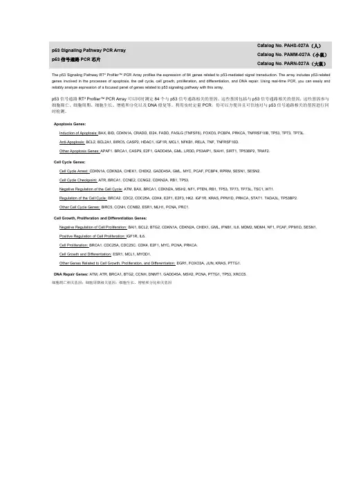
p53Signaling Pathway PCR Array p53信号通路PCR芯片Catalog No.PAHS-027A(人)Catalog No.PAMM-027A(小鼠)Catalog No.PARN-027A(大鼠)The p53Signaling Pathway RT²Profiler™PCR Array profiles the expression of84genes related to p53-mediated signal transduction.The array includes p53-related genes involved in the processes of apoptosis,the cell cycle,cell growth,proliferation,and differentiation,and DNA ing real-time PCR,you can easily and reliably analyze expression of a focused panel of genes related to p53signaling pathway with this array.p53信号通路RT²Profiler™PCR Array可以同时测定84个与p53信号通路相关的基因。
这些基因包括与p53信号通路相关的基因,这些基因参与细胞凋亡、细胞周期、细胞生长、增殖和分化以及DNA修复等。
利用实时定量PCR,你可以方便并且可信地对与p53信号通路相关的基因进行同时检测。
Apoptosis Genes:Induction of Apoptosis:BAX,BID,CDKN1A,CRADD,EI24,FADD,FASLG(TNFSF6),FOXO3,PCBP4,PRKCA,TNFRSF10B,TP53,TP73,TP73L.Anti-Apoptosis:BCL2,BCL2A1,BIRC5,CASP2,HDAC1,IGF1R,MCL1,NFKB1,RELA,TNF,TNFRSF10D.Other Apoptosis Genes:APAF1,BRCA1,CASP9,E2F1,GADD45A,GML,LRDD,P53AIP1,SIAH1,SIRT1,TP53BP2,TRAF2.Cell Cycle Genes:Cell Cycle Arrest:CDKN1A,CDKN2A,CHEK1,CHEK2,GADD45A,GML,MYC,PCAF,PCBP4,RPRM,SESN1,SESN2.Cell Cycle Checkpoint:ATR,BRCA1,CCNE2,CCNG2,CDKN2A,RB1,TP53.Negative Regulation of the Cell Cycle:ATM,BAX,BRCA1,CDKN2A,MSH2,NF1,PTEN,RB1,TP53,TP73,TP73L,TSC1,WT1.Regulation of the Cell Cycle:BRCA2,CDC2,CDC25A,CDK4,E2F1,E2F3,HK2,IGF1R,KRAS,PPM1D,PRKCA,STAT1,TADA3L,TP53BP2.Other Cell Cycle Genes:BIRC5,CCNH,CCNB2,ESR1,MLH1,PCNA,PRC1.Cell Growth,Proliferation and Differentiation Genes:Negative Regulation of Cell Proliferation:BAI1,BCL2,BTG2,CDKN1A,CDKN2A,CHEK1,GML,IFNB1,IL6,MDM2,MDM4,NF1,PCAF,PPM1D,SESN1.Positive Regulation of Cell Proliferation:IGF1R,IL6.Cell Proliferation:BRCA1,CDC25A,CDC25C,CDK4,E2F1,MYC,PCNA,PRKCA.Cell Growth and Differentiation:ESR1,MCL1,MYOD1.Other Genes Related to Cell Growth,Proliferation,and Differentiation:EGR1,FOXO3A,JUN,KRAS,PTTG1.DNA Repair Genes:ATM,ATR,BRCA1,BTG2,CCNH,DNMT1,GADD45A,MSH2,PCNA,PTTG1,TP53,XRCC5.细胞凋亡相关基因;细胞周期相关基因;细胞生长、增殖和分化相关基因。
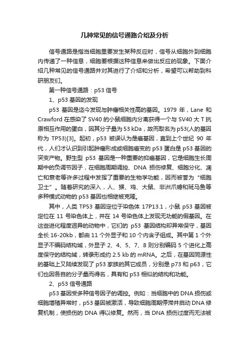
几种常见的信号通路介绍及分析信号通路是指当细胞里要发生某种反应时,信号从细胞外到细胞内传递了一种信息,细胞要根据这种信息来做出反应的现象。
下面介绍几种常见的信号通路并对其进行了介绍和分析,希望可以帮助到科研朋友们。
第一种信号通路:p53信号1、p53基因的发现p53基因是迄今发现与肿瘤相关性高的基因。
1979年,Lane和Crawford在感染了SV40的小鼠细胞内分离获得一个与SV40大T抗原相互作用的蛋白,因其分子量为53 kDa,故而取名为p53(人的基因称为TP53)[3]。
起初,p53被误认为是癌基因,直到上个世纪90年代,人们才认识到引起肿瘤形成或细胞癌变的p53蛋白是p53基因的突变产物。
野生型p53基因是一种重要的抑癌基因,它是细胞生长周期中的负调节因子,在细胞周期调控、DNA损伤修复、细胞分化、凋亡和衰老等许多过程中发挥了重要的生物学功能,因而被誉为“细胞卫士”。
随着研究的深入,人、猴、鸡、大鼠、非洲爪蟾和斑马鱼等多种模式动物的p53基因也相继被克隆。
其中,人类TP53基因定位于染色体17P13.1,小鼠p53基因被定位在11号染色体上,并在14号染色体上发现无功能的假基因。
在这些进化程度迥异的动物中,它们的p53基因结构却异常保守,基因全长16-20kb,都由11个外显子和10个内含子组成。
其中第1个外显子不编码结构域,外显子2、4、5、7、8则分别编码5个进化上高度保守的结构域,转录形成约2.5 kb的mRNA。
之后,在基因同源性的基础上又陆续发现了p53家族的其它成员,分别是p73和p63,它们也因各自的分子量而得名,具有和p53相似的结构和功能。
2、p53信号通路p53基因受多种信号因子的调控。
例如:当细胞中的DNA损伤或细胞增殖异常时,p53基因被激活,导致细胞周期停滞并启动DNA修复机制,使损伤的DNA得以修复。
然而,当DNA损伤过度而无法被修复时,作为转录因子的p53还可进一步激活下游促凋亡基因的转录,诱导细胞凋亡并杀死有DNA损伤的细胞。
P53及其信号通路在肿瘤耐药分子机制中的分析研究目的:讨论P53及其信号通路在肿瘤耐药分子機制中的应用,为日后的临床研究提供参考。
方法:在本次研究中,首先对P53自身稳定性与肿瘤耐药性展开分析,将得到的结果作为基础部分,深入开展P53及其信号通路在肿瘤耐药分子机制中的作用分析。
同时,对P53及其信号通路在肿瘤耐药分子机制中的应用进行相应的论述。
结果:P53自身稳定性与肿瘤耐药性集中在P53的自身降解、P53磷酸化等几个方面,经过大量的研究与分析,认为P53及其信号通路在肿瘤耐药分子机制中的作用非常重要。
结论:要想更好的治疗肿瘤患者,必须对肿瘤耐药分子机制展开研究。
经过长久的探究和分析,发现P53在肿瘤的发生发展中占有重要地位,研究P53及其信号通路在肿瘤耐药分子机制中的应用,具有较大的积极意义,日后可深入研究。
标签:P53分子;信号通路;降解;磷酸化;基因突变;肿瘤耐药性近年来,越来越多的医学人员注意到,P53对肿瘤细胞的耐药性具有较大的影响,通过了解P53及其信号通路在肿瘤耐药分子机制中的应用,可进一步制定抵抗耐药性的方案,帮助患者提升治疗效果,减少痛苦。
经过大量的医学研究,已经证实,半数以上的癌症,都存在P53的基因突变,这种现象很可能与P53参与的细胞周期调控以及凋亡有很大关系。
在此,本次研究主要对P53及其信号通路在肿瘤耐药分子机制中的应用展开分析,现做如下综述。
1资料与方法1.1一般资料在本次研究中,首先对P53自身稳定性与肿瘤耐药性展开分析,将得到的结果作为基础部分,深入开展P53及其信号通路在肿瘤耐药分子机制中的作用分析。
同时,对P53及其信号通路在肿瘤耐药分子机制中的应用进行相应的论述。
1.2方法在P53自身稳定性与肿瘤耐药性的研究方面,主要是通过研究P53的自身降解、P53磷酸化、P53基因突变等三个方面来完成。
而在P53及其信号通路在肿瘤耐药分子机制中的应用方面,主要是结合现阶段的医疗情况和需求来完成。
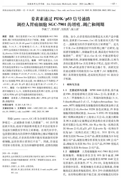
网络出版时间:2222-10-910:/3网络出版地址:httys://kos.oe/kcms/de/il/34.1225.R.0222308.4843.409.htmi姜黄素通过PI3K/p53信号通路调控人胃癌细胞SGC-3901的增殖、凋亡和周期李淑兰1,贺倩倩1,宫凯凯2,满玉清1摘要目的探讨姜黄素(Cur)对人胃癌细胞株SGC-7721增殖、凋亡和周期的影响及其分子机制。
方法采用不同浓度梯度Cur(2、12、22、42pmol/L)处理正常培养的SGC-7721细胞。
3-(4,5-二甲基噻唑A)-2,5-二苯基四氮唑漠盐(MTT)法检测在不同时间点(24、48、72h)细胞增殖能力变化;Hoechw33653荧光染色观察细胞凋亡状况;流式细胞术检测细胞凋亡水平及周期变化;Western blot检测PI3K/p53信号通路相关蛋白表达变化。
结果MTT结果显示,与对照组比较,Cur呈浓度依赖性抑制SGC-P72)细胞增殖;细胞荧光染色结果显示Cur处理组的细胞亮蓝色荧光强度减弱;流式细胞术结果显示,与对照组比较,不同浓度Cur处理组细胞凋亡率增加(P<2.25,P<2.211,65/61期细胞出现阻滞(P<2.21、;Western blot结果显示,与对照组比较,不同浓度Cau组细胞PTK蛋白表达量、Akt磷酸化水平均降低(P <2.21),而421和y53蛋白表达量升高(P<2.25,P< 2.21)o结论Cur能抑制SGC-7721细胞的增殖效应,通过诱导细胞发生G5/G1期阻滞进而促进其凋亡进程,这可能与其对PI3K/p53信号通路的调控密切相关。
关键词姜黄素;人胃癌细胞;增殖;凋亡;PI3K/y53信号通路;细胞周期中图分类号R285.5;R735.2文献标志码A文章编号302-392(2221)21-0243-26 doi:3.10425/(.c/hi.iss/1202-1299.029).4).409胃癌(gast/c cancer,GC)作为全球常见的恶性肿瘤之一,正威胁着全球人的健康3]。
CellResearch:浙⼤解析p53蛋⽩降解新信号通路来⾃浙江⼤学和新加坡科技研究局的科研⼈员发现了p53蛋⽩降解的新信号通路,这条途径并⾮依赖蛋⽩酶体降解途径,⽽是半胱氨酸蛋⽩酶Calpain3成了关键点。
由此他们提出了调控p53蛋⽩周转(turnover)的新途径即核仁信号通路,相关研究成果于2012年1⽉29⽇发表在CellResearch杂志上。
⾃浙江⼤学和新加坡科技研究局的研究⼈员在新研究中揭⽰了⼀条调控p53蛋⽩降解的新信号通路,相关论⽂“Def defines a conserved nucleolar pathway that leads p53 to proteasome-independent degradation”发表在1⽉29⽇的《细胞研究》(Cell research)杂志上通过泛素化途径的p53蛋⽩周转(turnover)是调控转录活性的⼀个重要机制,但⼈们对于有关蛋⽩酶⾮依赖途径的P53蛋⽩周转知之甚少。
消化器官的溶胀因⼦(Def) 蛋⽩对消化器官的发育⾄关重要。
丧失def基因的斑马鱼选择性地上调p53效应基因的表达,那么p53和Def之间有什么关系呢?此项研究中报道中证实了Def是⼀个核仁蛋⽩,def基因功能缺失导致p53蛋⽩上调,且累积在细胞核仁中。
⼴泛研究表明Def能介导p53蛋⽩的降解,这个途径并⾮依赖蛋⽩酶体途径,⽽是依赖Calpain3(半胱氨酸蛋⽩酶)。
总之,此项研究提出了调控p53蛋⽩周转(turnover)的新途径即核仁途径,这将增加⼈们对p53在器官和肿瘤发⽣中的作⽤的认知。
Cell Research:浙⼤解析p53蛋⽩降解新信号通路原⽂摘要:Def defines a conserved nucleolar pathway that leads p53 to proteasome-independentdegradationp53 protein turnover through the ubiquitination pathway is a vital mechanism in the regulationof its transcriptional activity; however, little is known about p53 turnover through proteasome-independent pathway(s). The digestive organ expansion factor (Def) protein is essential for the development of digestive organs. In zebrafish, loss of function of defselectively upregulates the expression of p53 response genes, which raises a question as to what is the relationshipbetween Def and p53. We report here that Def is a nucleolar protein and that loss of function ofdef leads to the upregulation of p53 protein, which surprisingly accumulates in the nucleoli. Our extensive studies have demonstrated that Def can mediate the degradation of p53 protein andthat this process is independent of the proteasome pathway, but dependent on the activity of Calpain3, a cysteine protease. Our findings define a novel nucleolar pathway that regulates the turnover function of p53, which will advance our understanding of p53's role in organogenesisand tumorigenesis.作者简介:彭⾦荣,教授,浙江⼤学动物科学学院副院长,功能基因组实验室主任研究领域:主要从事利⽤模式动物斑马鱼研究有关肝脏及其它消化道器官(如胰腺体,肠)在胚胎期发育的分⼦机理。
©La nd esBi o s ci en ce2004. N o tf or d i s t ri bu t i o n .[Cancer Biology &Therapy 3:2, 156-161, February 2004]; ©2004 Landes BioscienceGen Sheng WuCorrespondence to: Gen Sheng Wu; Program in Molecular Biology and Human Genetics; Karmanos Cancer Institute; Department of Pathology; Wayne State University School of Medicine; Room E216; The Prentis Building; 110 East Warren Avenue; Detroit, Michigan 48201 USA; Tel.: 313.833.0715x2328; Fax:313.831.7518; Email: wug@ Received 01/13/04; Accepted 01/13/04Previously published online as a Cancer Biology &Therapy E-publication:/journals/cbt/abstract.php?id=614KEY WORDSp53, MAPK , phosphatases, phosphorylation,transcriptional control, cell cycle, apoptosisACKNOWLEDGEMENTSI thank Drs. Larry H. Matherly and Ray Mattingly for critical reading of this manuscript.This work was supported by a grant from National Cancer Institute (1R01 CA100073-01 A1).I apologize to those colleagues whose work could not be cited due to space limitations.ReviewThe Functional Interactions Between the p53 and MAPK Signaling PathwaysABSTRACTThe p53 tumor suppressor protein exerts its growth inhibitory activity by activating and interacting with diverse signaling pathways. As a downstream target, p53 protein is phosphorylated and activated by a number of protein kinases in response to stressful stimuli.As an upstream activator, activated p53 acts as a transcription factor to induce and/or suppress a number of genes whose expression leads to the activation of diverse signaling pathways. p53 protein can also interact with a number of proteins, resulting in an increase or decrease in p53 activity itself. The activation of p53 leads to many outcomes in cells, including cell cycle arrest and apoptosis. It has become clear that the p53 protein can functionally interact with the mitogen-activated protein kinase (MAPK) pathways,including the stress-activated protein kinase [SAPK/c-Jun N-terminal protein kinase (JNK)],the p38 mitogen-activated protein kinase (MAPK), and the extracellular signal related kinase (ERK). Upon exposure to stressful stimuli, MAP kinases phosphorylate and activate p53, leading to p53-mediated cellular responses. Recent studies have suggested a role of p53 as an upstream activator to regulate MAPK signaling via the transcriptional acti-vation of members of the dual specificity phosphatase family. Because both the p53 and MAPK signaling pathways are altered in the majority of human tumors, understanding their functional interaction may provide new insights into the deregulated cell proliferation and survival that is characteristic of cancer.INTRODUCTIONp53 is the most commonly mutated gene in human cancer. The p53 tumor suppressor protein is a nuclear phosphoprotein with a short half-life that is regulated mainly through post-translational modifications. Upon stressful stimuli, such as DNA damage and oncogene activation, p53 protein is modified through multiple post-translational events, including phosphorylation and acetylation.1-3These modifications stabilize and activate p53 protein.In addition, a recent study suggested a transcriptional regulation of p53 by the interferon signaling pathway.4Once p53 is activated, it acts as a transcription factor for many genes that contain the consensus p53-binding sites in their promoters or intronic sequences.5p53 can also suppress the expression of a number of genes whose expression may favor cell survival. It is well accepted that the transcriptional activity of p53 plays a major role in tumor suppression, although transcription-independent activity is also suggested by its growth inhibitory activity. Activation of p53 triggers a number of signaling pathways that lead to cell cycle arrest, apoptosis, senescence, DNA repair and antiagiogenesis.The MAP kinase signaling pathways mainly consists of three subfamilies; the stress-activated protein kinase [SAPK/c-Jun N-terminal protein kinase (JNK)], the p38mitogen-activated protein kinase (MAPK ), and the extracellular signal related kinase (ERK)6,7(Fig. 1). Members of the MAPK family can be induced by multiple stimuli including growth factors and stresses, leading to diverse cellular responses. In the presence of stimuli, these MAPK s are activated through the reversible phosphorylation of both threonine and tyrosine residues of the TXY motif in the catalytic domain by upstream dual-specificity kinases. These activating kinases are called MAPK kinases and include MKK1/2 (selective for ERK1/2 activation), MKK3/6 (selective for p38 activation) and MKK-4/7 (selective for JNK activation)8-10(Fig. 1). Once MAP kinases are activated, they function as effector protein kinases to phosphorylate a number of substrates including p53, leading to many changes in cells, such as cell growth, differentiation and apoptosis.9Since the p53 signaling pathways including its activation by upstream kinases, regulation of its downstream targets, and the biological consequences of its activation are complex,this review will focus on the interactions between the p53 and MAPK pathways, including©La nd esBi o s ci en ce2004. N o tf or d i s t ri bu t i o n .MAPK-mediated p53 phosphorylation and p53-mediated transcrip-tional inhibition of MAPK signaling.PHOSPHORYLATION AND ACTIVATION OF p53BY THE MAP KINASESHuman p53 is a 393 amino acid protein that consists of four functional domains: an N-terminal transactivation domain, a proline rich domain, a DNA binding domain, and a C-terminal domain with a tetramerization and a nuclear localization signaling sequence (Fig. 2). There are a number of protein kinases that can phosphorylate p53 at a number of sites, which mainly reside in either the N-ter-minus or C-terminus. One such kinase is the ataxiatelangiectasia mutated (ATM) protein, a member of the phosphoinositide-3kinase-related kinase (PI 3K) superfamily. Upon DNA damage, such as that resulting from ionizing radiation, ATM phosphorylates p53at several sites including serines 9, 15, 20, and 46 directly or indi-rectly.11-13In addition, ATR (ATM-Rad3-related), another member of this PI 3K superfamily, has been shown to phosphorylate p53 at both serines 15 and 37.14,15Furthermore, several additional kinases have been shown to phosphorylate p53 at various sites upon expo-sure to stressful stimuli, including DNA-PK, casein kinases I and II,CDK-activating kinase (CAK), cdc2, cdk2, and protein kinase c.16-21Interestingly, the MAP kinases including p38, JNKs and ERKs have been shown to phosphorylate p53 in response to different stressfulstimuli, and such phosphorylation can initiate the p53 response,leading to cell cycle arrest and apoptosis.p38.The p38 pathway is activated by stressful stimuli and cytokines. Stressful stimuli and cytokines can lead to activation of several upstream kinases including MLKs, TAK-1 and Ask-1.8These activated kinases then phosphorylate and activate downstream kinases MK K 3 and MK K 6. Once MK K 3 and MK K 6 are activated, they phosphorylate and activate p38.22,23Activated p38 then phosphory-lates a number of cellular substrates,leading to diverse cellular responses,such as apoptosis and cell survival.8A number of studies have shown that p38 plays an important role in activation of p53 via a mechanism involving phos-phorylation. In normal human ker-atinocytes, UVB activates p38, which then phosphorylates p53 on serine 15.This p38-dependent phosphorylation leads to p53 accumulation in the cyto-plasm and thus prevents p53 from entering into the nucleus; this provides a protective mechanism against UV-induced cell death.24Consistent with this role of p38 to regulate p53, p38 phosphorylates p53 at serines 15 and 20 in response toUVB radiation in a mouse JB6 epidermal cell line.25,26It has also been shown that Gadd45a, a p53 target, regulates the stabilization of p53 that is induced by UVB through p38 because this induc-tion is abrogated in the presence of a p38 kinase inhibitor.27Inhibition of p38 activation correlates with the dereg-ulation of p53 activation and, moreover,p38 forms a complex with Gadd45 proteins.28In rabbit articularchondrocytes, nitric oxide-induced apoptosis involves p38-mediated p53 activation, and the blockade of p38 kinase activity correlates with reduced p53 accumulation and caspase-3 activity.29A further study shows that nitric oxide-induced activation of p38 kinase phosphory-lates p53 at serine 15, which results in accumulation of p53 protein and subsequent induction of apoptosis.30Further, in human prostate cancer cell line, methoxyestradiol-induced apoptosis involves Figure 1. Schematic diagram showing three major MAPK signaling cascades and their inhibition by members of the dual specificity phosphatase family.These three major MAPK pathways are the extracellular-signal regulated kinase/mitogen activated protein kinase (ERK/MAPK) signaling pathway,C-Jun amino-terminal kinase/stress-activated protein kinase (JNK/SAPK/MAPK) signaling pathway, and the p38/MAPK signaling pathway. All three pathways can be inactivated by members of the dual specificity phosphatase family including PAC1, MKP1 and DUSP5.Figure 2. A schematic diagram of the functional domains of p53 protein and its phosphorylation by MAP kinases. p53 protein consists of four functional domains: a transactivation domain (TA), a proline rich domain (PRD), a DNA binding domain (DBD), and a C-terminal region that contains a nuclear localization sequence (NLS) and a tetramerization domain (TETRA). Upon stressful stimuli, MAP kinases are activated and in turn, phosphorylate and activate p53, leading to p53-mediated cellular responses. Since MAPK-medi-ated p53 phosphorylation at certain residues is observed differently between mouse and human cells, thefollowing phosphorylated sites that occur either in human (h) or mouse (m) cells or both are indicated: p38can phosphorylate p53 at ser 15 (h and m), ser 20 (m), ser 33 (h and m), ser 37 and 46 (h and m), and ser 389 (m); JNKs can phosphorylate p53 at ser 15 (m), ser 20 (m), ser 34 (m) and thr 81 (h); and ERKs can phosphorylate p53 at ser 15 (h and m), thr 73 (m) and thr 83 (m). ser: serine; thr: threonine.©La nd esBi o s ci en ce2004. N o tf or d i s t ri bu t i o n .p53 activation through p38/JNK -dependent NF-κB/AP-1.31In neurons, p38-mediated p53 phosphorylation at serine 15 and subse-quent p53 induction is responsible for hypoxic neuronal death.32In addition to its phosphorylation of p53 at serines 15 and 20,p38 has been shown to phosphorylate p53 at several additional sites including serines 33, 37 and 46. For example, upon osmotic shock,p38 is activated and then phosphorylates p53 at serine 33 and subsequently causes a G1 arrest.33Interestingly, p38 is also involved in phosphorylating p53 at serine 33 in anticancer-drug-induced apoptosis.34p38 phosphorylates p53 at serines 33 and 46 in-vitro and inhibition of p38 activity during UV irradiation decreases phosphorylation of p53 at serines 15, 33 and 37 and reduces UV-induced p53-dependent apoptosis.35Finally, p38 has been shown to phosphorylate p53 at serine 389 in response to UV radia-tion.36Therefore, stress-induced p38 acts as a protein kinase to phosphorylate p53 at various serine residues, which, in turn, activatesp53, leading to p53-mediated cellular responses, such as the induction of apoptosis.JNKs.The stress-activated protein kinase (SAPK) pathway is alsocalled the Jun N-terminal kinase pathway as the kinase could phos-phorylate the NH2-terminus of the transcription factor c-JUN.37,38Like p38, JNK activation involves a phosphorylation cascade. Upon stressful stimuli, MEK K 1 is activated and subsequently activates MK K 4 and MK K 7.8Activated MK K 4/7 then phosphorylate and activate JNKs.39-41Once JNKs are activated, they can phosphorylate a number of substrates, leading to activation of many signaling pathways,8 including the p53 pathway.The main role of JNKs in regulating the p53 pathway is currently thought to be through their ability to phosphorylate p53 protein.Upon UVB radiation, JNKs are directly involved in phosphoryla-tion of p53 at serine 20, resulting in an increase in p53-dependent transcriptional activity. This response is suppressed in JNK1 or JNK2 knockout cells.26Under oxidative stress,JNKs can phosphorylate p53 at serine 15.42In addition,JNK phosphorylates p53 at threonine 81 in response to DNA damage and stress-inducing agents, and substitu-tion of this site (T81A) attenuates UV-and JNK-medi-ated p53 stabilization and subsequently impairs p53-mediated G1 arrest.43A further study has identified the amino-terminus of JNK as a functional inducer that activates p53 transcriptional activity.44In addition to phosphorylation of p53 upon DNA damage, JNK has been suggested to regulate p53 stability in nonstressed cells through an MDM2-independent mechanism.45Consistently, MEKK1, the upstream regulator of JNK,has been shown to regulate p53 stability.46Finally, it has been shown that JNK1 can phosphorylate p53 at serine 34 in murine cells.47Thus, like p38, JNKs function as upstream kinases to phosphorylate p53 at various sites in response to DNA damage and subsequently stabilize and activate p53 transcriptional activity.ERK1/2.Unlike p38 and JNK, ERK is generally acti-vated by growth actors (Fig. 1). Upon growth factor stimuli, the Ras/Raf pathway is activated and subse-quently activates MEK 1/2 kinases.48,49ActivatedMEK1/2 then activate ERK 1/2,50leading to activation of a number of transcription factors.8In spite of the fact that activation of p53 by growth factors has not been clearly established, phosphorylation of p53 by ERKs has been well observed in a number of experimental sys-tems. In ovarian cells, ERK1/2 have been shown to berequired for p53 phosphorylation at serine 15 and apoptosis inducedby cisplatin.51ERK also regulates p53 activation and subsequent cellcycle arrest induced by colcemid, and the selective MEK1 inhibitor PD098059 attenuates colcemid-induced p53 activation and G 1cell cycle arrest.52Furthermore, ERKs mediate resveratrol-induced acti-vation of p53 and apoptosis through phosphorylation of p53 at ser-ine 15.53Interestingly, in the murine system, ERK phosphorylates p53 at threonines 73 and 83, a proline-rich region that contains a transactivation domain of p53.54Since some of phosphorylated sites on p53 (e.g., thr 73 and 83) are detected only in mouse cells but not in human cells, it is believed that there is differential regulation of p53 phosphorylation between mouse and human cells.55Thus,ERKs act as upstream activators to phosphorylate p53 at serine and threonine residues, leading to p53 activation and subsequent cellular responses.REGULATION OF MAPK SIGNALING BY p53VIA A TRANSCRIPTIONAL MECHANISMIt is well known that the transcriptional activity of p53 plays a pivotal role in its tumor suppression activity.2There is a growing list of the genes that are transcriptionally regulated by p53, and activa-tion of these targets leads to distinct cellular responses in cells.3For example, p53-mediated growth arrest involves the activation of p21,56,57Gadd45,58and 14-3-3σ.59p53-induced apoptosis, on the other hand, involves the activation of several genes, which includeBax,60,61Fas/APO1,62KILLER/DR5,63,64Pigs,65IGF-BP3,66PERP ,67Noxa,68p53AIP1,69p53DINP1,70PUMA,71,72Bid 73and caspase 6.74It has also been shown that p53 can induce antiangio-genesis by upregulating Tsp1,75BaI1 and GD-AiF .76,77In addition,p53 induces MDM2, an oncogene that negatively regulates p53Figure 3. Models for the roles of phosphatases in p53-mediated cellular responses. DNAdamage activates p38 and, in turn, activates p53 via phosphorylation. Activated p53 can transcriptionally induce a number of genes, leading to apoptosis, which may be negatively regulated by p53-mediated induction of Wip1 because Wip1 can dephosphorylate p38and thus suppress its role in phosphorylating p53. It has been shown that DNA damage such as oxidative stress activates p53, leading to upregulation of PAC1 and DUSP5.Because ERK1/2 are believed to play a negative role in apoptosis in some cell types, inac-tivation of ERK1/2 by p53-mediated PAC1 and DUSP5 leads to induction of apoptosis. In the case of MKP1, activation of MKP1 can inhibit mitogen-mediated ERK activation and subsequently block re-entry of cells into the cell cycle. Therefore, it is believed that p53-mediated MKP1 induction controls re-entry of arrested cells into the cell cycle in response to mitogens in the absence of DNA damage, although it is not clear how p53 is activated in response to growth factor stimuli.©La nd esBi o s ci en ce2004. N o tf or d i s t ri bu t i o n .stabilization.78,79Interestingly, recent investigations have identified p53-regulated transcription of four phosphatases that are known to negatively regulate MAPK signaling: Wip1, MK P1, PAC1 and DUSP580-83(Fig. 3).Wip1.Wip1 was the first phosphatase to be identified as a tran-scriptional target of p53. Wip1 is a serine/threonine phosphatase belonging to the type 2 class that are insensitive to phosphatase inhibitors such as okadaic acid.80Wip1 is induced by ionizing radiation in a p53-dependent manner.80Induction of Wip1 by p53leads to p53-mediated G 1arrest in response to DNA damage.80A mechanistic study has revealed that Wip1 can inactivate p38 activity in response to UV radiation.84p38 can phosphorylate p53 at serines 33 and 46,35and p53-dependent induction of Wip1 could dephos-phorylate p38 and, in turn, downregulate UV-induced p53 phospho-rylation at these residues.35This study suggests that Wip1 may serve as a negative feedback loop to control the MAPK-p53 signaling path-way. Consistent with a role for Wip1 in the p53 pathway, fibroblasts derived from Wip1 knockout mice exhibit a premature senescence and a defect in entering mitosis.85MKP1.MKP1, also known as CL100, 3CH134, and Erp, is the founding member of the dual-specificity protein phosphatase fami-ly 86-88and found to be a new target of p53.82This family includes MKP1, MKP2, MKP3, MKP4, MKP5, VHR, Pac1, hVH2, hVH3,Pyst1 and Pyst2.89Because phosphorylation is required for the activation of MAP kinases, dephosphorylation by members of this family inactivates the MAP kinases and thus inhibits their signaling 90(Fig. 1). MKP1 was originally identified as an ERK-specific phos-phatase.88,91It is now known that MKP1 can also inhibit JNK and p38.92,93Constitutive MK P1 expression blocks G 1specific gene expression,94suggesting a role in the cell cycle. We have shown that MK P1 is induced in cells when p53 is conditionally induced or when cells are infected with p53 adenoviruses.82p53 regulates MKP1 induction through a p53-responsive element residing in the second intron of this gene.93Interestingly, overexpression of MKP1impairs re-entry of arrested cells into the cell cycle in response to growth factor stimuli.93This regulation appears to be DNA damage independent since p53 is generally induced and activated following DNA damage. Furthermore, inhibition of phosphatase activity by vanadate can partially relieve p53-mediated G 1arrest.93These data indicate that p53 may negatively control re-entry of arrested cells into the cell cycle in response to mitogens in the absence of DNA damage (Fig. 3).PAC1.Pac1 (phosphatase activated in cells 1), also known as DUSP2 (dual specificity phosphatase 2), is another member of the dual-specificity protein phosphatase family capable of inactivating all three major MAPK signaling pathways 89(Fig. 1). Using microarray technology, Pac1 was isolated as a p53 target.81Unlike other p53targets such as p21, Pac1 induction requires both functional p53 and serum starvation/oxidative damage.81Overexpression of Pac1 sensitizes cells to apoptosis induced by serum starvation and H 202treatment.Importantly, reduction of Pac1 by siRNA approach renders cells resistant to oxidative damage-induced apoptosis.81These data suggest that Pac1 may be involved in oxidative damage-induced p53-mediated apoptosis. Interestingly, p53 transcriptionally regulates Pac1 expression through a novel binding site residing in the promoter of the Pac1gene.81This binding site is distinct from the well-described p53consensus sequence.5Because Pac1 is able to inactivate the ERK 1/2-mediated MAPK pathway, and because p53 can induce Pac1 induction in response to oxidative stress, it is believed that p53-mediated apoptosis induced by oxidative stress is through Pac1-dependent inactivation of ERK pathways (Fig. 3).DUSP5.DUSP5 is the third member of the dual-specificity protein phosphatase family that has been shown to inhibit all three major MAPK pathways with preferential specificity 89(Fig. 1) and that has been recently identified as the new target for p53.83DUSP5is induced in cells by p53 overexpression.83Induction of DUSP5 by p53 appears to be mediated through a p53-binding site in the pro-moter of the DUSP5 gene.83Interestingly, a functional study sug-gested DUSP5 can preferentially dephosphorylate ERK s,83thus providing another link between the p53 and MAPK signaling pathways (Fig. 3).p53 REGULATES THE MAPK PATHWAYS AS A PROTECTIVE MECHANISM AGAINST CELLULAR INSULTSIn addition to phosphatases, there are several other genes that can functionally link the p53 to MAPK pathways. Using a p53-inducible cell system, Lee et al. found that inducible expression of p53 in human bladder cancer cells leads to an activation of MEK and its downstream kinase ERK, but not p38.95Activation of ERK by p53 requires transcriptionally active p53 because p53 mutants that lack transcriptional activity fail to do so.95This suggests that p53 can transcriptionally activate ERK/MAPK signaling. A mecha-nistic study of p53-mediated MAPK activation has led to the identi-fication of heparin-binding epidermal growth factor-like growth factor (HB-EGF) as a new p53 target.95,96Through binding to the EGF receptor, HB-EGF can activate the phosphatidylinositol 3-kinase (PI3K )/AK T pathway, a major cell survival pathway.Inhibition of HB-EGF activity can decrease p53-mediated MAPK activation, while overexpression of HB-EGF protects cells from oxidative damage-induced apoptosis.96These studies suggest that besides activation of the signaling pathways leading to growth arrest and apoptosis, p53 can also activate survival pathways in order to maintain the balance between life and death in response to DNA damage. This idea is supported by discovery of the involvement of discoidin domain receptor 1 (DDR1) in the p53 pathway.DDR1 was initially identified as a member of the receptor tyrosine kinase family. DDR1 was induced and tyrosine phosphorylated fol-lowing genotoxic stress in a p53-dependent manner.97Since blocking DDR1 expression by a means of dominant negative mutants or antisense constructs can sensitize cells to p53-mediated apoptosis induced by DNA damage, it is likely that DDR1 serves as another negative regulator that controls p53 responses via activation of the ERK pathway. These data are consistent with the role of the Ras/Raf/MAPK cascade in regulating p53-mediated cellular responses.98,99A recent study has identified cyclooxygense-2 (Cox2) as the third gene that can serve as a downstream effector to participate in the p53-MAPK pathway.100Cox2 is a key enzyme in the synthesis of prostaglandins and thromboxanes. Cox2 is highly induced in response to growth factors. It is known that the ERK/MAPK signal can activate Cox2 expression, that activation of Cox2 leads to cell survival, and that Cox2 is induced in a p53-dependent manner.100Interestingly, induction of Cox2 by p53 is not due to binding of p53to a p53 responsive element in the Cox2 genomic sequence, but rather by p53-induced activation of ERK/MAPK.100Like HB-EGF and DDR1, activation of Cox2 can inhibit p53-mediated apoptosis and chemosensitivity in response to genotoxic stress.100T ogether,these investigations have suggested that the outcomes of p53 activation are dependent on the balance between activation of death-related genes and survival-related genes.©La nd esBi o s ci en ce2004. N o tf or d i s t ri bu t i o n .CONCLUSIONSMuch of our understanding of the functional interactions between the p53-MAPK pathways has dealt with the role of MAP kinases in phosphorylating and activating p53 in response to DNA damage. Recent findings of the MAPK inhibitory phosphatases as p53 targets have provided a more complex and comprehensive picture of how p53 functionally interacts with the MAPK signaling pathways. On the one hand, MAPK-dependent p53 phosphoryla-tion following DNA damage can lead to p53-dependent cellular responses such as cell cycle arrest and apoptosis. On the other hand,activation of phosphatases by p53 suggests a mechanism by which p53 negatively regulates MAPK -dependent signaling. Although activation of p53, in most cases, causes growth arrest and apoptosis,identification of p53 target genes (e.g., DDR1) that are stimulatory components of the MAPK pathways suggests a role for p53 in cell survival. It is well known that the outcomes of p53 activation by stressful stimuli depend on many factors such as cell context and type and duration of stresses. This pattern of complex and integrated responses is also true in the case of the activation of the MAPK pathways. Therefore, it is conceivable that in response to stressful stimuli, the balance between the p53 and MAPK pathways will determine whether cells live or die.References1.Kastan MB, Onyekwere O, Sidransky D, Vogelstein B, Craig RW. Participation of p53 pro-tein in the cellular response to DNA damage. Cancer Res 1991; 51:6304-11.2.Vogelstein B, Lane D, Levine AJ. Surfing the p53 network. Nature 2000; 408:307-10.3.El-Deiry WS. Regulation of p53 downstream genes. Semin Cancer Biol 1998; 8:345-57.4.Takaoka ASHHY, Stoiber D, Negishi H, Kikuchi H, Sasaki S, Imai K, Shibue T, Honda K, Taniguchi T . Integration of interferon-alpha/beta signalling to p53 responses in tumour suppression and antiviral defence. Nature 2003; 424:516-23.5.El-Deiry WS, Kern SE, Pietenpol JA, Kinzler KW, Vogelstein B. Definition of a consensus binding site for p53. Nat Genet 1992; 1:45-9.6.Davis RJ. Signal transduction by the JNK group of MAP kinases. Cell 2000; 103:239-52.7.Johnson GL, Lapadat R. Mitogen-activated protein kinase pathways mediated by ERK,JNK, and p38 protein kinases. Science 2002; 298:1911-2.8.Pearson G, Robinson F , Beers Gibson T, Xu BE, Karandikar M, Berman K, Cobb MH.Mitogen-activated protein (MAP) kinase pathways: regulation and physiological functions,Endocr Rev 2001; 22:153-83.9.Chang L, Karin M. Mammalian MAP kinase signalling cascades. Nature 2001; 410:37-40.10.Kennedy NJ, Davis RJ. Role of JNK in tumor development. Cell Cycle 2003; 2:199-201.11.Banin S, Moyal L, Shieh S, Taya Y, Anderson CW, Chessa L, Smorodinsky NI, Prives C,Reiss Y, Shiloh Y, Ziv Y. Enhanced phosphorylation of p53 by ATM in response to DNA damage. Science 1998; 281:1674-7.12.Canman CE, Lim DS, Cimprich KA, Taya Y, Tamai K, Sakaguchi K, Appella E, KastanMB, Siliciano JD. Activation of the ATM kinase by ionizing radiation and phosphoryla-tion of p53. Science 1998; 281:1677-9.13.Saito S, Goodarzi AA, Higashimoto Y, Noda Y, Lees-Miller SP , Appella E, Anderson CW.ATM mediates phosphorylation at multiple p53 sites, including Ser(46), in response to ionizing radiation. J Biol Chem 2002; 277:12491-4.14.Tibbetts RS, Brumbaugh KM, Williams JM, Sarkaria JN, Cliby WA, Shieh SY, Taya Y,Prives C, Abraham RT. A role for ATR in the DNA damage-induced phosphorylation of p53. Genes Dev 1999; 13:152-7.kin ND, Hann BC, Jackson SP . The ataxia-telangiectasia related protein ATR mediatesDNA-dependent phosphorylation of p53. Oncogene 1999; 18:3989-95.16.Shieh S-Y, Ikeda M, Taya Y, Prives C. DNA damage-induced phosphorylation of p53 alle-viates inhibition by MDM2. Cell 1997; 91:325-34.17.Knippschild U, Milne DM, Campbell LE, DeMaggio AJ, Christenson E, Hoekstra MF ,Meek DW. p53 is phosphorylated in vitro and in vivo by the delta and epsilon isoforms of casein kinase 1 and enhances the level of casein kinase 1 delta in response to topoisomerase-directed drugs. Oncogene 1997; 15:1727-36.ne DM, Palmer RH, Meek DW. Mutation of the casein kinase II phosphorylation siteabolishes the anti-proliferative activity of p53. Nucleic Acids Res 1992; 20:5565-70.19.Bischoff JR, Friedman PN, Marshak DR, Prives C, Beach D. Human p53 is phosphory-lated by p60-cdc2 and cyclin B-cdc2. Proc Natl Acad Sci USA 1990; 87:4766-70.20.Price BD, Hughes-Davies L,Park SJ. Cdk2 kinase phosphorylates serine 315 of humanp53 in vitro. Oncogene 1995; 11:73-80.21.Baudier J, Delphin C, Grunwald D, K hochbin S, Lawrence JJ. Characterization of thetumor suppressor protein p53 as a protein kinase C substrate and a S100b-binding protein,Proc Natl Acad Sci USA 1992; 89:11627-31.22.Derijard B, Raingeaud J, Barrett T, Wu IH, Han J, Ulevitch RJ, Davis RJ. Independenthuman MAP-kinase signal transduction pathways defined by MEK and MKK isoforms.Science 1995; 267:682-5.23.Han J, Lee JD, Jiang Y, Li Z, Feng L, Ulevitch RJ. Characterization of the structure andfunction of a novel MAP kinase kinase (MKK6). J Biol Chem 1996; 271:2886-91.24.Chouinard N, Valerie K, Rouabhia M, Huot J. UVB-mediated activation of p38 mitogen-activated protein kinase enhances resistance of normal human keratinocytes to apoptosis by stabilizing cytoplasmic p53. Biochem J 2002; 365:133-45.25.She QB, Chen N, Dong Z. ERKs and p38 kinase phosphorylate p53 protein at serine 15in response to UV radiation. J Biol Chem 2000; 275:20444-9.26.She, QB, Ma WY, Dong Z. Role of MAP kinases in UVB-induced phosphorylation of p53at serine 20. Oncogene 2002; 21:1580-9.27.Jin S, Mazzacurati L, Zhu X, Tong T, Song Y, Shujuan S, Petrik KL, Rajasekaran B, Wu M,Zhan Q. Gadd45a contributes to p53 stabilization in response to DNA damage.Oncogene 2003; 22:8536-40.28.Bulavin DV , Kovalsky O, Hollander MC, Fornace AJ. Loss of oncogenic H-ras-inducedcell cycle arrest and p38 mitogen-activated protein kinase activation by disruption of Gadd45a. Mol Cell Biol 2003; 23:3859-71.29.Kim SJ, Ju JW, Oh CD, Yoon YM, Song WK, Kim JH, Yoo YJ, Bang OS, Kang SS, ChunJS. ERK-1/2 and p38 kinase oppositely regulate nitric oxide-induced apoptosis of chon-drocytes in association with p53, caspase-3, and differentiation status. J Biol Chem 2002;277:1332-9.30.Kim SJ, Hwang SG, Shin DY, Kang SS, Chun JS. p38 kinase regulates nitric oxide-inducedapoptosis of articular chondrocytes by accumulating p53 via NFkappa B-dependent tran-scription and stabilization by serine 15 phosphorylation. J Biol Chem 2002; 277:33501-8.31.Shimada, K., Nakamura, M., Ishida, E., Kishi, M., and Konishi, N. Roles of p38- and c-jun NH2-terminal kinase-mediated pathways in 2-methoxyestradiol-induced p53 induc-tion and apoptosis, Carcinogenesis. 24: 1067-1075, 2003.32.Zhu Y, Mao XO, Sun Y, Xia Z, Greenberg DA. p38 Mitogen-activated protein kinase medi-ates hypoxic regulation of Mdm2 and p53 in neurons. J Biol Chem 2002; 277:22909-14.33.Kishi H, Nakagawa K, Matsumoto M, Suga M, Ando M, Taya Y, Yamaizumi M. Osmoticshock induces G 1arrest through p53 phosphorylation at Ser33 by activated p38MAPK without phosphorylation at Ser15 and Ser20. J Biol Chem 2001; 276:39115-22.34.Sanchez-Prieto R, Rojas JM, Taya Y, Gutkind JS. A role for the p38 mitogen-acitvated pro-tein kinase pathway in the transcriptional activation of p53 on genotoxic stress by chemotherapeutic agents. Cancer Res 2000; 60:2464-72.35.Bulavin DV , Saito S, Hollander MC, Sakaguchi K, Anderson CW, Appella E, Fornace AJ.Phosphorylation of human p53 by p38 kinase coordinates N-terminal phosphorylation and apoptosis in response to UV radiation. EMBO J 1999; 18:6845-54.36.Huang C, Ma WY, Maxiner A, Sun Y, Dong Z. p38 kinase mediates UV-induced phos-phorylation of p53 protein at serine 389. J Biol Chem 1999; 274:12229-35.37.Hibi M, Lin A, Smeal T, Minden A, Karin M. Identification of an oncoprotein- and UV-responsive protein kinase that binds and potentiates the c-Jun activation domain. Genes Dev 1993; 7:2135-48.38.Derijard B, Hibi M, Wu IH, Barrett T, Su B, Deng T, Karin M, Davis RJ. JNK1: a proteinkinase stimulated by UV light and Ha-Ras that binds and phosphorylates the c-Jun activa-tion domain. Cell 1994; 76:1025-37.39.Sanchez I, Hughes RT, Mayer BJ, Yee K, Woodgett JR, Avruch J, Kyriakis JM, Zon LI.Role of SAPK/ERK kinase-1 in the stress-activated pathway regulating transcription factor c-Jun. Nature 1994; 372:794-98.40.Tournier C, Whitmarsh AJ, Cavanagh J, Barrett T, Davis RJ. Mitogen-activated proteinkinase kinase 7 is an activator of the c-Jun NH2-terminal kinase. Proc Natl Acad Sci USA 1997; 94:7337-42.41.Yao Z, Diener K , Wang XS, Zukowski M, Matsumoto G, Zhou G, Mo R, Sasaki T,Nishina H, Hui CC, Tan TH, Woodgett JP , Penninger JM. Activation of stress-activated protein kinases/c-Jun N-terminal protein kinases (SAPKs/JNKs) by a novel mitogen-acti-vated protein kinase kinase. J Biol Chem 1997; 272:32378-83.42.Cheng WH, Zheng X, Quimby FR, Roneker CA, Lei XG. Low levels of glutathione per-oxidase 1 activity in selenium-deficient mouse liver affect c-Jun N-terminal kinase activa-tion and p53 phosphorylation on Ser-15 in pro-oxidant-induced aponecrosis. Biochem J 2003; 370:927-34.43.Buschmann T , Potapova O, Bar-Shira A, Ivanov VN, Fuchs SY, Henderson S, Fried VA,Minamoto T , Alarcon-Vargas D, Pincus MR, Gaarde WA, Holbrook NJ, Shiloh Y, Ronai Z.Jun NH2-terminal kinase phosphorylation of p53 on Thr-81 is important for p53 stabi-lization and transcriptional activities in response to stress. Mol Cell Biol 2001; 21:2743-54.44.Buschmann T, Yin Z, Bhoumik A, Ronai Z. Amino-terminal-derived JNK fragment altersexpression and activity of c-Jun, ATF2, and p53 and increases H2O2-induced cell death. J Biol Chem 2000; 275:16590-6.45.Fuchs SY, Adler V , Buschmann T, Yin Z, Wu X, Jones SN, Ronai Z. NK targets p53 ubiq-uitination and degradation in nonstressed cells. Genes Dev 1998; 12:2658-63.46.Fuchs SY, Adler V , Pincus MR, Ronai Z. MEKK1/JNK signaling stabilizes and activatesp53, Proc Natl Acad Sci USA 1998; 95:10541-6.ne DM, Campbell LE, Campbell DG, Meek DW. p53 is phosphorylated in vitro andin vivo by an ultraviolet radiation-induced protein kinase characteristic of the c-Jun kinase,JNK1. J Biol Chem 1995; 270:5511-8.48.Kyriakis JM, App H, Zhang XF , Banerjee P , Brautigan DL, Rapp UR, Avruch J. Raf-1 acti-vates MAP kinase-kinase. Nature 1992; 358:417-21.49.Dent P , Haser W, Haystead TA, Vincent LA, Roberts TM, Sturgill TW. Activation of mito-gen-activated protein kinase kinase by v-Raf in NIH 3T3 cells and in vitro. Science 1992;257:1404-7.。
5 NLRSNOD 样受体(NOD like receptors, NLR)家族,广泛存在于人类的细胞胞浆内,识别相应配体之后能够激活NF-κ B (NOD1 和NOD2)信号途径,活化Caspase-1促进促炎因子IL-1β和IL-18(NLRP1/NALP1, NLRP3/NALP3/cryopyrin, and NLRC4/Ipaf )的产生,从而启动固有免疫和获得性免疫。
NOD 蛋白为胞浆内的模式识别受体,其结构包括:( 1 ) 中央的核苷酸结合寡聚化区域(NACHT),是NLRs 家族共同拥有的结构,对于NLRs 的寡聚化和活化非常重要;(2)N 末端效应结合区域,即N- 末端蛋白-蛋白相互作用的结构域,如半胱氨酸蛋白酶激活和募集结构域( caspase activation and recruitment domain,CARD). ( 3) C 末端富含亮氨酸的重复序列( LRRs ) ,可以识别受体。
在NOD家族成员中NOD2和NLRP3的突变均可引起炎症性疾病,因此是目前研究最多的NOD样分子。
NOD2突变可致Crohn’s病(CD)和Blau综合征(BS)的发生,而发生在NLRP3的编码基因CIAS1上的突变可导致家族性寒冷自发炎症综合征(FCAS)、Muckle-wells 综合征和慢性婴儿期皮肤关节综合征(CINCA)等自身炎症综合征。
NOD1和NOD2能够识别细菌肽聚糖(peptidoglycan,PGN)的不同降解产物,并以其CARD结构募集具有同样CARD结构的RIP2激酶(RICK),继而激活MAPK 和NF-κB两条信号途径。
Yang和Hiotsumatsu等学者认为cIAPS可与RIP2结合并使之泛素化,而与K63联接的RIP2多泛素化可招募TAK1分子进而激活IKK和MAPK。
研究证实NOD1和NOD2可在非依赖RIP2的情况下诱发自噬。
多种病原相关分子模式(PAMPs)内源性危险信号相关分子模式(DAMPs)均可诱发NLRP1,NLRP3以及NLRC4产生核转录信号复合物,即炎症小体。
P53信号通路,科研人的『心头好』首先,做个小预告吉凯网上商城9月全线上新shRNA克隆商品,吉凯ShRNA克隆适用于在各类细胞中抑制同源基因的表达,提供预设计的3个靶点,交付周期仅需5个工作日,价格仅1560元!p53细胞信号通路是如今医学界研究最热门的通路,在可预见的未来几年也是众多研究人员的『心头好』。
该通路涉及众多与肿瘤密切相关的基因,并且随着研究的深入,p53信号通路在肿瘤研究的过程中老大的地位愈发稳固。
P53信号转导通路是一直是众多医学科研人员研究得非常活跃的领域,从致癌基因到抑癌基因,p53基因功能的发现经历了一个漫长而奇妙的旅程。
P53通路中涉及很多关键基因,它们和肿瘤的发生和发展具有紧密的联系。
随着研究的深入,人们发现p53通路还有更多的秘密等着我们去发掘。
因此,以p53通路研究为主题的文章更是汗牛充栋,数不胜数。
即使是《nature》这样的重磅杂志,也经常见到p53通路的身影。
例如:2016年《nature》文章『Dual targeting of p53 and c-MYC selectively eliminates leukaemic stem cells.』不夸张的说,p53通路是所有从事肿瘤研究的科研人员或多或少都会涉及的通路,可见该通路的重要性,本文就是对该通路的简单介绍。
P53基因曾经被认定是致癌基因,随着研究的深入,人们发现p53是一个抑癌基因,并且发现了许多跟p53基因相互作用,发挥功能效用的基因。
其中一个就是MDM2基因。
MDM2基因MDM2基因是受p53蛋白调控的靶基因,MDM2蛋白可以通过多种方式抑制p53蛋白的作用。
比如,它可以与p53蛋白的反式激活结构域相结合并抑制其活性。
另外,MDM2蛋白还能起到E3泛素连接酶的作用,特异性地催化p53蛋白经泛素化途径降解。
p53蛋白诱导MDM2蛋白表达,然后MDM2蛋白促进p53蛋白降解,从而限制胞内p53蛋白活性。
p53 SignalingRT² Profiler™ PCR Arrayp53 Signaling Pathway PCR ArrayCellular Senescence PCR ArrayDNA Damage Signaling Pathway PCR ArrayCell Cycle PCR ArraySureSilencing RNAip53 Signaling Pathway Gene RNAiCellular Senescence Gene RNAiDNA Damage Signaling Pathway Gene RNAiCell Cycle Gene RNAiCignal™ Reporter Assaysp53 Pathway Reporter Assay KitE2F Reporter Assay KitEGR1 Reporter Kitp53 is a tumour suppressor protein that regulates the expression of a wide variety of genes involved in Apoptosis, Growth arrest, Inhibition of cell cycle progression, Differentiation and accelerated DNA repair or Senescence in response to Genotoxic or Cellular Stress. As a transcription factor, p53 is compos ed of an N-terminal Activation Domain, a central specific DNA Binding Domain, and a C-terminal Tetramerization Domain, followed by a Regulatory Domain rich in basic Amino acids. Having a short half-life, p53 is normally maintained at low levels in unstress ed mammalian cells by continuous ubiquitylation and subsequentdegradation by the 26S Proteasome. Nonphosphorylated p53 is ubiquitylated by the MDM2 (Mouse Double Minute-2) ubiquitin ligase. MDM2 binding inactivates p53 by two mechanisms. First, MDM2 binds to the transactivation domain of p53, precluding interaction with the transcriptional machinery. Second, this binding mediates the covalent attachment of ubiquitin to p53. Ubiquitylated p53 is then degraded by the Proteasome. Thus MDM2 acts as a major reg ulator of the tumor suppressor p53 by targeting its destruction. When the cell is confronted with stress like DNA damage, Hypoxia, Cytokines, Metabolic changes, Vi ral infection, or Oncogenes, however, p53 ubiquitylation is suppressed and p53 accumulates in the nucleus, where it is activated and stabilized by undergoing multiple covalent modifications including Phosphorylation and Acetylation (Ref.1 & 2).Phosphorylation of p53 mostly occurs in the N-terminal activation domain at the Ser6, Ser9, Ser15, Thr18, Ser20, Ser33, Ser37, Ser46, Thr55, and Thr81 residues, with some phosphorylation occurring in the C-terminal linker and basic regions at Ser315, Ser371, Ser376, Ser378, and Ser392. Phosphorylation on most of these sites is induced by DNA damage, with som e, such as Thr55 and Ser376, being repressed upon genotoxic stress. p53 phosphorylation is mediated by several cellular kinases including Chks (Checkpoint Kinases), CSNK1-Delta (Casein Kinase-1-Delta), CSNK2 (Casein Kinase-2), PKA (Protein Kinase A), CDK7 (Cyclin-Dependent Kinase-7), DNA-PK (DNA-Activated- Protein Kinase), HIPK2 (Homeodomain-Interacting Protein Kinase-2), CAK (CDK-Activating Kinase), p38 and JNK (Jun NH2-terminal kinase). Notably, phosphorylation at Ser15 by ATM (Ataxia Telangiectasia Mutated Gene)/ATR (Ataxia-Telangiectasia and Rad3 Related), either directly or through Chk1 (Cell Cycle Checkpoint Kinase-1)/Chk2 (Cell Cycle Checkpoint Kinase-2), or at Ser20 by Chk1/Chk2 has been shown to alleviate the inhibition or degradation of p53, leading to p53 stabilization and activation. The phosphorylation-induced p53 stabilization and activation are mediated through multiple mechanisms and may vary according to the cellular context or microenvironment. HIF-1Alpha (Hypoxia-Inducible Factor-1-Alpha) has been implicated to be involved in p53 stabilization, the precise mechanism by which HIF-1Alpha regulates p53-mediated function remains unknown. Recently, the interaction between p53 and HIF-1Alpha was reported to evoke HIF-1Alpha degradation. Members of the PIAS (Protein Inhibitor of Activated STAT) protein family have also been found to interact with p53. PIAS1 and PIAS-Gamma function as SUMO (Small Ubiquitin Related Modifier-1) ligases for p53. Moreover, the RING finger domain of PIAS1 binds to the C-terminus of the tumor suppressor p53 and catalyzes its sumoylation, a modification which represses p53 activity on a reporter plasmid containing consensus p53 DNA binding sites. PML (Promyelocytic Leukemia) also activates p53 by recruiting it to multiprotein complexes termed PML-nuclear bodies. PML is a tumor suppressor protein and the major component of multiprotein nuclear complexes that have been variably termed Kremer bodies, ND10, PODs (for PML Oncogenic Domains), and PML-NBs (PML-Nuclear Bodies). PML binds directly with p53 and recruits it to PML-NBs. Recruitment to PML-NBs activate p53 by bringing it in close proximity with CBP (CREB-Binding Protein) /p300. BRCA1 (Breast Cancer-1 Gene) and p53 can also physically associate, both in vitro and in vivo and function in a common pathway of tumor suppression. The ability of BRCA1 to biochemically modulate p53 function suggests that this may be afundamental role of BRCA1 in tumor suppression (Ref.3, 4 & 5).Another important modification of p53 is acetylation. p53 is specifically acetylated at Lys370, Lys372, Lys373, Lys381, and Lys382 by p300/CBP and at Lys320 by PCAF (p300/CBP-associated factor). Acetylation has been shown to augment p53 DNA binding, and to stimulate p53-mediated transactivation of its downstream target genes through the recruitment of coactivators. Acetylation may also regulate the stability of p53 by inhibiting its ubiquitination by MDM2. In vivo, acetylation at Lys320, Lys373, and Lys382 is induced by many genotoxic agents, including UV-radiation, IR (Ionizing Radiation), hypoxia, oxidative stress, and even depletion of ribonucleotide pools. p53 can also be deacetylated by HDAC1 (Histone Deacetylase-1) and SIRT1. Human SIRT1 is an enzyme that deacetylates the p53 tumor suppressor protein and has been suggested to modulate p53-dependent functions including DNA damage-induced cell death. p53 deacetylation has been suggested to down-regulate the activation of genes such as Bax and p21WAF1. Phosphorylation and acetylation are interdependent. In deed, phosphorylation at the p53N-terminus has been shown to enhance its interaction with acetylase p300/CBP and to potentiate p53 acetylation. Activated p53 functions effectively as a transcription factor and induces transcription of several genes. The D NA targets of p53 are consensussequences consisting of a 10-base pair repeat of 5'-PuPuPu-C(A/T)(T/A)GPyPyPy-3' (where Pu is a purine and Py is a pyrimidine). It also can bind to a palindromic site having a four or five-base pair inverted repeat of a similar sequence. Complete p53 is inactive for specific DNA binding unless activated by covalent and noncovalent modifications of the basic C-terminal domain. After p53 is activated it can be involved in cell-cycle inhibition, apoptosis, genetic repair, and inhibition of blood-vesselformation (Ref.5, 6 & 7).Cell cycle inhibition takes place when there is a block in cell-cycle division. p53 does this by stimulating the expression of p21 WAF1/CIP1 (Cyclin Dependent Kinase Inhibitor-p21). This protein is an inhibitor of CDKs (Cyclin-Dependent Kinases) that regulate the cell cycle via perturbation of their partner cyclin. Cyclins are involved to ensure successful transitions from S phase to G1. Since p21 WAF1/CIP1 inhibits CDKs it results in inhibition of both G1-to-S and G2-to-mitosis transitions by causing hypophosphorylation of Rb (Retinoblastoma) and preventing the release of E2F. Additionally p53 can stimulate 14-3-3, a protein that sequesters Cyclin B1-CDK1 complexes out of the nucleus. This results in a G2 block. Activated p53 may also initiate apoptosis and stop cell proliferation. p53 stimulates a wide network of signals that act through two major apoptotic pathways: Extrinsic Pathways and Intrinsic Pathways. The extrinsic pathway involves engagement of pa rticular `death' receptors that belong to the TNFR (Tumor Necrosis Factor Receptor) family and, through the formation of the DISC(Death-Inducing-Signaling-Complex), leads to a cascade of activation of Caspases, including Caspase8 and Caspase3, which in turn induce apoptosis. Most common death receptors involved in extrinsic apoptosis Fas, DR5 (Death Receptor-5) and PERP. The intrinsic apoptotic pathway is dominated by the Bcl2 (B-Cell CLL/Lymphoma-2) family of proteins, which governs the release of CytoC (Cytochrome-C) from the mitochondria. The Bcl2 family comprises anti-apoptotic (pro-survival) and pro-apoptotic members. The Bcl2 family is divisible into three classes: pro-survival proteins, whose members are most structurally similar to Bcl2, such as BclXL; pro-apoptotic proteins, BAX (Bcl2 Associated-X Protein) and BAK (Bcl2 Antagonist Killer-1), which are structurally similar to Bcl2 and BclXL and antagonize their pro-survival functions; and the pro-apoptotic `BH3-only' proteins. Intriguingly, a key subset of the Bcl2 family genes are p53 targets, including BAX, Noxa, PUMA (p53-Upregulated Modulator of Apoptosis) and the most recently identified, BID (BH3 Interacting Domain Death Agonist). p53 may also inhibit Bcl2 that is an inhibitor of apoptosis. p53 may also have a role in maintaining genetic stability by 'nucleotide-excision' repair of DNA, chromosomal recombination and chromosome segmentation. GADD45 (Growth Arrest- and DNA Damage-Inducible Gene-45) is a multifunctional protein that is regulated by p53 and that may play a role in DNA repair and cell cycle checkpoints. p53 can playa role in the inhibition of blood-vessel formation. In order for tumours to reach a large size, they must initiate the growth of nutrient-bringing blood vessels in their vicinity, the process of angiogenesis. p53 stimulates the production of genes that prevent this process from happening. p53 activates the expression of the Tsp1 (Thrombospondin-1), an anti-angiogenic factor, along with other angiogenesis inhibitor BAI1 (Brain-specific Angiogenesis Inhibitor-1) (Ref. 8, 9 & 10).In addition, p53 regulates MDM2 function in a negative feedback loop, because the MDM2 gene is a target for p53. Therefore, activation of p53 eventually leads to its own inactivation by switching on a pathway that leads to its destruction. MDM2 is subject to further regulation by direct binding of the ARF (Active Response Factor) protein, which prevents MDM2-mediated p53 proteolysis. PTEN (Phosphatase and Tensin Homolog), on the other hand inhibits MDM2-mediated p53 degradation. p53 can transcriptionally activate PTEN, which may further inhibit Akt activity. Therefore, inhibition of Akt by the inhibitors may trigger a positive feedback with perhaps additional anti-tumor effects. The c-Fos proto-oncogene is also a target for transactivation by the p53 tumor suppressor. Mutations in p53 are associated with genomic instability and increased susceptibility to cancer. It is the most frequently mutated protein in all cancer with an estimated 60% of all cancers having mutated forms that affect its growth suppressing activities. However some common tumours have a higher incidence such that 90% of cervical and 70% of colorectal are found to have p53 mutations. The p53 protein can be inactivated in several ways, in cluding inherited mutations that result in a higher incidence of certain familial cancers such as Li-Fraumeni syndrome. Certain DNA tumour viruses, such as the human adenovirus and the papilomavirus, bind to and inactivate the protein. Functional p53 is th ought to provide a protective effectagainst tumorigenesis (Ref.2, 11 & 12).。
P53-信号通路译文P53 信号通路P53是一个肿瘤抑制蛋白,调节各种各样基因的表达,包括细胞凋亡,生长抑制,抑制细胞周期进程,分化和加速DNA修复,基因毒性和细胞应激后的衰老。
作为一个转录因子,p53是N端激活域、DNA中央特定结合域和C-端四聚体化域的组成部分,而且其调控域富含碱性氨基酸。
p53半衰期很短,在26S蛋白酶体作用下,通过持续的泛素化和后期降解,p53在无刺激的哺乳类动物细胞中维持较低的含量。
去磷酸化的p53在MDM2(鼠双微体基因-2)泛素连接酶作用下被泛素化。
MDM2结合p53使其无活性是通过两种途径:第一,MDM2结合p53的转录激活域,阻止转录元件的相互作用。
第二,介导p53共价结合泛素蛋白,泛素化的p53被蛋白水解酶降解。
通过使p53的失活,MDM2扮演着p53抑癌基因的主要监视者。
当细胞面临着DNA 损伤、缺氧、细胞因子、代谢改变、病毒感染或者致癌基因等刺激时,导致p53泛素化被抑制和p53在细胞核内积累,通过多个共价修饰包括磷酸化和乙酰化,p53被激活并稳定存在。
p53的磷酸化大多数出现在N-末端激活域的Ser6,Ser9,Ser15,Thr18,Ser20,Ser33,Ser37,Ser46,Thr55和Thr81残基上,另外还有一些p53磷酸化出现在C-末端连接处和碱性区域的Ser315, Ser371, Ser376, Ser378, 和Ser392残基上。
大多数位点上的磷酸化是由DNA损伤诱发的,还有一些例如Thr55 and Ser376在基因毒性应激下被压抑。
P53磷酸化是由几个细胞激酶所介导,包括Chks,CSNK1-Delta,CSNK2,PKA,CDK7,DNA-PK,HIPK2,CAK,p38和JNK。
显然,由ATM/ATR作用在Ser15上磷酸化,直接作用或者通过Chk1/Chk2,在Chk1/Chk2作用下的Ser20磷酸化已经证实能够减缓抑制或减慢降解p53,导致p53稳定并活化。
P53信号通路
P53是一个肿瘤抑制蛋白,调节各种各样基因的表达,包括细胞凋亡,生长抑制,抑制细胞周期进程,分化和加速DNA修复,基因毒性和细胞应激后的衰老。
作为一个转录因子,p53是N端激活域、DNA中央特定结合域和C-端四聚体化域的组成部分,而且其调控域富含碱性氨基酸。
p53半衰期很短,在26S蛋白酶体作用下,通过持续的泛素化和后期降解,p53在无刺激的哺乳类动物细胞中维持较低的含量。
去磷酸化的p53在MDM2(鼠双微体基因-2)泛素连接酶作用下被泛素化。
MDM2结合p53使其无活性是通过两种途径:
第一,MDM2结合p53的转录激活域,阻止转录元件的相互作用。
第二,介导p53共价结合泛素蛋白,泛素化的p53被蛋白水解酶降解。
通过使p53的失活,MDM2扮演着p53抑癌基因的主要监视者。
当细胞面临着DNA损伤、缺氧、细胞因子、代谢改变、病毒感染或者致癌基因等刺激时,导致p53泛素化被抑制和p53在细胞核内积累,通过多个共价修饰包括磷酸化和乙酰化,p53被激活并稳定存在。
p53的磷酸化大多数出现在N-末端激活域的Ser6,Ser9,Ser15,Thr18,Ser20,Ser33,Ser37,Ser46,Thr55和Thr81残基上,另外还有一些p53磷酸化出现在C-末端连接处和碱性区域的Ser315,Ser371,Ser376,Ser378,和Ser392残基上。
大多数位点上的磷酸化是由DNA损伤诱发的,还有一些例如Thr55 and Ser376在基因毒性应激下被压抑。
P53磷酸化是由几个细胞激酶所介导,包括Chks,CSNK1-Delta,CSNK2,PKA,CDK7,DNA-PK,HIPK2,CAK,p38和JNK。
显然,由ATM/ATR作用在Ser15上磷酸化,直接作用或者通过Chk1/Chk2,在Chk1/Chk2作用下的Ser20磷酸化已经证实能够减缓抑制或减慢降解p53,导致p53稳定并活化。
磷酸化诱导的p53稳定和活化是通过多种机制介导以及很多细胞环境或微环境的改变所致。
HIF-1Alpha参与p53的稳定,HIF-1Alpha调节p53介导作用的详细机制依然是一个未知数。
最近,p53和HIF-1Alpha之间的作用已被报道能够引起HIF-1Alpha的降解。
PIAS蛋白家族也被发现能与p53发生相互作用。
PIAS1和PIAS-Gamma作用可作为p53的SUMO连接酶。
另外,PIAS1的环指域
结合p53抑癌基因的C-末端催化其类泛素化(sumoylation),这个修饰,能抑制报告质粒p53的活性,包含共同序列p53DNA的结合位点。
PLM通过吸收p53到多蛋白复合体(称为PLM核体)来激活p53。
PLM是一个肿瘤抑制蛋白,也是曾发生变异称为Kremer bodies蛋白核配合物(ND10, PODs and PML-NBs)的主要组成成分。
PLM直接和p53结合并进入PLM-NBs。
补充到PLM-NBs激活p53,通过把它引进到与CBP/p300极为贴近。
BRCA1和p53发生联合,在体内和体外是相同的肿瘤抑制途径。
BRCA1对于生化调节p53作用的能力暗示着在肿瘤抑制过程,这个可能是BRCA1一个基本的角色。
P53另一个重要的修饰是乙酰化作用。
在Lys370,Lys372, Lys373, Lys381, and Lys382 byp300/CBP and at Lys320残基上通过PCAF,p53发生特殊的乙酰化。
已证明,乙酰化可以增强p53DNA的结合、通过更新辅激活因子刺激p53介导的下游靶基因转录激活。
乙酰化可以通过MDM2抑制p53的泛素化来调节p53的稳定性。
在机体内,发生Lys320, Lys373, andLys382残基上的乙酰化是通过许多基因毒性介质所诱导的,包括紫外线、电离辐射、缺氧、氧化应激,甚至耗尽的核苷酸池。
P53同样也可以被HDAC1和SIRT1去乙酰化。
人类的SIRT1是一种酶,属于去乙酰化p53肿瘤抑制蛋白,能够被显示调节p53依赖作用,包括DNA诱导损伤的细胞死亡。
P53去乙酰化能够被应用到下调Bax and p21WAF1等基因的活性。
磷酸化和乙酰化是相互依赖的。
的确,p53N-末端的磷酸化被证实能够增强与乙酰化酶p300/CBP的相互作用和加强p53乙酰化。
激活p53功能实际上是一个转录因子和诱导一些基因的转录。
DNA靶基因p53是一个有十个5'-PuPu-C(A/T)(T/A)GPyPy-3'重复出现的共有序列。
它也可以结合到一个含有四到五个重复出现碱基对的相同序列的回文位点。
完整的p53是一个不活跃的特定DNA位点,除非C-末端域发生共价或非共价修饰。
激活后p53能够参与阻碍细胞周期、细胞凋亡、基因修复和抑制血管生成。
细胞周期的抑制发生在细胞分裂周期的一个时间点。
P53抑制细胞周期是通过刺激p21WAF1/CIP1的表达实现的。
这个蛋白是CDKs的抑制剂,CDKs调节细胞周期通路干扰细胞周期蛋白伴侣。
细胞周期蛋白参与保证S期向G1期的转变的成功。
p21WAF1/CIP1抑制CDKs通过引起Rb高度磷酸化和阻止E2F的释放,导致G1-S期、G2-分裂期的转变收到抑制。
此外,p53可以刺激14-3-3,14-3-3是
细胞核外细胞周期蛋白B1-CDK1复合体螯合剂蛋白。
这导致一个G2区。
激活的p53可能也启动细胞凋亡和中止细胞增殖。
p53能激活一个广泛的网络信号,通过两个主要的凋亡通路:
外部途径和内部途径。
外在途经包括参与特殊死亡受体,这个受体属于TNFR家族,通过DISC组成,导致一系列半胱天冬酶激活,包括
Caspase8andCaspase3,进而诱导细胞凋亡。
最常见的死亡受体参与外在细胞凋亡Fas,DR5和PERP。
内部细胞凋亡途径受Bcl2家族蛋白的控制,Blc2家族蛋白能控制CytoC从线粒体中释放。
Blc2家族由抗凋亡和促进凋亡两部分组成。
Blc2家族可以分为三类:
一类是促生存蛋白,它们在结构上和Blc2很相似,例如BclXL;一类是促凋亡蛋白,如BAX和BAK,它们的结构和Blc2和BclXL很相似,起到抵抗促生存蛋白作用;和‘BH3-only’促凋亡蛋白。
有趣的是,一个关键的Blc2家族基因的亚基是p53作用的靶点,包括BAX,Noxa,PUMA和最近发现的BID。
P53也可能抑制细胞凋亡抑制剂Blc2。
P53在维持遗传稳定性中扮演一个角色,通过修复DN
A、重组染色体、姐妹染色单体中核苷酸的缺失。
GADD45是一种多功能蛋白,能被p53调控,在DNA修复和细胞周期检控点扮演重要角色。
P53能抑制血管的生成,肿瘤为了大规模生成,他们必须启动吸收他们周围养分促进血管生长,这是血管生成的过程。
P53刺激基因产生防止这个过程的发生。
P53激活Tsp1的表达,Tsp1是一种抗血管生成因子,连同其它血管生成抑制剂BAI1。
此外,p53通过负反馈循环调节MDM2,因为MDM2基因是p53的靶基因。
因此,激活的p53最终导致自己失活,通过转换一个途径导致自己死亡。
MDM2受到进一步的控制,通过直接与ARF蛋白结合,它将防止MDM2介导
p53水解。
另一方面,PTEN抑制MDM2介导p53降解。
P53形成活化的PTEN,这样可以进一步抑制AKt的活性。
因此,通过抑制剂抑制的AKs可能引起一个积极的反馈作用,可能额外产生抗肿瘤的作用。
c-Fos原癌基因也是通过p53肿瘤抑制基因转录激活的目标。
突变的p53伴随着基因组的不稳定和增加肿瘤发生的概率。
在所有肿瘤中p53是最频繁突变的蛋白,据估计有60%的肿瘤有变异形式,影响其生长抑制活动。
然而一些常见的肿瘤有较高的发病率,90%的子
宫癌和70%的大肠癌发现p53发生突变。
p53蛋白可以通过几种方法失去活性,包括遗传突变,它导致某些家族性癌症的发病率较高,如Li-Fraumeni综合症。
某些肿瘤的DNA病毒,如人类腺病毒和乳头瘤病毒,结合并且抑制蛋白活性。
功能性的p53被认为具有对肿瘤发生的保护性作用。