浅表器官及组织的超声诊断
- 格式:ppt
- 大小:6.54 MB
- 文档页数:43
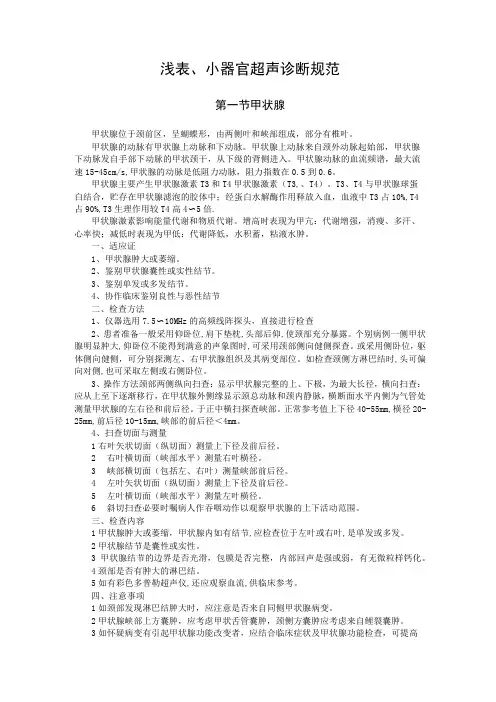
浅表、小器官超声诊断规范第一节甲状腺甲状腺位于颈前区,呈蝴蝶形,由两侧叶和峡部组成,部分有椎叶。
甲状腺的动脉有甲状腺上动脉和下动脉。
甲状腺上动脉来自颈外动脉起始部,甲状腺下动脉发自手部下动脉的甲状颈干,从下级的背侧进入。
甲状腺动脉的血流频谱,最大流速15-45cm/s,甲状腺的动脉是低阻力动脉,阻力指数在0.5到0.6。
甲状腺主要产生甲状腺激素T3和T4甲状腺激素(T3,、T4)。
T3、T4与甲状腺球蛋白结合,贮存在甲状腺滤泡的胶体中;经蛋白水解酶作用释放入血,血液中T3占10%,T4占90%,T3生理作用较T4高4〜5倍.甲状腺激素影响能量代谢和物质代谢。
增高时表现为甲亢:代谢增强,消瘦、多汗、心率快;减低时表现为甲低:代谢降低,水积蓄,粘液水肿。
一、适应证1、甲状腺肿大或萎缩。
2、鉴别甲状腺囊性或实性结节。
3、鉴别单发或多发结节。
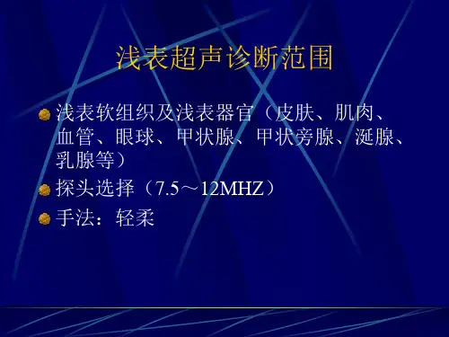
4、协作临床鉴别良性与恶性结节二、检查方法1、仪器选用7.5〜10MHz的高频线阵探头,直接进行检查2、患者准备一般采用仰卧位,肩下垫枕,头部后仰,使颈部充分暴露。
个别病例一侧甲状腺明显肿大,仰卧位不能得到满意的声象图时,可采用颈部侧向健侧探查。
或采用侧卧位,躯体侧向健侧,可分别探测左、右甲状腺组织及其病变部位。
如检查颈侧方淋巴结时,头可偏向对侧,也可采取左侧或右侧卧位。
3、操作方法颈部两侧纵向扫查:显示甲状腺完整的上、下极,为最大长径,横向扫查:应从上至下逐渐移行,在甲状腺外侧缘显示颈总动脉和颈内静脉,横断面水平内侧为气管处测量甲状腺的左右径和前后径。
于正中横扫探查峡部。
正常参考值上下径40-55mm,横径20-25mm,前后径10-15mm,峡部的前后径<4mm。
4、扫查切面与测量1右叶矢状切面(纵切面)测量上下径及前后径。
2右叶横切面(峡部水平)测量右叶横径。
3峡部横切面(包括左、右叶)测量峡部前后径。
4左叶矢状切面(纵切面)测量上下径及前后径。
5左叶横切面(峡部水平)测量左叶横径。


2023年11月 第9卷 第11期* 器材应用与技术研究 *不同超声频率探头对浅表软组织及小器官疾病的诊断价值研讨易怀红溧阳市人民医院超声科,江苏常州 213300摘要 目的 分析不同超声频率探头对浅表软组织及小器官疾病的诊断价值。
方法 随机选取2020年4月—2023年4月于溧阳市人民医院就诊的80例浅表软组织及小器官疾病患者作为研究对象,以组织病理学检查作为诊断的“金标准”,所有患者均分别进行高频率探头超声检查和低频率超声检查,对比两种频率探头超声检查结果。
结果 组织病理学检查结果显示,80例均为浅表软组织及小器官疾病患者;高频超声检查结果显示,共有78例浅表软组织及小器官疾病患者;低频超声检查结果显示,共有70例浅表软组织及小器官疾病患者。
组织病理学结果显示,浅表软组织疾病患者9例,男性外生殖器疾病患者21例,乳腺疾病患者32例,甲状腺疾病患者18例。
高频超声对浅表软组织疾病、男性外生殖器疾病的诊断符合率高于低频超声,差异有统计学意义(P <0.05)。
结论 高频超声诊断各类浅表组织及小器官疾病的准确性较高,安全性高。
关键词 浅表软组织;小器官;高频超声;低频超声;诊断价值中图分类号 R 445445..1 文献标志码 Adoi10.11966/j.issn.2095-994X.2023.09.11.15Discussion on the diagnostic value of different ultrasound frequency probes for su⁃perficial soft tissue and small organ diseasesYI HuaihongDepartment of Ultrasound, Liyang People's Hospital, Changzhou, Jiangsu Province, 213300 ChinaAbstract Objective To analyze the diagnostic value of different ultrasound frequency probes for superficial soft tissue and small organ dis⁃eases. Methods 80 patients with superficial soft tissue and small organ diseases who visited Liyang People's Hospital from April 2020 to April 2023 were randomly selected as the research subjects. Histopathological examination was used as the "gold standard" for diagnosis. All pa⁃tients underwent high-frequency probe ultrasound examination and low-frequency probe ultrasound examination, and the results of the two frequency probe ultrasound examinations were compared. Results The histopathological examination results showed that all 80 cases were pa⁃tients with superficial soft tissue and small organ diseases. The results of high-frequency ultrasound examination showed a total of 78 patients with superficial soft tissue and small organ diseases. The results of low-frequency ultrasound examination showed a total of 70 patients with superficial soft tissue and small organ diseases. The histopathological examination results showed that there were 9 patients with superficial soft tissue diseases, 21 patients with male external genital diseases, 32 patients with breast diseases, and 18 patients with thyroid diseases. The diagnostic accuracy of high-frequency ultrasound for superficial soft tissue diseases and male external genital diseases was higher than that of low-frequency ultrasound, and the differences were statistically significant (P <0.05). Conclusion High frequency ultrasound has high accuracy and safety in diagnosing various superficial tissue and small organ diseases.Key words Superficial soft tissue; Small organs; High frequency ultrasound; Low frequency ultrasound; Diagnostic value收稿日期:2023-09-09;修回日期:2023-09-29作者简介:易怀红(1979-),女,本科,副主任医师,研究方向为超声相关研究。


探讨浅表器官疾病超声诊断价值摘要:目的:分析临床上浅表器官疾病利用超声诊断的价值。
方法:就临床上常见浅表疾病采用超声进行诊断。
结果:浅表器官超声诊断可针对浅表较小的器官及其疾患。
结论:临床运用中将浅表器官超声诊断与一般高频超声及新技术配合使用,可提高诊断的精确性。
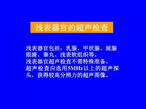
关键词:浅表器官;超声诊断;疾病临床上浅表器官超声诊断与腹部超声诊断及心血管超声诊断、妇产科超声诊断一起,大致涵盖超声医学在诊断领域的应用范围。
浅表器官超声诊断作为一种只针对浅表较小的器官及其疾患。
1浅表器官疾病超声检查过程临床上常用的超声为机械环阵式探头,探头前方有一水囊,聚焦点表浅,穿透深度为2.0cm,扫查范围为1.5cm~2.5cm,轴向分辨率80um,横向分辨率20um,因分辨率较高,减少了浅表伪像。
1.1确定病灶性质:病灶的性质需要利用超声技术进行确定,因浅表部的某些小囊肿体积小,部位表浅与低回声(极低回声区)采用常规的检测方法不可确定病症,临床上要对囊实性病灶做出鉴别,可通过超声提高分辨率,要对囊实性病灶作出判别,如对乳腺囊肿,乳腺纤维腺瘤及附睾囊肿通过超声探头辨别其性质。
不同病灶产生的检查现象是不一致的,皮肤局限性隆起的低回声区为黑色素瘤临床检查表现,其浅表及深部界限一般尚清楚,有时两侧边界不清,经采用彩色多普勒检查在其内部及其深部可见较多的短线状血流。
1.2界定病灶边界:肿块的边界、包膜在超声检测下可进行清晰辨认,如恶性肿瘤的细毛刺、蟹足样、锯齿样边界及良性肿瘤的包膜样回声及囊肿壁回声在超声下非常清晰、明确。
于二维声像图上良恶性淋巴结有一定特征,良性淋巴结呈现椭圆形,长短径比>2,其低回声的皮质区与强回声的髓质区分界清晰,皮质与髓质之比<2;而恶性淋巴结则呈圆形或不规则形状,长短径比<2,中央变细、变小或消失。
2浅表器官疾病超声检查的诊断价值2.1利用超声可提高浅表器官病变中微小钙化灶检出:经研究发现钙化颗粒小,为乳腺癌钙化的特征直径10~500μm,一般不超过1mm。
超声诊断学浅表器官部分课件整理眼一、眼球和眼眶的解剖概要二、探查方法及正常声像图(一)探查方法:仪器:眼科AB超、高频彩超。
1.直接探查法。
2.间接探查法。
运动试验、后运动试验、磁性试验。
(二)正常声像图:轴位切面依次显示眼睑、角膜、前房、晶状体、玻璃体、球壁、视神经及球后脂肪组织。
CDFI:视神经是选择血管的重要标志,视神经暗区中可以探测到视网膜中央动-静脉(取样点位于球后2-3mm)、脻状后动脉(取样点在视神经两侧,球壁后3-5mm)、眼动脉(取样点在球后15-25mm)。
三、眼球病变声像图表现(一)玻璃体病变:常见的玻璃体疾病有玻璃体积血、玻璃体浑浊等。
1、玻璃体积血:来自于周围组织,病因复杂,既可是全身性疾病所致,也可是眼局部疾病引起。
2、玻璃体混浊:玻璃体变性、玻璃体炎症等所致。
3、玻璃体后脱离:是很多玻璃体疾病的共同表现,为玻璃体的境界层与视网膜的。
目前,X线平片仍是眼内异物诊断的首选方法,但其对非金属异物不敏感,有时需CT明确诊断。
在异物定位上,超声优于其他检查,并可对继发病变进行诊断。
在眶性疼痛。
初期时多伴有上感表现。
2.慢性淋巴细胞性甲状腺炎:又称桥本氏甲状腺炎,是一种自身免疫性疾病,血清甲状腺微粒体抗体(TMAb)和甲状腺球蛋白抗体(TGAb)升高。
中、青年女性多见。
(三)甲状腺腺瘤:分滤泡状腺瘤和乳头状腺瘤,占甲状腺肿瘤的70%-80%,中青年女性多见。
呈类圆形,有包膜,质地软,半数以上有囊性变。
有10%可发生癌变,20%为高功能性,可引起甲亢。
(四)甲状腺癌:在甲状腺疾病中占的比例较小,占甲状腺肿瘤的 4.8%。
中老年女性多见。
50%-80%为乳头状腺癌,另外常见的有滤泡癌、髓样癌。
可侵犯淋巴结、血管、喉返神经。
甲状腺病变的鉴别诊断鉴别诊断:分弥漫性肿大和结节改变两大类。
弥漫性肿大见于亚甲炎、单纯性甲状腺肿、甲亢、桥本氏甲状腺炎。
结节样改变见于结甲、腺瘤、甲状腺癌。
甲状腺结节良恶鉴别要点(1)结节内性质:囊性、高回声、多发多为良性;实性、低回声、单发有恶性的可能。