三维电解剖标测系统(清晰详实)
- 格式:ppt
- 大小:5.97 MB
- 文档页数:81
“3Dbody解剖”是一套3D解剖教学系统平台,提供了男女二套全三维的数字模型,每套5000多个人体结构,是目前最完整全面的解剖学数据,涵盖了人体所有解剖系统,同时提供了骨性标志图、肌肉动作动画、肌肉起止点、针灸穴位、断层解剖、英文发音、注释等信息。
作为集大成者,不仅数据详实,而且操作功能强大,国内外高度领先,通过本软件实时三维操作,轻易获得层层解剖人体的机会。
其它内容:骨性标志图肌肉附着点(起止点)肌肉动作动画触发点(扳机点)3D针灸穴位主要功能特点:真正的3D效果不同于传统图片拼接技术,完全基于三维数字模型创建,立体直观,可以实现360度“任意”角度查看,随时为你展现所需要的视角。
显示、隐藏4000+解剖结构,“任何”一块可以显示或隐藏,只显示想要看到的结构,无限地自由拼接组合,一切由你来控制。
让鼠标成为解剖刀,实现逐层剥离,完全再现人体解剖的全过程。
免费软件交流联系softbase@ 或者pc-software@放大、缩小“无级”放大缩小,近距离观察细节,如在手中,近在咫尺。
高度清晰,前所未有。
透明“所有”部位都可以实现透明效果,而不是只有皮肤可以透明,透明度也可以调节,从0到100,深度掌控,随心所欲!画笔授课方便,突出重点。
即指即显鼠标划过某个结构,自动显示名称,方便易用。
搜索快速找到想要查看的结构。
全屏充分利用屏幕空间,最大限度的显示3D结构。
截图随时截屏,进一步制作幻灯片,供学习、授课等使用。
文字、图片和动画相关结构不仅有名称,还有详细的文字注释、图片或动画说明。
中英对照双语学习,更加方便。
中国血液流变学杂志.2020;30(3)311 doi:10.3969/j.issn.1009-881X.2020.03.010三维电解剖标测系统在指导射频消融治疗靶点邻近房室传导系统心律失常中的应用王小青,程亚敏,钱波,王卫明,邵山,杨玲(常州市第一人民医院心内科,江苏常州 213003)摘要:目的回顾射频消融治疗靶点邻近房室传导系统心律失常的病例,评价在三维电解剖标测系统(Carto3)指导下导管消融的有效性及安全性。
方法常州市第一人民医院心血管内科2010年02月—2020年07月经电生理检查及射频消融明确其消融靶点邻近房室传导系统的心律失常共942 例,包括房室结折返性心动过速(AVNRT)863 例,右侧间隔部旁道介导的房室折返性心动过速(AVRT)48 例,右侧间隔部房性心动过速(AT)9 例,邻近房室结和希浦系统的室性早搏(PVC)22 例。
于二维影像指导下标测消融定义为传统组,于Carto3指导下标测消融定义为三维组,比较两组之间消融治疗的有效性和安全性。
结果所有病例均完成射频消融,其中发生永久性Ⅲ°房室传导阻滞(A VB)4 例,发生率0.42%。
传统组576 例,发生永久性Ⅲ°A VB 4 例,发生率0.69%,其中A VNRT 3 例,左中间隔PVC 1 例。
三维组366 例,未发生永久性A VB。
结论靶点邻近房室传导系统的心律失常行射频消融时有发生A VB的风险,三维电解剖标测系统指导标测消融该类型心律失常不仅可提高有效性,亦有助于减少发生永久性A VB的风险。
关键词:三维电解剖标测系统;心律失常;射频消融;房室传导阻滞中图分类号:R563.1 文献标识码:A 文章编号:1009-881X(2020)03-0311-03 Application of Three-dimensional Electroanatomical Mapping System in Guiding Radiofrequency Catheter Ablation of Arrhythmias with the Targets Adjacent to the Atrioventricular Conduction SystemWANG Xiao-qing, CHENG Ya-min, QIAN Bo, WANG Wei-ming, SHAO Shan, YANG Ling (Department of Cardiology, Changzhou First People's Hospital, Changzhou, Jiangsu, 213003, China)Abstract:Objective To review the cases of arrhythmias with the targets adjacent to the atrioventricular conduction system treated by radiofrequency catheter ablation (RFCA) and to evaluate the efficacy and safety of RFCA under the guidance of the three-dimensional (3D) electrical anatomical mapping system (Carto3). Methods From February 2010 to July 2020, electrophysiological study (EPS) and RFCA were performed on 942 patients with arrhythmias and the targets were adjacent to the atrioventricular conduction system in cardiovascular department of Changzhou First People's Hospital, including 863 cases of atrioventricular node reentrant tachycardia (A VNRT), 48 cases of atrioventricular reentrant tachycardia (A VRT) mediated by right septal bypass, 9 cases of right septal atrial tachycardia (AT), and 22 cases of premature ventricular contraction (PVC) originating near the atrioventricular node and the His-Purkinje system. The conventional group was defined as 2-dimensional (2D) image- guided ablation and the 3D group as Carto3-guided ablation, thus comparing the efficacy and safety of the two groups. Results RFCA was performed in all cases. Permanent Ⅲ° atrioventricular block (AVB) occurred in 4 patients (3 patients were diagnosed with A VNRT and 1 with PVC originating from left ventricular 收稿日期:2020-08-29 作者简介:王小青(1977—),男,江苏金坛人,硕士,副主任医师,研究方向为心电生理与起搏。
右室流出道室性早搏的三维电解剖标测和导管消融陶海龙;龙德勇;张金盈;张力;王小芳;李晨【摘要】目的:探讨右室流出道(RVOT)室性早搏(室早)的三维电解剖特征及导管消融疗效。
方法:选择12例药物治疗无效的RVOT室早患者,药物及心室程序刺激诱发室早频繁发作后,在三维电解剖标测系统(Carto)指导下解剖重建右室流出道,进行室早的激动顺序标测和起搏标测,确定靶点后采用4 mm冷盐水灌注导管进行消融。
分析、总结RVOT室早局灶起源的解剖分布特点、电生理特征及消融即刻效果,观察消融后远期成功率。
结果:RVOT的激动顺序标测和起搏标测显示,室早的解剖分布主要位于RVOT间隔面(66.7%),少数起源于肺动脉瓣上(16.7%),其靶点电生理记录在窦性心律和室早时具有不同特征。
平均放电(2.3±0.5)次可完全终止室早发作。
远期随访复发率较低(16.7%)。
结论:室早多起源于RVOT间隔部位,也可起源于肺动脉瓣上;即刻消融成功率高。
%Aim: To investigate the characteristics of three-dimension electroanatomy and efficacy of catheter ablation for premature ventricular contracts( PVCs) from right ventricular outflow tract( RVOT) . Methods: After PVCs induction under medicine and program stimulation, 12 patients with refractory PVCs from RVOT were employed electroanatomic reconstruction for RVOT. PVCs foci were localized by activation mapping and pacing mapping. Catheter ablation was performed under Carto three cardiac electroanatomic guidance with saline-irrigation. Analyze the characteristics of three-dimension electroanatomy for PVCs from RVOT, acute and long-term efficacy of catheter ablation. Results: All patients were successfully performed activation mapping and pacing mapping. The foci of PVCs weremainly distributed at the septal plane of RVOT(66. 7% ) , while exceptional cases above the pulmonary artery valve( 16. 7% ) with a novel characteristics during sinus rhythm and PVCs onset. After (2. 3 ±0. 5) firing tries, PVCs was terminated completely with a benign follow-up results. Conclusion: PVCs of RVOT are mainly localized at septal plane of RVOT while rare cases above pulmonary artery valve. Catheter ablation can effectively eliminate PVCs of RVOT.【期刊名称】《郑州大学学报(医学版)》【年(卷),期】2011(046)004【总页数】4页(P547-550)【关键词】右室流出道;室性早搏;电解剖标测;导管消融【作者】陶海龙;龙德勇;张金盈;张力;王小芳;李晨【作者单位】郑州大学第一附属医院心内科,郑州450052;北京安贞医院心内科,北京100029;郑州大学第一附属医院心内科,郑州450052;郑州大学第一附属医院心内科,郑州450052;郑州大学第一附属医院心内科,郑州450052;郑州大学第一附属医院心内科,郑州450052【正文语种】中文【中图分类】R541.7室性早搏(室早)是最常见的心律失常,可长期存在并呈间断发作,引起心悸、胸闷等不适。

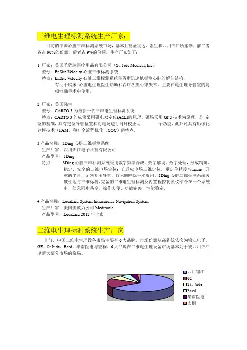
三维电生理标测系统生产厂家:目前的中国心脏三维标测系统市场,基本上被圣犹达、强生和四川锦江所垄断,前二者各占90%的份额,后者占9%的份额。
生产厂家如下:1 厂家:美国圣犹达医疗用品有限公司(St. Jude Medical, Inc)型号:EnSite Velocity心脏三维标测系统特点:EnSite Velocity心脏三维标测系统能清晰迅速地标测心脏的解剖结构,有助于临床心脏电生理医生诊断和治疗各类心律失常,主要在电生理导管室的射频消融手术中使用。
2 厂家:美国强生型号:CARTO 3为最新一代三维电生理标测系统特点:CARTO 3的成像采用磁电双定位(ACL)的原理。
磁场采用GPS技术为原理,是定位的基础,具有定位导管位置和对电场进行时时校正两个功能,此外还具有影像化建模技术(FAM)和)全流程优化(COC)的特点。
3产品名称:3Ding心脏三维标测系统生产厂家:四川锦江电子科技有限公司产品型号:3Ding特点:3Ding心脏三维标测系统采用数字频率合成、数字解调、数字处理,形成精确、稳定、安全的三维电场定位;自适应电场三维定位,重定位精度≤1mm;开放的平台,无须专用导管,较大的降低手术费用。
3Ding心脏三维标测系统突破性地将三维标测、完备的二维电生理标测及内置程控刺激仪结合在一个系统中,信息同步共享、操作方便、功能完善、性能稳定。
4产品名称:LocaLisa System Intracardiac Navigation System生产厂家:美国美敦力公司Medtronic产品型号:LocaLisa 2012年上市二维电生理标测系统生产厂家目前,中国二维电生理设备市场主要有6大品牌,市场份额从高到低依次为锦江电子、GE、St.Jude、Bard、华南医电与宏桐。
6大品牌在二维电生理设备市场基本处于被四川锦江垄断大部分市场的格局。


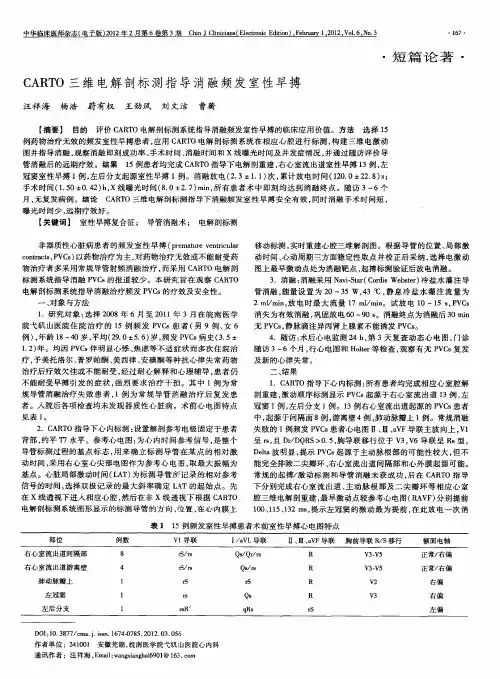
CARTO3三维标测系统快速解剖建模在阵发性心房颤动射频消融术中的应用田野;杨龙;郑亚西;刘晓桥;刘志琴;范寿年;殷跃辉【摘要】Objective: To investigate the safety and efficacy of CARTO3 fast anatomical mapping during radiofrequency ablation in patients with paroxysmal atrial ifbrillation (PAF). Methods: A total of 120 PAF patients treated in our hospital from 2013-01 to 2015-07 were enrolled. All patients received CARTO3 system for mapping and they were randomly divided into 2 groups: Control group, the patients had selective pulmonary vein angiography, followed by conventional point by point method to reconstruct left atrial model for guiding the ablation of PFA and Treatment group, the patients had selective pulmonary vein angiography followed by fast anatomical mapping to build left atrial model for guiding the ablation of PFA; the rest operational steps such as trans-septal and circumferential pulmonary vein ablation were the same.n=60 in each group. The times of operation, X-ray exposure and the rates of success, complication occurrence were compared between 2 groups. Results: All patients were successfully completed radiofrequency ablation for PAF. Compared with Control group, Treatment group had increased modeling time (8.5 ± 3.6) min vs (5.2 ± 2.3) min, while decreased pulmonary vein ostium determing time (12.0 ± 5.6) min vs (25.0 ± 8.4) min, circumferential pulmonary vein ablation time (95.0 ± 22.0) min vs (115.0 ± 25.0) min and X-ray exposure time (15.0 ± 6.3) min vs (24.0 ±5.5) min, allP<0.05. The rates of success andcomplication occurrence were similar between 2 groups, P>0.05. Conclusion: CARTO3 fast anatomical mapping is safe and effective for guiding radiofrequency ablation in PAF patients, it may decrease the X-ray exposure time and operation time which were important for treating the relevant patients.%目的:探讨CARTO3三维标测系统快速解剖建模在阵发性心房颤动(房颤)射频消融术中的安全性及有效性。
3D解剖学图谱用户手册
3D解剖学图谱能帮您更好地了解男性和女性不同的解剖结构。
您可以通过平板电脑和手机端轻松查阅人体15个不同的解剖层次。
3D解剖学图谱具有如下独特功能。
性别选择
通过屏幕左下方男女性别图标,选择男性或女性的解剖结构视图。
旋转3D全视角
移动屏幕底部的光标,即可围绕人体360°进行3D全视角观察。
缩放
3种方式实现缩放功能:
➢使用鼠标滚轮
➢使用屏幕右下方的“+”和“ - ”进行缩放
➢用两个手指在触摸屏缩放
移动
2种方式移动屏幕上的身体:
➢点击并拖动
➢在触摸屏上用手指拖动
解剖图层
涵盖15个解剖图层:
-皮肤
-复合表层-复合深层-肌肉
-深层肌肉-器官
-呼吸系统-消化系统-动脉
-静脉
-动脉和静脉-神经
-淋巴
-骨骼
-韧带
点击屏幕右侧的图标,查看相应解剖图层。
锁定皮肤层和/或骨骼层
点击左侧图示的按钮,结合皮肤和/或骨骼查看解剖图层。
部位名称
点击身体各个部位,即可看到相应部位描述.
其他功能
返回医纬达主页
关于3D解剖学图谱及其版权
保存或打印
让人体重新对准屏幕中心。
众茂医疗解剖学系列-人体3D系统解剖学
人体3D系统解剖学是医学生和医学专业人员学习人体解剖学的重要工具。
通过3D技术,我们能够以更加直观和生动的方式展示人体各个系统的解剖结构,帮助学习者更好地理解和掌握人体解剖学的知识。
众茂医疗-人体3D系统解剖学功能如下:
1.立体的视觉效果:3D人体解剖的最大特点是其立体的视觉效果。
通过3D 位置和相互关系。
2.全方位的视角:3D人体解剖提供了全方位的视角,让医学生可以从任意角度观察人体结构。
这有助于他们更好地理解人体构造,掌握复杂的解剖学知识。
3.可视化的互动:3D人体解剖软件通常提供可视化的互动功能,让用户可以实时操作并查看不同部位的组织结构。
这种互动性使得学习过程更加有趣、直观。
众茂医疗-人体3D解剖以其独特的立体视觉效果、全方位视角以及高精度等特点,为医学生、医生和研究人员提供了一种全新的学习方式。
它不仅提高了学习效率,还为诊断、手术规划以及医学研究提供了有力的支持。
随着技术的不断进步和应用领域的拓展,我们有理由相信,众茂医疗-人体3D解剖将在未来的医学教育和研究中发挥越来越重要的作用。