先天性血管环(congenital_vascular_rings)(研究运用)
- 格式:ppt
- 大小:6.27 MB
- 文档页数:26
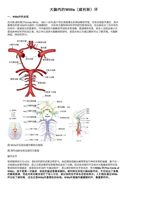
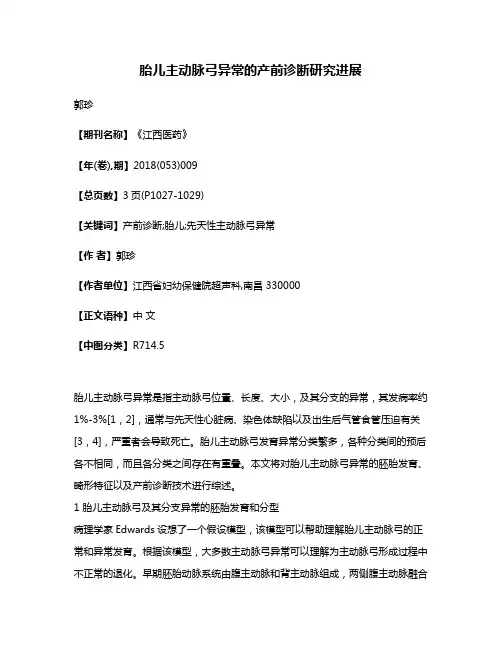
⼤脑内的Willis(威利斯)环⼀、Willis环的发现托马斯.威利斯(Thomas Willis,1621–1675)是17世纪英国著名的神经解剖学家,写有多部医学著作,其中最著名的是1664年出版的《⼤脑解剖》,对后来⼤脑和神经科学的研究影响深远,在出版后近⼆百多年的时间中⼀直被视为权威著作。
书中描述的⼤脑解剖学结构⾮常准确,图谱堪称完美,他本⼈也因此被认为是临床神经科学的创⽴者。
他之所以选择⼤脑解剖的研究,是因为他认为通过解剖可以了解灵魂。
⼤脑解剖起,神经科学兴。
图 Willis环及前后循环解剖⽰意图图颅内动脉名称及缩写⽰意图脑内五环他将脑神经分为10对,前6对的研究成果沿⽤⾄今。
他后期发现偏头痛常常始于神经末梢的抽搐,基于这⼀点他提出⾎管学假说,即认为是⾎管舒张刺激神经造成了头痛。
但这些成就均不及他对⼤脑基底部环状⾎管结构的详细描述,就是现在所说的“⼤脑动脉环”,是以威利斯的名字命名的,称为Willis 环(The Circle of Willis),他不是第⼀次描述,但他的描述是最准确的。
威利斯在发现⼤脑动脉环后,不仅给出了准确的解剖图谱,⽽且对其功能也进⾏了深⼊讨论,故以他的名字命名也恰如其分。
⼈们现在通过Willis 环记住了威利斯,这也正是Willis环重要性的体现。
Willis环是脑内最耀眼的环,最重要的环。
⼆、Willis环,形如五环⽣活中圆形、环状的东西有很多,我们的⼤脑是圆的,现在你还知道,其实⼤脑⾥⾯有最著名的Willis环,它就像闪亮红星⼀样,散散发光,特别耀眼。
Willis环,⼜被称为⼤脑动脉环,是指供应脑组织的动脉在脑底形成的环状结构。
它是颅内最重要的侧⽀循环途径,将双侧⼤脑半球和前、后循环联系起来。
由双侧⼤脑前动脉始段、双侧颈内动脉末端、两侧⼤脑后动脉及前、后交通动脉连通⽽成。
⽣理意义Willis环可以对供应脑组织的动脉进⾏⾎液调配,防⽌脑⾎液循环的过剩或不⾜。
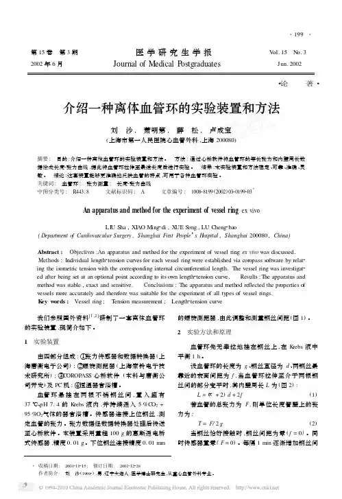
第15卷 第3期 医学研究生学报 V ol.15 N o.3 2002年6月 Journal of Medical P ostgraduates Jun.2002・论 著・介绍一种离体血管环的实验装置和方法刘 沙, 萧明第, 薛 松, 卢成宝(上海市第一人民医院心血管外科,上海200080)摘要: 目的:介绍一种离体血管环的实验装置和方法。
方法:通过心桥软件将血管环的等长张力和内壁周长数据绘成长度2张力曲线,据此将血管环拉伸至最适长度后进行实验。
结果:本实验装置和方法稳定、可靠、准确、灵敏。
结论:这套装置能够更准确地反映血管的特点,可用于各种血管环实验。
关键词: 血管环; 张力测量; 长度2张力曲线中图分类号: R443.8 文献标识码: A 文章编号: 100828199(2002)0320199203ΞAn apparatus and method for the experiment of vessel ring ex vivoLI U Sha,XI AO Ming2di,X UE S ong,LU Cheng2bao(Department o f Cardiovascular Surgery,Shanghai Fir st People’s Hospital,Shanghai200080,China)Abstract: Objectives:An apparatus and method for the experiment of vessel ring ex vivo was discussed. Methods:Individual length2tension curves for each vessel ring were established via coropass s oftware by relat2 ing the is ometric tension with the corresponding internal circum ferential length.The vessel ring was investigat2 ed after being set at an optional point according to its own length2tension curve. Results:The apparatus and method was stable,exact and sensitive. Conclusions:The apparatus and method reflected the properties of vessels m ore accurately and therefore was suitable for the experiment of all types of vessel rings.K ey w ords: Vessel ring; T ension measurement; Length2tension curve 我们参照国外资料[1,2]研制了一套离体血管环的实验装置,现简介如下。

胎儿主动脉弓异常的产前诊断研究进展郭珍【期刊名称】《江西医药》【年(卷),期】2018(053)009【总页数】3页(P1027-1029)【关键词】产前诊断;胎儿;先天性主动脉弓异常【作者】郭珍【作者单位】江西省妇幼保健院超声科,南昌 330000【正文语种】中文【中图分类】R714.5胎儿主动脉弓异常是指主动脉弓位置、长度、大小,及其分支的异常,其发病率约1%-3%[1,2],通常与先天性心脏病、染色体缺陷以及出生后气管食管压迫有关[3,4],严重者会导致死亡。
胎儿主动脉弓发育异常分类繁多,各种分类间的预后各不相同,而且各分类之间存在有重叠。
本文将对胎儿主动脉弓异常的胚胎发育、畸形特征以及产前诊断技术进行综述。
1 胎儿主动脉弓及其分支异常的胚胎发育和分型病理学家Edwards设想了一个假设模型,该模型可以帮助理解胎儿主动脉弓的正常和异常发育。
根据该模型,大多数主动脉弓异常可以理解为主动脉弓形成过程中不正常的退化。
早期胚胎动脉系统由腹主动脉和背主动脉组成,两侧腹主动脉融合成主动脉囊,背主动脉尾融合成降主动脉。
正常的主动脉弓发育过程中,左侧主动脉弓、动脉导管持续发育,而右侧锁骨下动脉与降主动脉之间的背主动脉、动脉导管退化。
最终,右侧主动脉弓近端发育为分叉成右侧颈总动脉、锁骨下动脉的头臂动脉。
正是由于发育过程中,6对鳃动脉弓的异常退化变异,才形成各种类型的主动脉弓先天发育异常。
主动脉弓发育异常胚胎学起源复杂,分类繁多,预后各不相同,并且各分类之间还存有重叠。
2017年李胜利等[5]在Stewart分型基础上,在Ⅳ组主动脉弓畸形中新增补了12个类型,总结分析主动脉弓异常的类型多达30多种,了解这些胚胎学发育概念和分类,对于理解先天性主动脉弓异常的发育有较大帮助[6]。
2 胎儿主动脉弓异常的畸形特点单纯主动脉弓缩窄胎儿在出生术后,健康状态良好。
而胎儿主动脉弓缩窄、右位主动脉弓常累计全身各器官,合并各种心外畸形。


血管环先天性血管环畸形胎儿发育早期由成对的主动脉组成的血管环未能正常地向单一主动脉过渡,其右背侧主动脉退化吸收不全或主动脉弓其他各段发育异常,使小儿主动脉弓依然保留完全或不完全的环形结构,而行走在血管环内的食管和气管受到不同程度的压迫,这种主动脉弓各段组合方式的异常称为血管环畸形。
血管环畸形的发生率在东方国家较低,畸形血管包绕食管和器官,对它们产生压迫,引起临床症状,从而需要外科处理,以缓解症状。
一、病理解剖及病理生理胎儿主动脉弓的发育是由环形主动脉弓向单一主动脉弓的发育过程。
胎儿血管环最初是由连接与主动脉囊和背侧主动脉弓之间的六对主动脉弓所形成,以后1、2、5对主动脉弓相继吸收,第3、4对主动脉弓之间的背侧主动脉中断,最后只剩下第4对和第6 对主动脉弓构成血管环。
此时主动脉囊分隔为升主动脉和肺动脉干,升主动脉和第4对主动脉弓相连,肺动脉干和第6对主动脉弓相连。
以后随着血管环的缩短变形,背侧主动脉的第1、2节间动脉段完全吸收,3—7节间动脉平面缩短,只有两例相当于第8节间的背侧主动脉才构成血管环侧缘。
正常情况下右第6弓远端和右背主动脉第8节间段吸收消失,血管环的右缘断裂、吸收,形成左侧单一的主动脉弓。
因此血管环畸形不外是由于右侧第8节间段背侧主动脉吸收不全或左侧动脉弓的异常吸收所致。
最常发生畸形的部位是在两侧第4弓和第8节间段的背主动脉的背主动脉。
血管环畸形的分类方法颇多,其中常见的有以下几种。
(一)双主动脉弓双主动脉弓是血管环的最常见类型,是因胚胎发育时左、右第4主动脉弓同时存在,其二个主动脉弓均发自升主动脉,从气管、食管两侧绕过背部汇入降主动脉,形成一个真正的环。
通常右弓(背部)发出右颈总和右锁骨下动脉,而较小的左弓(前部)发出左颈总和左锁骨下动脉。
约73%的病人以右弓为主,仅20%的病人以左弓为主,还有2%的病人左、右大小相仿。
(二)右主动脉弓并有左侧动脉韧带胚胎发育时如果左第4弓退化可产生右位主动脉弓,根据左弓退化中断的部位和左颈总功脉、左锁骨下动脉和动脉导管分支的模式,使右主动脉弓可产生多种类型的血管环畸形。
临床医学研究与实践2021年6月第6卷第17期DOI :10.19347/ki.2096-1413.202117044作者简介:王曦曦(1980-),女,汉族,北京人,主治医师,硕士。
研究方向:超声诊断学。
*通讯作者:王丽梅,E -mail :lmwangtrhos@.Prenatal ultrasound diagnosis and image analysis of fetal congenital vascularringWANG Xixi 1,WANG Limei 2*(1.Image Department,Beijing Yayuncun Amcare Women's and Children's Hospital,Beijing 100101;2.Ultrasound Department,Beijing Obstetrics and Gynecology Hospital,Capital Medical University,Beijing 100026,China)ABSTRACT:Objective To investigate the types of fetal congenital vascular ring (CVR)and the characteristics of prenatal ultrasound images.Methods A total of 17cases of CVR fetuses were found in pregnant women undergoing systematic prenatal ultrasound examination in our hospital from December 2012to June 2020.The sonographic data of the fetuses were retrospectively analyzed and the fetuses were followed up.Results Among 17cases of CVR diagnosed by prenatal ultrasound,there were 13cases of complete CVR (12cases of U-shaped CVR and 1case of O-shaped CVR);there were 4cases of partial CVR (3cases of C-shaped CVR and 1cases of pulmonary artery sling).Among the 17fetuses,6cases were complicated with other congenital heart diseases (2cases with tetralogy of Fallot,1caseswith absence of proximal leftpulmonary artery,2cases with ventricular septal defect,1cases with complete endocardial cushion defect and multiple organ malformation),and the remaining 11cases were not complicated with other malformations.Conclusion Three vessels and trachea view (3VT)and three vessels and pulmonary arterial branches view (3VP)planes are the most importantsections for diagnosing CVR in prenatal ultrasound examination.When vascular ring malformation is found,it is suggested to investigate the chromosomal abnormalities of the fetus clinically,and the fetus should be closely observed and followed up after birth.KEYWORDS:prenatal ultrasound diagnosis;congenital vascular ring;double aortic arch;pulmonary artery sling胎儿先天性血管环的产前超声诊断及图像分析王曦曦1,王丽梅2*(1.北京亚运村美中宜和妇儿医院影像科,北京,100101;2.首都医科大学附属北京妇产医院超声科,北京,100026)摘要:目的探讨胎儿先天性血管环(CVR )的类型与产前超声图像特征。
先天性血管环的外科诊断与治疗作者:李小兵王蓓妮沈立等来源:《中国医药导报》2016年第05期[摘要] 目的总结先天性血管环患儿的临床资料和外科手术治疗经验。
方法回顾性分析2011年3月~2015年10月在上海市儿童医院接受手术治疗的35例先天性血管环患儿临床资料,34例合并不同程度气管狭窄,临床表现为反复肺炎、慢性咳嗽、喘鸣、呼吸窘迫、吞咽困难和反复呕吐。
22例肺动脉吊带、6例双主动脉弓和1例右位主动脉弓伴左侧动脉导管患儿采用在非体外循环下手术治疗,6例肺动脉吊带患儿采用在体外循环下手术治疗。
所有患儿气管狭窄术中均未处理。
术后观察呼吸道症状、消化道症状、生长发育情况和气管狭窄的变化。
结果 35例患儿均顺利完成手术,术后呼吸机辅助时间为(47.80±133.19)h,监护室时间为(116.42±190.02)h,住院时间为(19.22±13.07)d;6例患儿体外循环时间为(125.16±29.99)min;3例患儿主动脉阻断时间为(70.00±13.00)min。
1例患儿术后并发气管内肉芽生成造成气道狭窄加重,脱离呼吸机困难,术后10 d行气管内支架置入手术,术后顺利脱离呼吸机。
2例患儿拔出气管插管后呼吸困难,再次气管插管呼吸机辅助,经积极处理后顺利撤离呼吸机。
1例患儿术后42 d仍不能脱离呼吸机,最终家长放弃治疗。
全组34例患儿顺利出院,1例术后放弃治疗。
术后随访1个月~4年,患儿术后呼吸道症状完全消失17例,喉喘鸣症状明显好转14例,反复呕吐消失2例。
患儿生长发育改善,气管狭窄随访中。
结论先天性血管环患儿易出现呼吸道和消化道症状,临床上出现反复肺炎、呼吸困难、喘鸣、吞咽困难和反复呕吐症状的患儿应怀疑血管环的可能,心脏超声和多排螺旋CT血管造影检查可确诊,早发现、早期手术治疗效果良好,合并心内畸形和局限性的气管狭窄并不需要同时纠治。
[关键词] 先天性;血管环;双主动脉弓;肺动脉吊带[中图分类号] R726.2 [文献标识码] A [文章编号] 1673-7210(2016)02(b)-0144-04Surgical diagnosis and treatment for congenital vascular ringLI Xiaobing WANG Beini SHEN Li XIE Yewei Z HANG Rufang▲Department of Cardiothoracic Surgery, Children's Hospital of Shanghai Children's Hospital Affiliated to Shanghai Jiaotong University, Shanghai 200062, China[Abstract] Objective To summarize the clinical experience of surgical treatment and the clinical information of children diagnosed of congenital vascular ring. Methods The clinical information of 35 patients diagnosed of congenital vascular ring treated in Children's Hospital of Shanghai from March 2011 to October 2015 were analyzed retrospectively, among whom 34 patients suffered from different degree of tracheostenosis, clinical manifestation were repeated pneumonia, chronic cough, asthma wheezing, respiratory distress, dysphagia and repeated vomiting. 22 cases ofpulmonary artery sling, 6 cases of double aortic archard and 1 case of right aortic arch with left artery catheter accepted vascular repairation without extracorporeal circulation. 6 patients of pulmonary artery sling completed vascular repairation undergoing extracorporeal circulation. The tracheostenosis weren't dealt with in the operation. Respiratory symptoms, enteron symptoms,physical development and the change of tracheostenosis were observed postoperatively. Results All 35 patients received the surgery successfully. The mean mechanical ventilation time was(47.80±133.19) h, the mean ICU observing time was (116.42±190.02) h, the mean length of stay was (19.22±13.07) d. The mean time of 6 cases of extracorporeal circulation was(125.16±29.99) min. The mean time of 3 cases of aortic clamp was (70.00±13.00) min. 1 patient suffered from endotrachealgranulation tissue formation which aggravated the tracheostenosis. The patient accepted endotracheal metallic stents placement 10 d after surgery and weaned from ventilator after the placement. 2 patients suffered from dyspnea when weaned from ventilator, they accepted intubation immediately and were able to the remove the trachea cannula successfully. 1 patient wasn't able to be weaned from ventilator 42 d after surgery and the parents gave up treatment.34 patients were discharged successfully and 1 case gave up treatment after operation. Postoperative follow-up lasted for 1 month to 4 years. In 17 cases, respiratory symptoms subsided completely. In 14 cases, asthma wheezing subsided. In 2 cases, recurrent vomitting subsided. Pediatric physical were improved and the tracheostenosis was under follow-up visit. Conclusion Patients suffered from congenital vascular ring are easily to have respiratory and enteron symptoms. Patients with manifestation of recurrent pneumonia, dyspnea, asthma wheezing, dysphagia and recurrent vomiting should be considered to be congenital vascular ring. Echocardiography and MSCTA can confirm the diagnosis. The outcome of surgery can be good if diagnosed early. Congenital vascular ring associated with intracardiac anomalies and focal limited tracheostenosis do not need to be treated simultaneously.[Key words] Congenital; Vascular ring; Double aortic arches; Pulmonary artery sling先天性血管环属于先天性主动脉弓血管畸形,尽管有些畸形并不形成一个完整的环,但都可形成血管包绕和压迫气管或食管。
先天性血管环ngenialvasularrings-V1
先天性血管环(ngenial vasularrings),也叫喉部动脉环,是一种
喉部周围的先天性血管畸形。
它是由于出生时喉部的血管过度生长和
分支导致的。
在正常情况下,这些血管会缩小并消失,但是在出生时
它们没有缩小,导致畸形。
先天性血管环的症状和表现多种多样。
有些患者没有症状,但其他患
者可能会出现吞咽困难、喉咙疼痛、呼吸困难等问题。
在一些情况下,这种血管环可能会压迫气管和食管,导致呼吸道和消化道问题。
目前,先天性血管环的确切病因不清楚。
但有一些因素可能增加患者
发展这种畸形的风险,例如细胞变异、遗传基因、毒物等。
如果发现了先天性血管环,患者可以接受多种治疗方式。
有些患者可
能只需要观察,因为没有症状。
其他患者可能需要手术或介入治疗,
这通常使用内科导管控制它。
更复杂的治疗可能包括在喉部植入支架
或手术切除血管环。
总的来说,先天性血管环是一种很常见的畸形,但它的病因和症状都
各不相同。
对于患者来说,重要的是及时检查和治疗,以避免任何潜
在的并发症。
先天性视盘前血管襻的临床分析刘求红;余杨桂;詹宇坚【摘要】Objective A retrospective study of congenital prepapillary vascular loops 9 cases of clinical data, analysis and summary of its clinical features. Methods Statistical analysis patients age, sex, which eye, visual acuity,chief complaint first visit. According to the ophthalmoscope eye examination, fundus photography and fundus fluorescence vascular imaging, analysis of the blood vessels in the loop part of the optic disc, the nature of vascular ( artery or vein ), the number of the loop, the vitreous and the retina situation, with the exception of blood vessels outside the loop of the other eye Complications. Results A total of 9 cases of 9 eyes , 5 right eye, 4 left eye, is single eye ; to the nature of angiogenesis : arterial vascular loops have 7 eyes, the vein vascular loops have 2 eyes; loop-like blood vessels and the optic disc : over side Temporal and often on a total of 6 eyes , 3 eyes below. Vascular loop to a single number 6, a loop 2, 3 loop more than 2 to a single common. Initial chief complaint ( symptoms ): 6 eyes id the floaters, as the 2, ophthalmic examination found a chance to floaters ( in front of fluttering shadow ) of the most common. Fundus examination revealed : vitreous opacity 5 , vascular hemorrhage loop 3 , a branch vein occlusion. Cases have been bleeding Chinese medicine treatment, branch vein occlusion with laser add TCM treatment, were clinically cured. Conclusion Congenital prepapillary vascular loops 9 cases clinical features are summarized as follows: Syruptoms of the disease isfloaters, and other disease as a result of eye examination can also be found. We found that the 9 cases are young adults , and have no difference hetween of men and women. the disease may happen in single eye or two eyes , the eyes suffer from these diseases in this group isMonocular,Monocular should be common. Vascular loop for more than artery, on the surface near the edge of the ahove ( the temporal,on ) as often.%目的回顾性研究先天性视盘血管襻9例(9只眼)的临床资料,分析并总结其临床特点.方法统计分析患者的年龄、性别、眼别、视力、初诊主诉.根据检眼镜眼底检查、眼底照像和荧光素眼底血管造影(FFA),分析血管襻在视盘的部位、血管性质(动脉性或静脉性),襻的数目,玻璃体及视网膜状况,除血管襻外的其它眼部合并症.结果共9例(9只眼),其中右眼5例,左眼4例,均为单眼性;以血管性质分:动脉性血管襻7只眼,静脉性2只眼;襻状血管与视盘关系:以上方及颞上多见共6只眼,下方3只眼.血管襻数目为单个6只眼,2襻1只眼,3襻以上2只眼,以单个多见.初诊主诉(自觉症状):飞蚁症6只眼,视矇2只眼,眼科检查中偶然发现1只眼,以飞蚁症(眼前飘动性黑影)最常见.眼底检查发现:玻璃体混浊5只眼,血管襻出血3只眼,分枝静脉阻塞1只眼.出血病例均经中医辨证内服中药治疗,分枝静脉阻塞者行激光光凝配合中医辨证治疗,均获临床治愈.结论本病初诊自觉症状(主诉)以飞蚁症最常见,视矇(视力下降)及因其它病眼底检查中也可发现.发现时年龄多为青壮年,无男女差别.本病单眼、双眼均可发病,本组病例均为单眼,可见单眼应常见.血管襻多为动脉性,多在视盘表面或盘缘附近,以上方(颞上、上)为多见.【期刊名称】《临床眼科杂志》【年(卷),期】2011(019)003【总页数】3页(P243-245)【关键词】先天性视盘前血管襻;临床分析【作者】刘求红;余杨桂;詹宇坚【作者单位】510405,广州中医药大学第一附属医院眼科;510405,广州中医药大学第一附属医院眼科;510405,广州中医药大学第一附属医院眼科【正文语种】中文先天性视盘前血管襻(congenital prepapillary vascular loops)又称视乳头血管襻或玻璃体内血管襻。
文/罗 凯 上海交通大学医学院附属上海儿童医学中心先天性血管环相关知识随着优生优育观念及产前诊断技术的不断提高,越来越多先天性畸形在胎儿期即可通过心脏彩超或心脏磁共振明确诊断,其中尤以血管畸形的检出率最高。
本文将介绍先天性血管环的发生与治疗。
化学物质接触史;孕妇有糖尿病、高血压等慢性病史,或者有吸烟、饮酒等不良嗜好;夫妻双方存在先天性心脏病家族史等。
对于存在以上高危因素的孕妇,一般建议在孕18~28周内至专科医院完善胎儿超声心动图检查。
限制,引起相关临床症状:管狭窄,主要症状包括喘鸣、气促、打鼾、仰头呼吸及频发呼吸道感染等;为吞咽困难、误吸及食欲不振等。
由于常规的线片、超声心动图及CT等影像学检查对血管环成像较为容易,因此临床诊断并不困难,胎儿期即可诊断,且检出率及准确率均较高。
如何治疗先天性血管环?由于先天性血管环本身不存在自愈可能,因此手术是唯一的治疗手段。
多数血管环患儿因出现气管或食管压迫症状而就诊,症状的严重程度与受压程度相关。
一般来说,出现因血管环导致青春护航Youth Escort的临床症状即存在手术适应证,手术时机的选择:①单纯的血管环手术年龄多选择在6个月至2岁之间,大部分此年龄段的患儿已经出现初发症状,但血管环对气管的压迫尚在可逆范围内(长期压迫会造成不可逆的气管切迹),且胸腔空间较浅,手术操作难度不大;②约15%~20%的血管环会同时合并其他先天性心血管畸形,手术时机可选择与其他畸形同时进行治疗。
先天性血管环的手术原则为在保证主动脉及其分支供血正常的情况下,离断血管环,解除其对气管及食管的压迫。
单纯血管环手术目前多采用侧开胸小切口的方式,根据解剖结构的不同选择不同入路:①右侧开胸;②左侧开胸。
对于合并其他心血管畸形需同时处理者,则需选择正中开胸切口完成手术。
部分仅需离断动脉导管韧带的患儿也可实施胸腔镜或机器人手术。
手术成功率方面,近30年,单纯血管环已无手术死亡率,合并其他心血管畸形的手术成功率达到95%以上,患儿术后生活质量与正常人无异。
血管环模型的研究进展李秋影【摘要】Main study on vascular ring could depart in two aparts,one for vascular contractility,the other for angiogenesis.The study for vascular contractility was used in variety fields including hypertension, cerebrovascular insufficiency syndrome pulmonary hypertension congestive heart failure shock etc.And anti-turner research is the main field of angiogenesis study.Vascular ring study play an important role in new drug founding and in basic research.%血管环模型在新药筛选和病理生理基础研究中占有重要地位.目前血管环的研究类型主要包括两大类:血管收缩活性研究和血管生成活性研究.血管收缩活性研究涉及的疾病领域宽广,包括高血压、脑供血不足、充血性心力衰竭等.血管生成活性研究则主要集中在抗肿瘤研究和动静脉吻合的研究上.随着新技术和新思路的引进,血管环实验在相关领域的新药研发、基础研究等方面发挥着重要的作用.【期刊名称】《医学综述》【年(卷),期】2011(017)003【总页数】4页(P339-342)【关键词】血管环;血管收缩;血管生成【作者】李秋影【作者单位】天津中医药大学,天津,300193;天津天士力研究院药理毒理所,天津,300410【正文语种】中文【中图分类】R965.2血管环研究起始于1980年,Robert F.Furchgott教授把血管条改成血管环模型,发现内皮细胞可释放一种使血管舒张的因子,即一氧化氮(nitric oxide,NO)。
先天性血管环12例诊断与治疗分析崇梅;贺彦;李伟;韩玲;刘迎龙;金梅;顾虹;程沛;苏俊武;耿斌【期刊名称】《中国循证儿科杂志》【年(卷),期】2012(7)3【摘要】Objective To study the clinical features, diagnosis and treatment of congenital vascular ring. Methods The clinical data(age, gender, age of onset, clinical manliestations, laboratory examinations ), imaging examinations (X-ray, echocardiography and multi-detector CT scan ), pulmonary function, bronchoscopy, diagnostic methods, treatment, surgery according to the pathologic types and follow-up of children identified as congenital vascular rings at Beijing Anzhen Hospital from July 2008 to December 2011 were retrospectively reviewed. Results Twelve cases diagnosed as congenital vascular rings were included into the study, including 3 males and 9 females, aged from 1 month to 21 years ( 7 cases less than 1 year of age ). Two cases were correctly diagnosed at the first hospital visit and 9 cases were misdiagnosed. Eleven cases had a history of recurrent cough and wheezing, 2 cases had difficulty in feeding, 3 cases had cyanosis, 10 cases had heart murmurs and 9 cases had growth retardation. The results of echocardiography were double aorta arch in 4 cases, right aortic arch with left ductus arteriosus in 1 case, pulmonary artery sling in 7 cases. Ten cases were combined with other heart malformations. Ten cases were given multi-detector CT airwayreconstruction and showed tracheobronchial stenosis in 9 cases ( 5 cases with trachea, 3 cases with right principle bronchus, 1 case with left principle bronchus ), esophagus stenosis in 2 cases. Two cases were given bronchoscopy to confirm the stenosis of trachea in 1 case, right principle bronchus in 1 case. Seven patients underwent surgery according to the pathologic types including 2 cases of cutting off the small arch, 1 case of cutting off the left patent ductus arteriosus and 4 cases of reimplanting of the left pulmonary artery. One of them died. The respiratory symptomsand growth in 6 cases gradually were improved in 3 to 6 months after operation. Conclusions The children with recurrent respiratory symptoms such as chronic cough and wheeze should be examined for the possible presence of congenital vascular rings. Echocardiography as a noninvasive technique combined with multi-detector CT can clearly show the anatomy of vascular rings. Early surgical management to remove the tracheal stenosis is vital for survival.%目的探讨先天性血管环的临床特征、诊断与治疗.方法回顾性分析2008年7月至2011年12月在首都医科大学附属北京安贞医院诊断为先天性血管环患儿的临床特征、影像学检查、诊断和治疗.结果 12例先天性血管环患儿进入分析,男3例,女9例.就诊年龄1个月至21岁,<1岁7例.反复咳喘11例,喂养困难2例,青紫3例,心脏杂音10例.12例超声心动图示主动脉双弓4例、右位主动脉弓伴左位动脉导管未闭1例、肺动脉吊带7例,其中10例伴其他心脏畸形.10例64排CT气道重建示主气管狭窄5例、左主支气管狭窄1例,右侧气管狭窄3例,食管狭窄2例;2例纤维支气管镜示主气管狭窄1例,右主支气管狭窄1例.7例行手术治疗,根据血管环的病理类型选择术式,包括2例切断双主动脉弓中的次弓、1例切断右位主动脉弓伴随的左位动脉导管、4例左肺动脉移植术.1例术后死亡,6例术后3~6个月呼吸道症状逐渐好转,生长发育改善.结论儿童中反复出现原因不明的咳喘等呼吸道症状者,应考虑到先天性血管环的可能,超声心动图联合多排CT可明确诊断,及早手术解除气管狭窄是改善预后的关键.【总页数】5页(P205-209)【作者】崇梅;贺彦;李伟;韩玲;刘迎龙;金梅;顾虹;程沛;苏俊武;耿斌【作者单位】首都医科大学附属北京安贞医院儿童心脏中心,北京,100029;首都医科大学附属北京安贞医院儿童心脏中心,北京,100029;首都医科大学附属北京安贞医院儿童心脏中心,北京,100029;首都医科大学附属北京安贞医院儿童心脏中心,北京,100029;首都医科大学附属北京安贞医院儿童心脏中心,北京,100029;首都医科大学附属北京安贞医院儿童心脏中心,北京,100029;首都医科大学附属北京安贞医院儿童心脏中心,北京,100029;首都医科大学附属北京安贞医院儿童心脏中心,北京,100029;首都医科大学附属北京安贞医院儿童心脏中心,北京,100029;首都医科大学附属北京安贞医院儿童心脏中心,北京,100029【正文语种】中文【相关文献】1.成人先天性巨输尿管的诊断与治疗(附12例报告) [J], 汪官富;陈安屏;章灵芝;方夏玲;卢思保;王仙友2.先天性弧立肾的诊断与治疗附12例临床分析 [J], 陈荣山;纵斌;崔飞轮;徐获荣;董刚3.儿童先天性血管环41例多排螺旋CT诊断的回顾性分析 [J], 毕璐琳;杨辉4.单中心60例先天性血管环回顾性分析 [J], 范国清;张智睿;黎媛;陈应富;李静5.先天性输尿管瓣膜症16例诊断与治疗分析 [J], 朱大元因版权原因,仅展示原文概要,查看原文内容请购买。
胎儿血管环的超声诊断特点与检查技巧胎儿血管环的超声诊断特点与检查技巧本文关键词:超声,胎儿,血管,诊断,检查胎儿血管环的超声诊断特点与检查技巧本文简介:摘要:先天性血管环是一种罕见的弓动脉发育不良畸形,其胚胎学发育机制复杂,表现形式多样,部分血管环畸形可合并染色体异常,产前超声治疗困难。
超声核查心动图作为临床常用的胎儿心脏检查方法,在先天性之中心脏病的筛查和诊断中发挥重要作用。
本文就胎儿血管环的二维及三维超声诊断三维特征、检查技巧,以及该病作出的鉴别诊断等进行综述。
胎儿血管环的超声诊断特点与检查技巧本文内容:摘要:先天性血管环是一种罕见的弓动脉畸形,其胚胎学发育机制复杂,表现形式多样,个别血管环畸形可合并染色体异常,产前超声诊断困难。
超声心动图作为临床常用的胎儿心脏检查方法,在展现出先天性心脏病的筛查和诊断中发挥重要作用。
本文就胎儿脊髓血管环内的二维及三维超声诊断特点、检查技巧,以及该病的鉴别诊断等进行综述。
关键词:超声心动描记术; 弓动脉畸形; 血管环; 胎儿;Abstract:Congenital vascular ring is a rare malformation of aortic arch anomalies. The embryonic development of vascular rings is complex with diverseforms.In addition, vascular rings may associate with chromosome abnormalities. It is challenging for obstetric sonographers to make a correct diagnosis. As ly used incardiac examination, fetal echocardiography plays a key rolein the screening and diagnosis of congenital heartdisease.This paper summaries the prenatal diagnostic features and scanning skills of vascular ring by two and three-dimensional echocardiography, and the differential diagnosis and prognosis of this rare abnormality are also commented.Keyword:Echocardiography; Aortic arch anomaly; Vascular ring; Fetus;先天性血管环是弓动脉的畸型,临床罕见,占胎儿先天性心脏病的0.8%~1.3%[1]。
血管环和血管带第一节血管环血管环是大动脉(主动脉弓及其分支)的畸形,压迫气管和 / 或食管。
【简史】1737年, Hommel首次描述双主动脉弓畸形(后为Turner引证),一个世纪以后Von Siebold再次描述。
1939年,Wolman报道双主动脉弓压迫气管和食管综合征。
1794年,Bayford 发表了一篇据他认为由于迷走右锁骨下动脉引起吞咽困难的报道,但他描绘的血管走行于食管和气管之间,而不是食管后方的实际位臵。
1945年,Gross施行双主动脉弓矫治手术,成为现代外科治疗的首次尝试。
此后,他开创了血管环外科治疗的其他多种术式。
血管环X线诊断方法最初是由Neuhauser 报告的。
1922年,Congdon观察了人体胚胎期主动脉弓的复杂发育过程,但是直到Gross开创手术疗法以后,才得到临床应用。
1948年,Edwards提出双主动脉弓病因的假说,形成了主动脉弓多种复杂畸形的理论,Stewart、Kincaid 和Edwards进一步作了阐述。
1951年,Barry 对Congdon的基础理论进行了透彻的总结和回顾。
【形态学】血管环病人升主动脉、主动脉弓和降主动脉及其分支排列的变异很多。
有几种变异形式可使气管或食管,甚至两者都受压,并具有外科意义。
可分类为:完全性血管环;-部分性血管环;?无名动脉或左颈总动脉压迫。
一、完全性血管环(一)双主动脉弓双主动脉弓病人的升主动脉起源正常,但在出心包后分成两个分支,即左、右主动脉弓,两弓向后汇合成降主动脉。
左前弓经气管前延伸至气管正常位臵的左侧,与动脉导管(动脉韧带更多见)汇成降主动脉。
右后弓越过食管右后方汇入左侧降主动脉,从而形成血管环可加重喘鸣。
颈部伸展时常使喘鸣缓解。
婴儿哭声嘶哑,没有症状明显的喘鸣和呼吸响声,常有持续性犬吠样咳嗽。
可发生窒息、严重紫绀和意识丧失。
气管梗阻严重时,有明显的吸气凹陷。
常反复发生呼吸道感染,并加重呼吸道梗阻。
梗阻较轻时,只有在呼吸道感染时有明显的梗阻症状。