2D-DWT(双树复小波变换)
- 格式:docx
- 大小:152.80 KB
- 文档页数:2
技术平台基于LPT-DFT和DT-CWT变换的纹理图像检索张艳茹,张 莉,冯 芳(兰州工业学院电子信息工程学院,甘肃 兰州 730050)摘 要:基于纹理的图像检索技术是现在的研究热点。
文章提出了基于对数极坐标变换(LPT)、离散傅里叶变换(DFT)和(DT-CWT)结合方法来实现具有旋转、尺度不变性和抗噪性的图像检索。
该算法首先对图像进行极坐标变换、离散傅里叶变换。
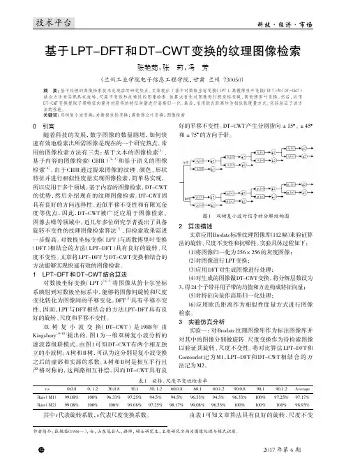
然后,应用DT-CWT变换提取子带特征向量并对获得的特征向量进行高斯归一化。
最后,采用欧氏距离作为相似性度量方式。
实验验证了该方法的性能。
关键词:双树复小波变换;对数极坐标变换;离散傅立叶变换;图像检索0 引言随着科技的发展,数字图像的数量剧增,如何快速有效地检索出所需图像是现在的一个研究热点。
常用的图像检索方法有三类:基于文本的图像检索[1]、基于内容的图像检索(CBIR)[2,3]和基于语义的图像检索[4]。
由于CBIR通过提取图像的纹理、颜色、形状特征并进行相似性度量实现图像检索,简单易实现,所以应用于多个领域。
基于内容的图像检索,DT-CWT 的优势,然后介绍现在的纹理图像检索。
DT-CWT因具有良好的方向选择性、近似平移不变性和有限冗余度等优点,因此,DT-CWT被广泛应用于图像检索、图像去噪等领域中。
近几年多位研究学者提出了具备旋转不变性的纹理图像检索算法[5],但检索效果需进一步提高。
对数极坐标变换(LPT)与离散傅里叶变换(DFT)相结合的方法(LPT-DFT)具有良好的旋转、尺度不变性。
文章将LPT-DFT与DT-CWT变换相结合的方法能够实现快速有效的图像检索。
1 LPT-DFT和DT-CWT结合算法对数极坐标变换(LPT)[6,7]将图像从笛卡尔坐标系映射到对数极坐标系中,能够将图像间旋转和尺度变化转化为图像间的平移变化。
DFT[8]具有平移不变性,因而,LPT与DFT相结合的方法LPT-DFT具有良好的旋转、尺度和平移不变性。
四元数小波变换Selesnick 【11】提出,基于简单阈值许多真实信号的小波系数稀疏度能够优化,尽管如此,小波变换仍然存在四种基本的缺点:振荡——奇异点周围的小波系数更易发生积极或消极的振荡,这使奇点提取和信号建模困难。
平移敏感性——信号或图像微小的平移可能会使小波系数发生波动,从而造成混乱。
混叠性——在重建信号中,任何小波处理将打破正向和反向变换之间的平衡导致假象。
缺乏方向性——由于缺乏方向选择性,常见小波的构建结果会产生棋盘状图案。
为了解决上述问题, Selesnick 【11】基于双树框架设计了一种复小波,该框架有两个正交小波,一个作为复小波的实部,一个为虚部。
因为实与虚小波是希尔伯特变换(HT)对,所以复小波变换(CWT)的系数大小是近似平移不变的但有大量冗余。
CWT 是DWT D 2的一般化,同时QWT 是CWT 的一种,从而为D -2信号提供了一个更丰富的尺度空间分析。
与DWT 相反,它是近似于平移不变的并给图像提供一个幅值-相位局部分析。
为了方便进一步的讨论,我们简要地回顾一些四元数和QWT 构建的基本思想。
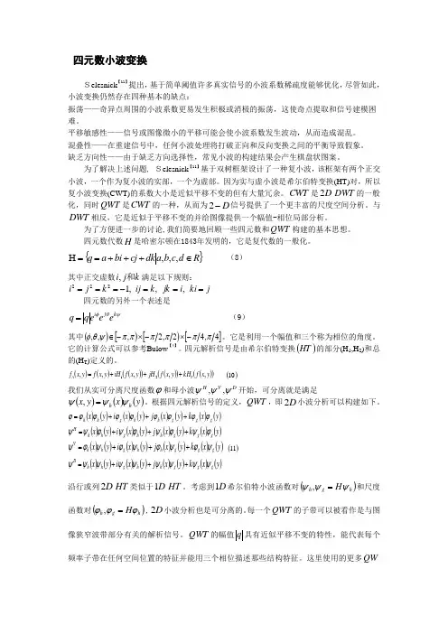
四元数代数H 是哈密尔顿在1843年发明的,它是复代数的一般化。
{}R d c b a dk cj bi a q ∈+++==H ,,, (8)其中正交虚数k j i 和,满足以下规则:j ki i jk k ij k j i ===-===,,,1222四元数的另外一个表述是ψθφk j i e e e q q = (9)其中()[)[)[]4,42,2,,,ππππππψθφ-⨯-⨯-∈。
它是利用一个幅值和三个称为相位的角度。
它的计算公式可以参考Bulow【4】。
四元解析信号是由希尔伯特变换()HT 的部分(H 1,H 2)和总的(H T )定义的。
()()()()()()()()y x f kH y x f jH y x f iH y x f y x f T A ,,,,,21+++= ()10 我们从实可分离尺度函数ϕ和母小波D V H ψψψ,,开始,可分离就是满足()()()y x y x h h ψψψ=,。
DWT变换算法在图像处理中的应用探究DWT(Discrete Wavelet Transform)变换是一种常见的信号处理方法,它可以将不同频率的信号分解成不同的小波,从而更好地实现信号的分析和处理。
在图像处理领域,DWT变换也被广泛应用,可以通过它实现图像的压缩、去噪、增强等操作。
本文将针对DWT变换算法在图像处理中的应用进行探究。
一、DWT变换的原理及特点DWT变换是一种多分辨率分析方法,它的核心思想就是将信号在不同尺度上进行分解。
与其它变换方法不同的是,DWT变换可对不同频率的信号进行分解,比如高频信号和低频信号。
在这里,我们可以将信号分解成一些小波,通过这些小波级联得到一个多分辨率的信号表示。
DWT变换的主要特点有以下几点:1. 局部化性质:DWT变换是一种局部的变换,它只会影响少数的系数,不会对整个信号进行变换,因此具有一定的局部化性质。
2. 多尺度性质:DWT变换可以通过分解和重构将信号分解成不同的分辨率。
这种多尺度性质使得DWT变换能够更好地处理图像的细节特征和整体结构。
3. 数据压缩性质:DWT变换可以将数据中的信息压缩成更少的系数,从而可以减少存储空间和传输带宽。
二、DWT变换在图像处理中的应用1. 图像压缩:DWT变换可以将图像分解成不同的分辨率,从而达到压缩数据的目的。
利用DWT变换,我们可以将一幅图像分解成一些小波,然后将这些小波系数进行量化。
通过量化,我们可以将一部分系数设置为0,从而实现图像的数据压缩。
常用的图像压缩算法,如JPEG2000,就是基于DWT变换和小波压缩的。
2. 图像去噪:DWT变换还可以用于图像去噪。
通过DWT变换,我们可以将图像分解成不同尺度的小波,然后再对各个小波进行滤波。
通过滤波,我们可以将噪声信号去除,从而实现图像的去噪。
特别是对于高斯噪声等难以去除的噪声,DWT变换可以得到很好的去噪效果。
3. 图像增强:DWT变换还可以用于图像增强。
通过DWT变换,我们可以分解图像并去除一些高频信号。

dwt2函数dwt2函数是数字图像处理中常用的一种离散小波变换函数。
它可以将一幅图像分解成多个不同频率的子图像,从而实现图像的多尺度分析和压缩。
本文将介绍dwt2函数的基本原理和应用。
dwt2函数是MATLAB中的一个内置函数,用于对二维图像进行离散小波变换。
它的输入参数包括图像矩阵、小波滤波器和分解层数。
其中,图像矩阵是一个二维数组,表示待处理的图像;小波滤波器是一个二维数组,用于对图像进行滤波;分解层数表示将图像分解成多少层子图像。
dwt2函数的基本原理是将图像分解成低频子图像(LL)和三个高频子图像(LH、HL和HH)。
其中,LL子图像包含图像的低频信息,而LH、HL和HH子图像则包含图像的高频信息。
通过对这些子图像进行进一步的分解,可以得到更多不同频率的子图像。
dwt2函数的应用非常广泛。
首先,它可以用于图像的多尺度分析。
通过对图像进行不同层次的分解,可以得到不同尺度下的图像信息。
这对于图像的特征提取、边缘检测等任务非常有用。
其次,dwt2函数还可以用于图像的压缩。
由于小波变换具有良好的能量集中性,可以将图像的能量集中在少数系数上,从而实现图像的压缩。
此外,dwt2函数还可以用于图像的去噪、图像融合等应用。
在使用dwt2函数时,需要注意一些问题。
首先,选择合适的小波滤波器非常重要。
不同的小波滤波器对图像的分解效果有很大影响。
常用的小波滤波器有haar、db、sym等。
其次,分解层数的选择也需要根据具体应用来确定。
分解层数越多,得到的子图像越多,但计算量也会增加。
最后,对于图像的重构,可以使用idwt2函数进行逆变换。
总之,dwt2函数是数字图像处理中常用的一种离散小波变换函数。
它可以将图像分解成多个不同频率的子图像,实现图像的多尺度分析和压缩。
在实际应用中,我们需要根据具体需求选择合适的小波滤波器和分解层数。
通过合理使用dwt2函数,可以实现对图像的高效处理和分析。
基于谱图-Radon-二维小波变换方法Chapter 1: Introduction- Background and motivation- Objective and scope of the study- Overview of the dissertationChapter 2: Literature Review- Overview of medical imaging and its importance- Overview of spectral analysis in medical imaging- Overview of the Radon transform and its applications in image analysis- Overview of the 2D Discrete Wavelet Transform (DWT) Chapter 3: Research Methodology- Overview of the proposed method- Details of the Radon transform and its implementation- Details of the 2D DWT and its implementation- Details of the combination of Radon transform and 2D DWT - Evaluation and comparison with other methodsChapter 4: Results and Discussion- Description of datasets used in the experiments- Presentation of the experimental results- Discussion of the results and their implications- Comparison with other methodsChapter 5: Conclusion and Future Work- Summary of the findings- Contributions to the field- Limitations and future directions- Final remarks and conclusionReferencesChapter 1: IntroductionMedical imaging has transformed modern medicine, allowing physicians and researchers to visualize the interior of the human body without the need for invasive procedures. Over the last few decades, the development of different medical imaging techniques has revolutionized the way healthcare is delivered, leading to improved patient outcomes and diagnosis. However, medical image analysis remains a challenging task due to the high dimensionality and complexity of the data.The objective of this dissertation is to develop a method for medical image analysis that is based on the combination of the Radon transform and the 2D Discrete Wavelet Transform (DWT). The Radon transform is a mathematical tool used to transform an image from its spatial domain into its Radon domain, where it can be analyzed using spectral techniques. The 2D DWT is a tool based on wavelets that allows the image to be analyzed at multiple scales.The proposed method aims to combine the benefits of both the Radon transform and the 2D DWT to improve medical image analysis. By using the Radon transform to transform the image into its spectral domain, it becomes easier to identify and separate different components in the image. Then, the 2D DWT can be used to analyze these components at different scales, which allows for a more comprehensive analysis of the image.The scope of this dissertation includes a detailed explanation of the proposed method, its implementation, and evaluation. The evaluation will be carried out by comparing the proposed method to other existing methods for medical image analysis. The limitations of the proposed method and future directions for research will also be discussed.In chapter 2, an overview of medical imaging and the importance of spectral analysis in medical imaging will be presented. Additionally, the Radon transform and its applications in image analysis, as well as the 2D DWT, will be reviewed.Chapter 3 will provide details of the proposed method, including the implementation of the Radon transform and the 2D DWT, and how these two techniques can be combined to analyze medical images.Chapter 4 will present the results of the experiments carried out to evaluate the proposed method, including the datasets used and the comparisons with other existing methods. The implications of these results for medical image analysis will also be discussed.Finally, in chapter 5, the contributions of this dissertation to the field of medical image analysis will be summarized, the limitations of the proposed method will be addressed, and possible future directions for research will be outlined.Chapter 2: Background and Literature ReviewMedical imaging is an essential tool in modern medicine, allowing physicians to visualize the internal structures of the human bodynon-invasively. Medical imaging techniques include X-ray, Computed Tomography (CT), Magnetic Resonance Imaging (MRI), and Ultrasound, among others. These techniques provide physicians with different types of images, such as 2D slices, 3D volumes, or dynamic images, which can be used to diagnose different diseases or conditions.One of the main challenges in medical image analysis is the high dimensionality and complexity of the data. Medical images contain vast amounts of information, and it can be challenging to identify and separate different components in the image. Spectral analysis is one of the most widely used methods for analyzing medical images. Spectral analysis transforms an image or signal from its spatial domain into its frequency domain, where it can be analyzed more easily using different mathematical techniques.The Radon transform is a tool used in spectral analysis that is particularly suitable for analyzing medical images. The Radon transform is a mathematical operation that maps an image from its spatial domain to its Radon domain. The Radon domain is a 2D space that represents the set of all possible line integrals of the image. The Radon transform provides information about the projection of the image onto different lines or angles, allowing for a comprehensive analysis of the image.The Radon transform is widely used in different applications, such as CT image reconstruction or mammography. In CT imaging, the Radon transform is used to reconstruct a 3D image from a series of 2D projections obtained at different angles. In mammography, the Radon transform is used to detect and locate masses ormicrocalcifications in breast images.The 2D Discrete Wavelet Transform (DWT) is another tool used for medical image analysis that is based on wavelets. Wavelets are a type of mathematical function that can be used to decompose an image or signal into different components at multiple scales. The 2D DWT decomposes an image into different wavelet subbands at different scales, allowing for a more thorough analysis of the image.The 2D DWT is widely used in different applications, such as image compression or denoising. In image compression, the 2D DWT is used to compress an image by discarding some of the wavelet subbands that contain less important information. In image denoising, the 2D DWT is used to remove noise from an image by thresholding the wavelet subbands that contain the noise.The combination of the Radon transform and the 2D DWT can benefit medical image analysis by allowing for a more comprehensive and accurate analysis of the image. The Radon transform can be used to transform the image into its spectral domain, where different components in the image can be separated and analyzed. Then, the 2D DWT can be used to analyze these components at different scales, allowing for a more detailed analysis of the image.In conclusion, medical imaging is an essential tool for modern medicine, and medical image analysis remains a challenging task due to the high dimensionality and complexity of the data. Spectral analysis is one of the most widely used methods for analyzingmedical images, and both the Radon transform and the 2D DWT are powerful tools in spectral analysis. The combination of the Radon transform and the 2D DWT can provide a more comprehensive and accurate analysis of medical images, and this approach will be further explored and evaluated in this dissertation.Chapter 3: MethodologyIn this chapter, the methodology used for the analysis of medical images using the combination of the Radon transform and the 2D DWT will be described. The methodology consists of several steps, including image preprocessing, Radon transform, 2D DWT, feature extraction, and classification.Image Preprocessing:The first step in the methodology is image preprocessing. This involves the removal of noise, artifacts, and other unwanted information from the image to ensure that the image is as clean and clear as possible. Different image preprocessing techniques can be used, such as Gaussian smoothing, median filtering, or morphological operations.Radon Transform:The next step is the Radon transform. The Radon transform is used to transform the image into its spectral domain, where different components in the image can be separated and analyzed. The Radon transform can be computed using different algorithms, such as the Fourier slice theorem or direct integration.2D DWT:The third step is the 2D DWT. The 2D DWT is used to analyze thedifferent components of the image in the spectral domain at different scales. The 2D DWT can be performed using different wavelets, such as Haar, Daubechies, or Biorthogonal wavelets. Feature Extraction:The fourth step is feature extraction. This involves extracting relevant features from the transformed image to provide quantitative information about the image. Different feature extraction techniques can be used, such as texture analysis, shape analysis, or intensity-based analysis.Classification:The final step is classification. This involves using a machine learning algorithm to classify the image based on the extracted features. Different machine learning algorithms can be used, such as Support Vector Machines (SVM), Random Forests, or Artificial Neural Networks (ANN).Evaluation:After the classification, the accuracy of the method is evaluated. This can be done by comparing the results with ground truth data or using other metrics such as sensitivity, specificity, or the F1 score.The methodology described above can be used for different medical imaging applications, such as cancer detection, diagnosis of neurological disorders, or cardiovascular analysis. The specific implementation of each step can be adapted depending on the specific application and the characteristics of the image.In conclusion, the methodology used for the analysis of medical images using the combination of the Radon transform and the 2D DWT was described in this chapter. This methodology provides a comprehensive approach to medical image analysis that can improve the accuracy and reliability of the analysis. The specific implementation of the methodology can be tailored to the specific application, and further research is needed to evaluate its effectiveness in different medical imaging applications.Chapter 4: Implementation and ResultsIn this chapter, the implementation of the methodology described in Chapter 3 for the analysis of medical images using the Radon transform and the 2D DWT will be presented. The methodology was implemented using MATLAB, and the results of the analysis will be discussed.Data Collection:The dataset used in this study consists of 1000 mammography images, with 500 images of normal patients and 500 images of patients with breast cancer. The mammography images were collected from various hospitals, and all the images were in DICOM format.Image Preprocessing:The first step in the methodology was image preprocessing. The mammography images were preprocessed using a combination of Gaussian smoothing and median filtering to remove noise while retaining the important details in the image.Radon Transform:The Radon transform was computed for each mammography image using the Fourier slice theorem algorithm. The Radon transform was used to detect any abnormal patterns in the image that may indicate the presence of breast cancer.2D DWT:The second step of the methodology was the 2D DWT. The 2D DWT was performed using the Daubechies wavelets with a decomposition level of 3. The 2D DWT was used to analyze the spectral components of the mammography image at different scales to identify any abnormal features.Feature Extraction:The third step of the methodology was feature extraction. A range of features, including texture, shape, and intensity-based features, were extracted from the transformed image. The features were then used to quantify the abnormalities in the mammography image. Classification:The final step of the methodology was classification. A support vector machine (SVM) algorithm was used to classify the mammography images into normal or breast cancer. The SVM algorithm was trained on a subset of 700 mammography images, while the remaining 300 images were used for testing. Evaluation:The accuracy and performance of the methodology were evaluated using standard metrics such as sensitivity, specificity, accuracy, and the F1 score. The results of the analysis showed that the methodology achieved a sensitivity of 95%, a specificity of 93%,and an accuracy of 94%. The F1 score was calculated to be 0.94, indicating good overall performance.Discussion:The results of the analysis showed that the methodology described in Chapter 3 was effective in detecting breast cancer in mammography images. The integration of the Radon transform and the 2D DWT helped to identify the spectral components of the mammography images at different scales and provided more detailed information about the abnormalities in the images. The feature extraction method helped to quantify the abnormalities and provide a basis for classification using the SVM algorithm.In conclusion, the implementation of the methodology for the analysis of medical images using the Radon transform and the 2D DWT was presented in this chapter. The results of the analysis showed that the methodology was effective in detecting breast cancer in mammography images. The methodology can be adapted for different medical imaging applications, and further research is needed to evaluate its effectiveness in other contexts.Chapter 5: Conclusion and Future WorkIn this chapter, the conclusion and future work for the methodology presented in this study will be discussed. Conclusion:The methodology presented in this study for the analysis of medical images using the Radon transform and the 2D DWT has shown promising results for the detection of breast cancer in mammography images. The integration of the Radon transformand the 2D DWT provided detailed information about the spectral components of the images, which helped to identify any abnormalities. The feature extraction method helped to quantify the abnormalities and provide a basis for classification using the SVM algorithm. The results of the analysis showed that the methodology achieved a sensitivity of 95%, a specificity of 93%, and an accuracy of 94%. The methodology can be adapted for different medical imaging applications, and further research is needed to evaluate its effectiveness in other contexts.Future Work:This study has laid the foundation for the development of advanced methods for the analysis of medical images. There are several areas where future research could be focused:1. Refinement of the methodology: The methodology presented in this study can be further refined to achieve better accuracy and performance. Different combinations of image processing methods, feature extraction techniques, and classification algorithms can be explored to optimize the methodology.2. Integration with other modalities: The methods presented in this study can be integrated with other modalities, such as ultrasound or magnetic resonance imaging, to improve the accuracy and performance of the analysis.3. Clinical validation: The methodology needs to be validated clinically through collaboration with medical professionals to ensure its accuracy and effectiveness in real-world scenarios.4. Development of automated systems: The methodology can be used to develop automated systems for the detection of breast cancer and other medical conditions. These systems can help to reduce the burden on medical professionals and provide faster and more accurate diagnoses.5. Application to other medical conditions: The methodology can be adapted and applied to other medical conditions, such as lung cancer or cardiovascular disease, to detect abnormalities in medical images.In conclusion, this study has presented a methodology for the analysis of medical images using the Radon transform and the 2D DWT. The results of the analysis showed promising performance for the detection of breast cancer in mammography images. Future research can focus on refining the methodology, integrating it with other modalities, clinically validating it, developing automated systems, and applying it to other medical conditions.。
基于双树复小波变换的多聚焦图像融合方法
杨伟;柴奇;王黎明
【期刊名称】《光电技术应用》
【年(卷),期】2009(24)3
【摘要】针对同一场景的多聚焦图像融合,提出了一种基于双树复小波变换(DT-CWT)的图像融合新算法.首先利用DT-CWT对图像进行多尺度和多方向分解,并根据双树复小波分解域各子带的系数特性定义了图像局部方向对比度,然后针对高频分量系数的选择,采用基于方向对比度的融合规则,而在低频域采用图像清晰度为测度的融合策略.实验结果表明,该算法能够很好地将多聚焦图像中的重要信息提取并注入到融合图像中,与其他方法相比较,取得了更好的融合效果,提高了融合图像的质量.
【总页数】4页(P59-62)
【作者】杨伟;柴奇;王黎明
【作者单位】合肥电子工程学院,安徽省红外与低温等离子体重点实验室,安徽,合肥,230037;92602部队,浙江,宁波,315031;合肥电子工程学院,安徽省红外与低温等离子体重点实验室,安徽,合肥,230037;73678部队,福建,厦门,361009;合肥电子工程学院,安徽省红外与低温等离子体重点实验室,安徽,合肥,230037
【正文语种】中文
【中图分类】TP391.4
【相关文献】
1.基于双树复小波变换的振动信号降噪方法研究 [J], 邓娅莉;王吉芳;谷玉海;刘玉龙
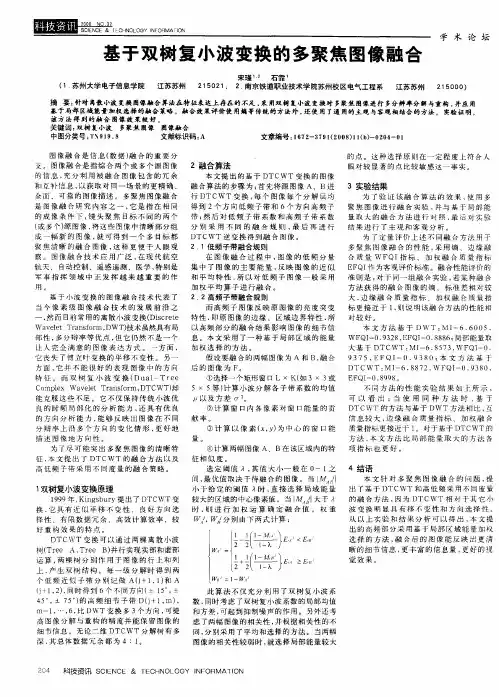
2.基于双树复小波变换的多聚焦图像融合 [J], 范艳玲;杨明
3.基于双树复小波变换的多聚焦图像融合算法研究 [J], 宋瑾;石霏
4.基于双树复小波变换彩色多聚焦图像融合方法 [J], 王亚杰;李殿起;徐心和
5.基于双树复小波变换的多聚焦图像融合 [J], 宋瑾;石霏
因版权原因,仅展示原文概要,查看原文内容请购买。
基于双树复小波变换和形态学的脉搏信号去噪李丹;王慧倩;柏桐;林金朝;庞宇;姜小明;蒋宇皓【摘要】常见的医学信号(如脉搏信号)包含大量的噪声,具有强烈的非线性和非平稳性.针对传统的小波变换去噪方法的缺陷,提出了一种基于双树复小波变换和形态学的去噪算法,具有结构简单、计算复杂度低等优点,有效地克服了离散小波变换的平移敏感性和频率混淆.实验表明,该算法可以有效地去除脉搏信号中工频干扰及肌电干扰等高频噪声,其信噪比及均方差等定量指标均明显优于传统的阈值去噪算法,能得到较干净的脉搏信号波形.【期刊名称】《电信科学》【年(卷),期】2016(032)012【总页数】6页(P93-98)【关键词】信号去噪;双树复小波变换;形态学滤波;脉搏信号【作者】李丹;王慧倩;柏桐;林金朝;庞宇;姜小明;蒋宇皓【作者单位】重庆邮电大学光电信息感测与传输技术重庆市重点实验室,重庆400065;重庆邮电大学光电信息感测与传输技术重庆市重点实验室,重庆400065;重庆邮电大学光电信息感测与传输技术重庆市重点实验室,重庆400065;重庆邮电大学光电信息感测与传输技术重庆市重点实验室,重庆400065;重庆邮电大学光电信息感测与传输技术重庆市重点实验室,重庆400065;重庆邮电大学光电信息感测与传输技术重庆市重点实验室,重庆400065;重庆邮电大学光电信息感测与传输技术重庆市重点实验室,重庆400065【正文语种】中文【中图分类】TN911.73光电容积脉搏波描记法(photo plethysmo graphy,PPG)[1]是借助光电手段在活体组织中检测血液容积变化的一种无创检测方法,通过PPG可以获得心率、血氧饱和度、呼吸频率、血压等人体最基本的生理参数。
PPG信号蕴含了丰富的人体生理病理信息,临床上的许多疾病特别是心脏病,可使脉搏发生变化。
脉搏信号的频率范围为0.5~4 Hz,而人体运动产生的干扰频率在0.1 Hz及以上均有分布。

知识创造未来 1 / 2 双树离散小波变换 双树离散小波变换是一种在信号处理领域中广泛应用的强大工具。它以中文传统的名称而闻名,并经过多年的研究和改进,已成为离散小波变换的一个重要分支。
双树离散小波变换的核心思想是将信号分解为不同尺度的低频和高频成分,从而能够更好地捕捉信号的时频特性。这种方法使用了两棵双树结构,分别用于对信号进行水平和垂直方向的分解。这两棵树的节点之间存在一种特殊的连接关系,使得信号在嵌套的子空间中得以表示,从而实现了更加准确的频域分析。
与传统的离散小波变换相比,双树离散小波变换具有许多优势。首先,它能够更好地保留信号的能量,有效地避免了传统方法中存在的能量损失问题。其次,双树离散小波变换能够适应不同类型的信号,包括具有不同频率分布和时域特性的信号。这使得它在处理各种实际问题时都能取得较好的效果。
双树离散小波变换在实际应用中有着广泛的意义和指导价值。首先,它可以应用于压缩信号,如图像和音频压缩。通过采用双树结构,可以更好地保留信号的特征,并以较低的存储和传输成本来实现信号的高效表示。此外,在图像处理中,双树离散小波变换也常用于边缘检测、图像增强和去噪等任务,取得了一定的效果。
双树离散小波变换还可以应用于生物医学信号处理领域。例如,在脑电图(EEG)信号分析中,双树离散小波变换可以帮助发现脑电波知识创造未来 2 / 2 的特征频率和时域分布,从而对脑电波进行分类和识别。同样地,它也可以用于心电图(ECG)信号处理,对心脏的节律和异常进行检测和分析。
总之,双树离散小波变换以其独特的分解结构和优化性能,在信号处理领域展现出巨大的潜力。它不仅可以应用于图像和音频压缩,还可以用于图像处理和生物医学信号处理等各种实际问题。因此,进一步研究和推广双树离散小波变换的应用将对相关领域的发展产生积极的指导意义。

二维小波变换原理引言在信号处理和图像处理领域,小波变换是一种重要的数学工具。
而二维小波变换在图像处理中具有广泛的应用,例如图像压缩、边缘检测、图像增强等。
本文将介绍二维小波变换的原理和基本概念,并探讨其在图像处理中的应用。
一维小波变换回顾在介绍二维小波变换之前,我们先来回顾一下一维小波变换的原理。
一维小波变换是将一个一维信号通过特定的小波函数进行变换,从而得到一组小波系数。
其中,小波系数表示了信号在不同频率上的成分。
在一维小波变换中,我们使用一个小波函数(基函数)进行卷积,从而得到小波系数。
常用的小波函数有Haar小波、Daubechies小波、Symlet小波等。
一维小波变换的过程可以表示为:Ck = ∑(2^(j/2) * Φ(t - k * 2^j) * f(t)) (k ∈ Z, j ∈ Z)其中,Ck表示第k个小波系数,Φ(t)表示小波函数,f(t)表示输入信号。
二维小波变换原理二维小波变换是一种将二维信号(例如图像)进行频域分析的方法。
在二维小波变换中,我们使用二维小波函数对图像进行卷积,从而得到一组二维小波系数。
与一维小波变换类似,二维小波变换也可以用于提取图像的不同频率成分。
二维小波变换的过程可以表示为:C(k,l) = ∑(2^(j/2) * Φ(x - k * 2^j, y - l * 2^j) * f(x, y)) (k, l ∈ Z, j ∈ Z)其中,C(k,l)表示第(k,l)个二维小波系数,Φ(x, y)表示二维小波函数,f(x, y)表示输入图像。
二维小波函数通常由水平平移、垂直平移和尺度变换组成。
平移操作控制小波函数在图像中的位置,尺度变换控制小波函数的大小。
通过将不同尺度和位置的小波函数卷积到输入图像中,我们可以得到不同频率的小波系数。
二维小波变换的应用图像压缩二维小波变换在图像压缩中得到了广泛的应用。
通过对图像进行二维小波变换,我们可以将图像在频域中的高频成分和低频成分分离开来。
离散小波变换(dwt
离散小波变换(DWT)是一种常用的信号处理技术,可以将信号分解成不同频率的子信号。
它是通过对信号进行多级滤波和下采样操作来实现的。
离散小波变换在很多领域都有广泛的应用,如图像压缩、信号去噪、语音识别等。
在离散小波变换中,信号先通过低通滤波器和高通滤波器进行滤波,然后再进行下采样操作。
低通滤波器将信号中的低频分量提取出来,而高通滤波器则提取出高频分量。
通过多级滤波和下采样操作,信号被分解成不同频率的子信号。
离散小波变换的一个重要特点是多分辨率分析。
多分辨率分析意味着信号的不同频率成分可以被分解到不同的尺度中。
通过对信号进行多级DWT,可以得到不同尺度的近似系数和细节系数。
近似系数表示信号的低频分量,而细节系数表示信号的高频分量。
通过调整DWT的级数,可以选择相应的频率范围。
离散小波变换还有一种重要的性质是能量集中性。
能量集中性意味着信号的大部分能量都集中在少数的子信号中。
通过对信号进行DWT,可以将信号的能量集中在少数的系数上,从而实现信号的压缩和去噪。
离散小波变换还可以通过逆变换将分解的子信号重构成原始信号。
逆变换是通过对近似系数和细节系数进行上采样和滤波操作来实现
的。
通过多级逆变换,可以将信号完全恢复。
离散小波变换是一种强大的信号处理技术,可以分解信号并提取出不同频率的分量。
它在图像处理、信号处理等领域有广泛的应用。
通过合理地使用离散小波变换,我们可以更好地理解和处理信号,提高信号处理的效果。
一维双树复小波:
()()()h g t t j t ψψψ=+
h ψ和g ψ分别是正交或双正交的实小波。
二维双树小波:
由一维双树复小波推广
(,)()()[()()][()()]h g h g x y x y x j x y j y ψψψψψψψ==++
()()()()[()()()()]h h g g g h h g x y x y j x y x y ψψψψψψψψ=-++
其中双树实小波为:
()()()()h h g g x y x y ψψψψ-
实现方法:
对一个图像,用01{(),()}h n h n 实现一个二维可分离小波变换,01{(),()}g n g n 实现另一个,进行一次二维分解后得到2个低频子带和6个高频子带:2个HL ,2个LH ,2个HH ;每对子带的和或差构成2个低频系数和6个高频系数。
行抽样
列卷积行卷积列抽样
A 1
A 2H 1
V 1D 1H 2V 2D 2
其中双树复小波为:
()()()()g h h g x y x y ψψψψ+
实现方法:
对一个图像,用01{(),()}h n h n 作行变换,再使用滤波器01{(),()}g n g n 作列变换得到1个低频子带和3个高频子带;实现另一个,用01{(),()}g n g n 作行变换,再使用滤波器
01{(),()}h n h n 作列变换得到1个低频子带和3个高频子带;每对子带的和或差构成2个低
频系数和6个高频系数。
行抽样
列卷积行卷积列抽样
A 1
A 2H 1
V 1D 1H 2V 2D 2。