肝素诱导的血小板减少症诊断与治疗
- 格式:pdf
- 大小:370.62 KB
- 文档页数:4

肝素诱导的血小板减少症诊断及治疗研究进展刘中洁,李莹重庆医科大学附属第一医院,重庆400016摘要:肝素诱导的血小板减少症(HIT)是使用普通肝素及低分子肝素过程中出现的一种药物免疫介导并发症,其特点是血小板计数下降,以体外循环设备反复凝血及动静脉内痿血栓形成为主要表现,严重时影响透析生命线,甚至危及患者生命。
由于既往诊断金标准限制,患者诊断较困难并018年我国HIT专家共识提出4Ts评分M6分及酶联免疫吸附试验阳性,临床即可诊断为HIT o HIT患者必须停止使用肝素类药物,使用替代抗凝药物(阿加曲班等)、新型口服抗凝药物(利伐沙班等、、免疫球蛋白和血浆置换等方式进行治疗。
关键词:肝素诱导的血小板减少症;血液透析;肝素;血小板计数;4Ts评分;阿加曲班;利伐沙班doi:10.3969/j.issn.1002-266X.2020.33.029中图分类号:R558R692.5文献标志码:A文章编号:1002-266X(2020)33-0108-04对于尿毒症患者来说,肝素类药物可防止血液在体外透析通路中凝固,保证透析充分性,从而减少尿毒症并发症发生,提高透析患者生存质量。
随着肝素类药物的广泛使用,人们也逐渐认识到其使用过程中存在的出血、血小板减少等药物不良反应。
肝素诱导的血小板减少症(HIT)是使用普通肝素及低分子肝素过程中出现的一种药物免疫介导并发症,其特点是血小板计数下降,出血风险较低反而体内高凝,易形成血栓⑴。
HIT患者以 体外循环设备反复凝血及动静脉内痿血栓形成为主要表现,严重时影响透析生命线,甚至危及患者生命。
HIT发病率不高,主要原因是HIT的诊断金标准是血小板功能检测,其需在特定的实验室内完成,检测难度较大并临床未能普及,致使确诊病例较少,临床医生对其重视不足,认为只要无肝素抗凝即可,忽视了预后抗凝方式的选择及血栓并发症的处理,致使患者没有及时有效的治疗,影响生活生存质量。
近年来,有关HIT诊治的研究报道较多,现综述如下。
肝素诱导性血小板减少症(肝素诱发的血小板减少症)肝素诱导性血小板减少症是一种常见的药物相关性疾病,通常由肝素引起。
肝素是一种抗凝血药物,用于预防和治疗血栓形成。
然而,一些患者在接受肝素治疗期间会出现血小板减少的并发症,这就是肝素诱导性血小板减少症。
病因肝素诱导性血小板减少症的具体病因尚不完全清楚,但目前认为主要有以下几个方面:1.免疫反应:肝素可能会引发机体免疫反应,导致血小板破坏,从而引起血小板减少症。
2.骨髓抑制:肝素可能对骨髓造成抑制,抑制血小板的生成,导致血小板减少。
3.其他因素:个体差异、遗传因素等也可能影响肝素引起的血小板减少。
临床表现患者患上肝素诱导性血小板减少症后,常常表现为以下症状:•流血倾向:患者出现皮肤、黏膜、消化道等不明原因的出血。
•瘀斑:皮肤或黏膜上出现紫癜、瘀痕等。
•血小板减少:患者血小板计数降低,可能伴有出血时间延长等。
诊断肝素诱导性血小板减少症的诊断通常通过以下方式进行:1.病史:了解患者用药史、过敏史等,排除其他原因引起的血小板减少。
2.实验室检查:包括血小板计数、凝血功能检查等。
3.肝素致敏试验:通过肝素致敏试验判断患者是否对肝素过敏。
治疗对于肝素诱导性血小板减少症,治疗主要包括以下方面:1.停用肝素:停止使用肝素治疗,尝试其他抗凝药物。
2.支持疗法:对症治疗,包括输血、止血药物等。
3.免疫治疗:对免疫相关性的患者可考虑使用免疫抑制剂、皮质类固醇等治疗。
预后肝素诱导性血小板减少症的预后通常较好,停用肝素后血小板计数会逐渐恢复正常。
然而,部分患者可能会出现再次发作,因此需密切监测患者的血小板计数和症状变化。
结语肝素诱导性血小板减少症是一种重要的临床问题,尤其在使用肝素治疗的患者中需要引起重视。
及时识别并处理肝素诱导性血小板减少症对患者的预后至关重要。
希望本文能够帮助读者对这一疾病有更深入的了解。
以上就是关于肝素诱导性血小板减少症的相关介绍,希望对您有所帮助。
肝素引起的血小板减少1. 介绍肝素是一种常用的抗凝药物,广泛应用于临床上预防和治疗血栓性疾病。
然而,尽管肝素的疗效显著,但它也有一些不良反应,其中之一就是引起血小板减少。
本文将详细探讨肝素引起的血小板减少的机制、临床表现、诊断方法和处理策略。
2. 机制肝素引起的血小板减少主要通过两种机制发生:免疫介导和非免疫介导。
2.1 免疫介导机制免疫介导的肝素引起的血小板减少是指肝素与体内抗体结合形成复合物,进而激活免疫系统,导致血小板被消耗。
这种机制通常发生在既往没有接触过肝素的患者中。
2.2 非免疫介导机制非免疫介导的肝素引起的血小板减少是指肝素直接影响骨髓中血小板的生成和释放,导致血小板减少。
这种机制通常发生在长期使用肝素的患者中。
3. 临床表现肝素引起的血小板减少可以表现为以下症状:•容易出血:皮肤、黏膜出现紫癜、瘀斑等出血点。
•出血时间延长:常规止血时间延长。
•鼻出血、牙龈出血:常见的局部出血症状。
•月经过多:女性患者可能出现月经过多的情况。
4. 诊断方法肝素引起的血小板减少的诊断主要依靠以下方法:4.1 血小板计数通过检测患者的全血细胞计数,可以确定患者是否存在血小板减少。
4.2 凝血功能检测肝素引起的血小板减少还会导致凝血功能异常,可以通过检测患者的凝血功能指标来辅助诊断。
4.3 肝素抗体检测对于免疫介导机制引起的肝素引起的血小板减少,可以通过检测患者的肝素抗体来确认诊断。
5. 处理策略肝素引起的血小板减少的处理策略主要包括以下几个方面:5.1 停用肝素对于免疫介导机制引起的肝素引起的血小板减少,停用肝素是最关键的处理方法。
一旦诊断明确,应立即停止使用肝素并选择其他合适的抗凝药物。
5.2 补充血小板对于严重的血小板减少患者,可以考虑进行血小板输注来快速增加患者体内的血小板数量。
5.3 对症治疗根据患者具体情况,可以采取一些对症治疗措施,如止血药物、促进骨髓生成等。
结论肝素引起的血小板减少是一种常见但重要的不良反应。


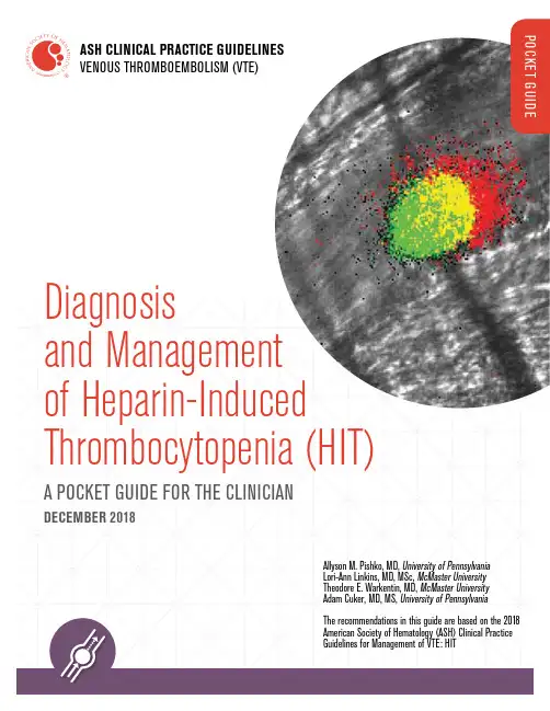
Diagnosis and Management of Heparin-Induced Thrombocytopenia (HIT)A POCKET GUIDE FOR THE CLINICIANDECEMBER 2018ASH CLINICAL PRACTICE GUIDELINES VENOUS THROMBOEMBOLISM (VTE)Allyson M. Pishko, MD, University of Pennsylvania Lori-Ann Linkins, MD, MSc, McMaster University Theodore E. Warkentin, MD, McMaster University Adam Cuker, MD, MS, University of Pennsylvania The recommendations in this guide are based on the 2018 American Society of Hematology (ASH) Clinical Practice Guidelines for Management of VTE: HITEvaluating the Clinical Probability of HITIn patients with suspected HIT, the ASH guideline panel recommends using the 4Ts score to estimate the probability of HIT rather than a gestaltTHE 4Ts: A CLINICAL PROBABILITY SCORING SYSTEM1,2(≤ 3 points)Missing or inaccurate information may lead to a faulty 4Ts score and inappropriate management decisions. Every effort should be made to obtain accurate and complete information necessary to calculate the 4Ts score. If key information is missing, it may be prudent to err on the side of a higher 4Ts score. Patients should be reassessed frequently. If there isa change in the clinical picture, the 4Ts score should be recalculated.1 Adapted from Lo et al., J Thromb Haemost 2006;4:759 and Warkentin et al., J Thromb Haemost 2010;8:1483.2 The 4Ts score may be used as a guide for clinicians but should not substitute for clinical judgment. Laboratory DiagnosisIn patients with a low-probability 4Ts score, the ASH guideline panel recommends againstuncertainty about the 4Ts score (for example, due to missing data).If there is an intermediate- or high-probability 4Ts score, the ASH guideline panel recommendssuggests a functional assay.platelet factor 4; SRA, serotonin release assay1 Includes chemiluminescence assay, lateral flow immunoassay, latex agglutination assay, particle gel immunoassayDifferent immunoassays and functional assays are available. The choice of assay maybe influenced by diagnostic accuracy, availability, cost, feasibility, and turnaround time.If an ELISA is used, a low threshold is preferred over a high threshold. Diagnostic and Initial Treatment Algorithm1 If the patient has an intermediate-probability 4Ts score, has no other indication for therapeutic-intensity anticoagulation, and is judged to be at high risk for bleeding, the panel suggests treatment with a non-heparin anticoagulant at prophylactic intensity rather than therapeutic intensity. If the patient has an intermediate-proba-bility 4Ts score and is not judged to be at high risk for bleeding or has another indication for therapeutic-intensity anticoagulation, the panel suggests treatment with a non-heparin anticoagulant at therapeutic intensity rather than prophylactic intensity. In a patient with a high-probability 4Ts score, the panel recommends treatment with a non-heparin anticoagulant at therapeutic intensity.2 For all intermediate/high-probability patients with a positive immunoassay, including those who were receiving prophylactic-intensity treatment with a non-heparin anticoagulant prior to the availability of the immunoassay result, the panel recommends treatment with a non-heparin anticoagulant at therapeutic intensity.3 In some settings, a functional assay may not be available and decisions may need to be made based on the results of the 4Ts and immunoassay. A functional assay may not be necessary in patients with a high 4Ts score and a strongly positive immunoassay (e.g., an ELISA > 2.0 optical density units).4 Most patients with a negative functional assay do not have HIT and may be managed accordingly. However, depending on the type of functional assay and the technical expertise of the laboratory, false-negative results are possible. Therefore, a presumptive diagnosis of HIT may be considered for some patients with a negative functional assay, especially if there is a high-probability 4Ts score and a strongly positive immunoassay. This is represented in the figure by a dashed line.TreatmentIn patients with acute HIT complicated by thrombosis (HITT) or acute HIT without thrombosis (isolated HIT), the ASH guideline panel recommends discontinuation of heparin and initiation of a non-heparinsuggests argatroban, bivalirudin, danaparoid, fondaparinux, or a direct oral anticoagulant (DOAC) .2 Dosing for treatment of acute HIT is not well-established. Suggested dosing is extrapolated from venous thromboembolism and based on limited published evidence Selecting a Non-Heparin AnticoagulantChoice of agent may be influenced by drug factors (availability, cost, ability to monitor the anticoagulant effect, route of administration, half-life), patient factors (kidney function, liver function, bleeding risk, clinical stabili-ty), and experience of the clinician.TRANSITIONING TO AN ORAL AGENTIn patients with HIT initially treated with a parenteral agent, the ASH guideline panel suggests transitioning to a DOAC rather than warfarin . The ASH guideline panel recommends against initiation of warfarin prior to platelet count recoveryClinical Practice Guidelines for Management of VTE: HIT.Clinically stableFondaparinux or a DOAC may bepreferred due to ease of administration,lack of need for lab monitoring, andfeasibility of outpatient useLife or limb threateningthrombosisArgatroban, Bivalirudin, Danaparoid,or Fondaparinux may be preferredbecause few such patients have beentreated with a DOACModerate or severehepatic dysfunction(Child-Pugh Class B and C)Avoid Argatroban or use a reduceddose. Avoid DOACs.Heparin Re-Exposure in Patients with a History of HITCARDIAC AND VASCULAR SURGERYIn patients with a history of HIT, HIT laboratory testing may be used to define the phase of HIT and an appropriate strategy for intraoperative anticoagulation.1If heparin is used, exposure should be limited to the intraoperative setting and avoided before and after surgeryCARDIAC CATHETERIZATION/PERCUTANEOUS CORONARYINTERVENTION1If heparin is used, exposure should be limited to the intraprocedural setting and avoided before and after the procedureSCREENING FOR ASYMPTOMATIC DVT AND DURATION OF ANTICOAGULATIONIn patients with acute HIT without clinically apparent thrombosis, the ASH guideline panel suggests bilateral lower extremity compression ultrasonography to screen for asymptomatic proximal DVT .If an upper extremity central venous catheter is present, the ASH guide-line panel suggests ultrasonography of the limb with the catheter to screen for asymptomatic DVT .In patients with acute HIT without clinically apparent thrombosis and no asymptomatic DVT identified by screening ultrasonography, the ASH guideline panel suggests that anticoagulation be continued, at a minimum, until platelet count recovery (usually a platelet count of ≥150 × 109/L). The ASH guideline panel suggests against continuing treat-ment for more than three months unless the patient has persisting HIT without platelet count recovery . In patients with HIT complicated by thrombosis and no other indication for anticoagulation, anticoagulation is typically given for 3-6 months.PLATELET TRANSFUSIONIn patients with acute HITT or acute isolated HIT who are at average bleeding risk, the ASH guideline panel suggests against routine plate-let transfusion . Platelet transfusion may be an option for patients with active bleeding or at high bleeding risk.Renal Replacement in Patients with a History of HIT1Citrate is not appropriate for patients with acute HIT, who require systemic rather than regional anticoagulation .American Society of Hematology 2021 L Street NW, Suite 900 Washington, DC 20036 © 2018 American Society of HematologyAll rights reserved. No part of this publication may be reproduced, stored in a retrieval system, or transmitted in any form or by any means, electronic or mechanical, includingphotocopy, without prior written consent of the American Society of Hematology.For expert consultation on HIT and other hematologic diseases, submit a request to the ASH Consult a Colleague program at /consult(ASH members only).Strength of Recommendations and Quality of EvidenceThe methodology for determining the strength of each recommendation and the quality of the evidence supporting the recommendations was adapted from GRADE: an emerging consen-sus on rating quality of evidence and strength of recommendations. Guyatt GH, et al; GRADE Working Group. 2008;336(7650):924–926. More details on this specific adaptation of the GRADE process can be found in the 2018 ASH Clinical Practice Guidelines for Management of VTE: HIT.1How to Use This Pocket GuideASH pocket guides are primarily intended to help clinicians make decisions about diagnostic and treatment alternatives. The information included in this guide is not intended to serve or be construed as a standard of care. Clinicians must make decisions on the basis of the unique clinical presentation of an individual patient, ideally through a shared process that considers the patient’s values and preferences with respect to all options and their possible outcomes. Decisions may be constrained by realities of a specific clinical setting, including but not limited to institutional policies, time limitations, or unavailability of treatments. ASH pocket guides may not include all appropriate methods of care for the clinical scenarios described. As new evidence becomes available, these pocket guides may become obsolete. Following the guid-ance in these pocket guides cannot guarantee successful outcomes. ASH does not warrant or guarantee any products described in these pocket guides.The complete 2018 ASH Clinical Practice Guidelines for Management of VTE: HIT 1 include additional remarks and contextual information that may affect clinical decisionmaking. To learn more about these guidelines, visit /vte .Drs. Pishko, Linkins, Warkentin, and Cuker declare no competing financial interests.1Cuker A, Arepally GM, Chong BH, et al. American Society of Hematology 2018 guidelines for management of venous thromboembolism: heparin-induced thrombocytopenia. Blood Adv . 2018;2(22). In press.。


肝素诱导血小板减少抗体的检验方法肝素诱导血小板减少抗体(Heparin-Induced Thrombocytopenia Antibodies, HIT)是一种由于对肝素类药物的过敏反应而导致的血小板减少现象。
HIT抗体通常是由治疗中使用的肝素或其他肝素类药物引起的,而这些药物是用于治疗血栓性疾病和预防血栓形成的。
检测肝素诱导血小板减少抗体的方法对于临床诊断和治疗至关重要。
下面将介绍一些常见的检验方法:1.血小板因子检测法:这是最常用的检测方法之一,它基于病人的血小板被肝素诱导的抗体所激活后的凝集现象。
医生会将病人的血样与肝素加入的血小板因子混合,然后观察血小板是否发生凝聚的现象。
这种方法简单易行,但可能存在一定的误诊率。
2.血浆因子检测法:这种方法是通过特定的检测试剂盒测定血浆中是否存在肝素诱导的抗体。
这种方法相对准确,但需要较长的测试时间和专业的实验室操作。
3.免疫阻断试验法:这是一种新型的检测方法,它基于抗体与相应抗原结合的原理。
医生会将病人的血样与已知抗原进行反应,然后观察是否产生抗体与抗原结合的免疫阻断现象。
这种方法对于特异性和敏感性都有很高的要求,但在实验室条件允许的情况下,可以作为辅助诊断手段。
4.血小板活化试验法:这是一种较为先进的检测方法,它基于病人的血浆与血小板共同反应后的凝集现象。
医生通过特殊的试剂盒将病人的血浆与血小板共同反应,然后观察血小板是否发生凝聚的现象。
这种方法较为复杂,需要专门的实验条件和设备。
总的来说,检测肝素诱导血小板减少抗体的方法有很多种,每种方法都有其自身的优缺点。
临床医生在选择适当的检测方法时,需要结合病人的具体情况和实验室条件来进行综合考虑。
同时,病人在接受检测前也需要咨询医生,了解适合自己的具体检测方法,并按照医嘱进行检测。
希望通过这些检测方法,可以及时发现并治疗肝素诱导血小板减少抗体的问题,从而保障患者的健康。

肝素诱导的血小板减少症一、简介HIT 多见于肝素治疗第 5~14 天,血小板计数相对值下降 50%或绝对值降至50~80 × 109/L,停药后 4~14 天恢复正常。
若合并血栓形成,称为肝素诱导血小板减少症伴血栓形成(HITT),可致深静脉血栓形成、肺栓塞、肢体坏疽、急性心梗、脑卒中等致命性并发症。
二、分型HIT 分为两种类型:Ⅰ 型为非免疫介导的,无需停用肝素制剂,血小板即可自行恢复。
Ⅱ 型即临床上通常所说的 HIT,为免疫介导的抗体反应。
除非特别说明,目前文献中、临床上和共识中所指的 HIT 是HIT Ⅱ 型。
三、诊断HIT 的诊断首先基于病史及临床表现,如既往或正在使用肝素、血小板计数明显下降,伴或不伴血栓栓塞并发症。
目前,国际上对于 HIT 的主流诊断思路是,在4T’S 评分和血小板数量动态监测基础上,联合 HIT 抗体检测和(或)血小板功能试验进行排除诊断和确诊。
(一)4T’S 评分4T's 评分是由血小板减少的数量特征、血小板减少的时间特征、血栓形成类型以及是否存在其他导致血小板减少的原因四个要素构成,四项评分相加,根据得分多少确定 HIT 的临床可能性:≤ 3 分为低度、4~5 分为中度和 6~8 分为高度临床可能性。
建议疑似 HIT 患者首先使用 4T's 评分进行临床危险度分层,为排除诊断和早期临床干预提供初步依据。
循证证据表明,4T's 评分诊断 HIT 的敏感性较高,具有较高的阴性预测值,低度临床可能性患者可以排除 HIT,不需进一步行 HIT 抗体检测和连续监测血小板计数;对于 4T's 评分为中、高度临床可能性患者,推荐检测 HIT 抗体,并持续监测血小板计数。
(二)实验室诊断●5-羟色胺释放试验(SRA)具有高度灵敏性和特异性,是目前公认的参考标准。
●肝素诱导的血小板活化试验(HIPA)与 SRA 相近的灵敏性和特异性,缺点是检测过程复杂、成本高、耗时长,且结果判读依赖检测人员主观,使结果可靠性降低。
ECMO持续治疗下肝素诱导的血小板减少症1例
摘要:肝素是ECMO治疗中常用的抗凝剂,但其使用可能会导致血小板减少症。
本文报告了一例ECMO持续治疗下肝素诱导的血小板减少症患者的临床特点、诊断和治疗过程,旨在提高对该疾病的认识和处理能力。
关键词:ECMO治疗;肝素;血小板减少症;抗凝治疗
病例报告:一例62岁男性患者,因双肺感染合并多器官功能衰竭入院。
入院后予以抗感染治疗、机械通气和体外膜氧合治疗。
ECMO治疗中使用肝素作为抗凝剂。
治疗初期患者血小板计数正常,但在ECMO治疗后第2天血小板开始逐渐下降。
第3天时,患者发生明显的出血现象,同时血小板计数降至30×10^9/L。
根据临床症状和实验室检查结果,诊断为肝素诱导的血小板减少症。
治疗过程:在确诊后,立即停止肝素的使用。
为了替代抗凝剂的需要,使用阿加曲班(Argatroban)替代抗凝剂,同时给予血小板输注支持治疗。
停止肝素后,患者的血小板计数开始逐渐上升,并最终恢复到正常范围。
出血现象得到了控制,并在ECMO治疗的第10天完成了撤离。
对于肝素诱导的血小板减少症,最重要的治疗措施是立即停止肝素的使用。
同时需要给予血小板输注支持治疗,以增加血小板计数。
对于需要抗凝治疗的患者,可以考虑使用替代抗凝剂,如阿加曲班等。
肝素诱导性血小板减少症的治疗肝素诱导性血小板减少症(HIT)是一种严重的并发症,常见于使用肝素治疗的患者。
HIT是指在接受肝素治疗期间,患者体内产生了抗肝素抗体,从而导致血小板减少和血小板功能异常。
因此,HIT的治疗需要综合考虑患者的病情和临床表现进行个体化治疗。
首先,治疗HIT的首要措施是停止使用肝素。
这是因为肝素是引起HIT的主要原因之一,停用肝素可以阻断抗肝素抗体的进一步产生和进一步损害患者的血小板。
停用肝素后,一般可以观察到患者血小板数量逐渐恢复正常。
停用肝素后,需要鉴别和排除HIT引起的其他并发症,尤其是血栓形成。
因为HIT患者具有较高的血栓形成风险,治疗过程中需要密切观察患者的凝血功能。
如果患者存在血栓的情况,可能需要使用抗凝剂来防止进一步的血栓形成。
在HIT的治疗中,需要给予患者以较高的个体化治疗。
根据患者的具体情况,可能需要考虑使用其他的抗凝剂,如阿哌沙班或比伐卢定。
这些抗凝剂不会导致HIT,能够有效地预防和治疗血栓形成。
然而,使用新的抗凝剂也要谨慎,因为它们可能带来新的副作用和并发症。
HIT患者同时需要进行密切的临床监测。
定期监测血小板数量和凝血功能可以帮助评估治疗的效果,并及时发现和处理并发症。
另外,需要密切观察患者的病情变化和系统功能,以及评估他们对治疗的耐受性。
除了药物治疗,HIT患者还需要注意日常生活的调整。
他们应该减少身体活动量,避免剧烈运动和重物提拿。
合理安排作息时间和饮食,避免过度疲劳和饥饿。
此外,需要注意个人卫生,避免感染和其他的危险因素。
最后,HIT患者在治疗过程中需要得到充分的支持和关怀。
他们经历了严重的疾病和治疗,不仅身体上需要医学上的帮助,心理上也需要关怀和理解。
医务人员应该与患者建立良好的沟通和信任,关注他们的需求和安排合理的康复计划。
综上所述,HIT的治疗需要综合考虑患者的病情和临床表现进行个体化治疗。
停用肝素、鉴别并排除血栓形成、个体化药物治疗、密切临床监测、日常生活调整和心理支持都是治疗HIT 的重要措施。
肝素诱导性血小板减少症,肝素诱导性血小板减少症的症状,肝素诱导性血小板减少症治疗【专业知识】疾病简介肝素诱导性血小板减少症(heparin-induced thrombocytopenia),肝素所致的血小板减少的程度与肝素的剂量、注射的途径和既往有无肝素接触史等并无明确的关系,但是,与肝素制剂的来源有关。
疾病病因一、发病原因各种剂型的肝素均可诱发血小板减少症,实验研究表明高分子量的肝素更易于与血小板相互作用,导致血小板减少症,这与临床中所观察到的使用低分子量肝素治疗的患者血小板减少症发生率较低的结果一致。
二、发病机制肝素诱导性血小板减少症可能与免疫机制有关,部分患者体内可以出现一种特异性抗体IgG,该抗体可以与肝素-PF4(血小板4因子)复合物结合,PF4又称肝素结合阳离子蛋白。
由血小板α颗粒分泌,然后结合于血小板和内皮细胞表面。
由血小板α颗粒分泌,然后结合于血小板和内皮细胞表面。
抗体-肝素-PF4形成1个3分子复合物,再与血小板表面的Fcγ Ⅱa受体结合,免疫复合物可以激活血小板,产生促凝物质,是肝素诱导性血小板减少症伴发血栓并发症的可能机制。
而其他药物所致的血小板减少症一般没有血栓并发症,可以作为鉴别。
免疫复合物通过与血小板表面的FcγR Ⅱa分子交联而激活血小板。
FcγR Ⅱa分子氨基酸链第131位点的His/Arg多态性能影响其与IgG结合的能力,从而可以作为一个预测因素来预测肝素诱导性血小板减少症的个体危险性。
症状体征一、症状根据临床上应用肝素治疗后所诱发的血小板减少症的病程过程,可以分为暂时性血小板减少和持久性血小板减少。
1、暂时性血小板减少大多数发生在肝素治疗开始后,血小板立即减少,但是,一般不低于50×109/L。
可能与肝素对血小板的诱导聚集作用有关,肝素可以导致血小板发生暂时性的聚集和血小板黏附性升高,血小板在血管内被阻留,从而发生短暂性血小板减少症。