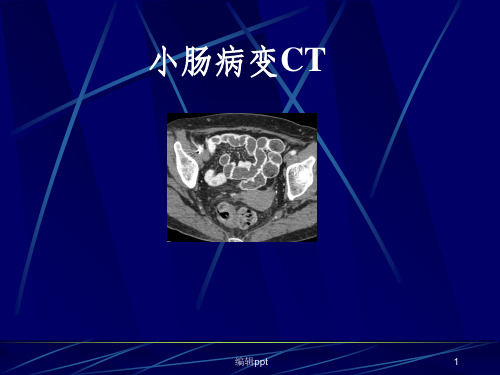
小肠肿瘤的CT诊断和影像学表现
- 格式:ppt
- 大小:2.78 MB
- 文档页数:65


小肠影像学新进展与临床应用摘要小肠是胃肠道最长的器官,因其走行弯曲,肠管常互相重叠,传统的胃肠道内镜检查仅单纯观察消化道腔内结构,不能很好显示肠壁和腔外结构。
多层螺旋CT和磁共振成像具有快速扫描和三维后处理能力,能清晰反映肠壁和肠腔外的病变。
新的影像技术,如CT小肠造影、MR小肠造影具有良好的软组织对比度及三维成像能力,不仅可以观察黏膜,同时能够分析肠管周围的改变,提高了人们对小肠疾病的影像诊断。
关键词多层螺旋CT 磁共振成像小肠疾病诊断小肠造影小肠在消化道中最长,走行弯曲,活动度大。
小肠疾病的发病率近年来有增加的趋势,而在诊断上存在一定的困难。
传统的小肠疾病诊断方法主要是口服钡剂小肠造影和小肠插管灌肠,能较好地显示肠壁黏膜和肠管形态,但其病变检出率低、灵敏度不高,且后者操作复杂,目前已很少使用。
普通内镜难以观察全部小肠,近年来临床开始采用的胶囊内镜也只能观察其腔内情况,且价格昂贵、易在肠管狭窄处嵌顿,故在不少小肠疾病的诊断中应用受限。
同时,所有上述检查方法都不能直接显示肠壁全层和腔外结构,远不能满足临床对疾病的定性、分期和并发症诊断的需要。
近年来迅速发展的多层螺旋CT(multidetector-row computed tomography,MDCT)和磁共振成像(magnetic resonance imaging,MRI),以其快速、薄层扫描和强大的后处理功能,在临床上得到了广泛的应用。
对小肠疾病而言,MDCT 和MRI能够清晰显示小肠壁和肠管外病变,能够对病变范围、性质和分期作出全面准确评价,将小肠疾病的诊断提高到一个新水平。
口服法MDCT小肠造影(MDCT enterography,MDCTE)和磁共振小肠造影(MR enterography,MRE)作为小肠病变新的检查方法由此产生,并在临床上得到更为广泛的应用。
1 影像学检查方法无论采用何种影像学技术,理想的肠道扩张、静脉内阳性对比剂的应用及薄层扫描是较好检测小肠的前提条件[1-2]。
CT小肠造影在诊断小肠肿瘤中的应用价值徐莹;陈业媛;舒虹;肖香佐【摘要】目的:探讨多层螺旋CT小肠造影(MSCTE)在小肠肿瘤诊断中的应用价值。
方法回顾性分析经手术病理证实的45例小肠肿瘤的MSCTE影像表现,总结其影像学表现特点。
结果45例小肠肿瘤中,良性肿瘤9例,其中脂肪瘤6例,影像表现为腔内含脂肪低密度肿块影;腺瘤2例,表现为腔内均质的软组织肿块;回肠系膜淋巴管瘤1例,表现为不规则囊性病灶。
常见的恶性肿瘤34例,其中腺癌14例,表现为肠壁局限不规则的环形增厚,中度不均匀强化;间质瘤11例,表现为较大的软组织肿块,以腔外生长为主,密度不均匀,中度或明显强化,常伴有囊变、坏死;淋巴瘤9例,肠壁不规则增厚或形成软组织肿块,累及肠段较长,均匀轻中度强化,常伴有肠系膜、腹膜后淋巴结肿大。
少见的恶性肿瘤2例,转移瘤1例,表现为多发肠壁不规则增厚,明显强化,腹壁出现转移性结节;类癌1例,右下腹不规则肿块影。
结论 MSCTE在小肠肿瘤诊断与鉴别诊断中有较好的临床应用价值。
%Objective To investigate application prospects of Multi-slice computed tomography Enterography(MSCTE) in diag-nosing small intestine tumor (SIT). Methods MSCTE findings were retrospectively analyzed in 45 cases of SIT diagnosed patho-logically after surgery and were summarized in terms of imaging features. Results Of all 45 cases of SIT,9 were benign ones and 6 were lipoma,characterized by low-density mass shadow in intestinal cavity;2 were adenoma,characterized by homogeneous soft tissue mass in intestinal cavity;1 belonged to ileac mesentery lymphangioma,characterized by irregular cyst-like focus;34 were the most common type of malignancies,14 of which wereadenocarcinoma,characterized by localized ring-formed thickening of in-testinal wall with moderate heterogeneous enhancement;11 were mesenchymoma,characterized by soft tissue mass relatively big in measurement,which primarily grew outside the intestinal cavity,with heterogeneous density,moderate or distinct enhancement ac-companied frequently by cystic degeneration and necrosis;9 werelymphoma,characterized by irregular thickening of intestinal wall or formation of soft tissue mass,which affected intestine in relatively greater length,and which took on homogeneous slight-moder-ate enhancement accompanied frequently by enlargement of retroperitoneal lymph nodes and those in mesentery. 2 were rare ma-lignancies and 1 was metastatic tumor,characterized by multiple irregular chickening of intestinal wall with distinct enhancement and presence of metastatic nodules in abdominal wall;1 was carcinoid tumor characterized by irregular mass in right lower quad-rant. Conclusion It suggests that MSCTE has good prospects of clinical application in diagnosis and differential diagnosis of SIT.【期刊名称】《江西医药》【年(卷),期】2015(000)005【总页数】4页(P387-390)【关键词】小肠肿瘤;多层螺旋CT小肠造影【作者】徐莹;陈业媛;舒虹;肖香佐【作者单位】南昌大学第一附属医院影像科,南昌 330006;南昌大学第一附属医院影像科,南昌 330006;南昌大学第一附属医院影像科,南昌 330006;南昌大学第一附属医院影像科,南昌 330006【正文语种】中文【中图分类】R816.5原发性小肠肿瘤发病率较低,仅占所有胃肠道肿瘤的3%-6%[1]。