斑马鱼的发育历程19页PPT
- 格式:ppt
- 大小:1.90 MB
- 文档页数:19


清华大学毕业设计论文摘要斑马鱼,作为发育生物学研究的最佳脊椎动物模型,为人类的相关研究提供了重要的参考依据。
本文主要讨论了斑马鱼幼体的软骨,特别是脊椎软骨的发育过程及其软骨的超微结构特点。
为转基因、基因突变鱼类的研究提供了对比依据。
本文主要讨论了斑马鱼脊椎软骨的发育进程,以及软骨细胞特别是脊索旁软骨的排列,形貌,大小及其显微结构特点和中胚层分化的显微形貌。
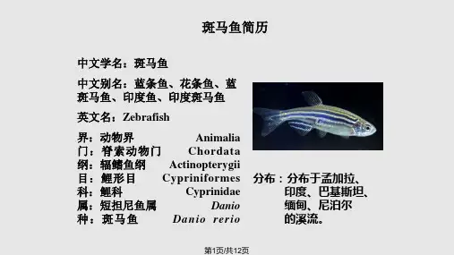
斑马鱼脊椎软骨在配卵后2~3天开始发育,围绕在脊索周围,并逐渐向神经管和血管方向延伸。
斑马鱼脊椎软骨沿体节发育,腹部以前的体节在6天已基本分化为软骨细胞,并且随着时间的推移,沿体节向尾部方向的生骨节逐步分化为软骨细胞,在1个半月左右的时间,软骨组织的分化基本完成。
斑马鱼的软骨组织排列整齐,大多数细胞大小在5-20μm范围内。
斑马鱼的软骨组织为透明软骨,胶原纤维为II型胶原,没有典型的周期结构,含量很少。
在TEM的观察中发现,斑马鱼的肌肉组织分化较早,在3天已基本分化完成。
而体干部的脊索旁仍为中胚层的间叶细胞,为软骨细胞的原基,在分化成软骨细胞的过程中,首先是在形状和排列上趋于整齐和规则。
斑马鱼的骨化方式为软骨骨化。
软骨细胞的细胞质中富含大量的线粒体和内质网。
在细胞质中还有大量直径为100-400nm的囊泡,紧密排列在一起,起传输物质的作用。
此外,脊髓处的细胞,其细胞核较大,细胞质很少,有别于软骨细胞。
并且该处细胞有部分已死亡,被其它细胞所代替。
关键词:软骨细胞,发育,显微结构- 1 -清华大学毕业设计论文AbstractZebrafish provides an important referenced foundation for the relative research as the best pattern of vertebrate in developing biology. In this paper the process of development and the ultrastructure of the cartilage especially the parachordal cartilage in zebrafish larva are investigated . And that supply the contradistinctive basis for the research about transgenic and gene mutation fishes.Here we investigate the developing process of parachordal cartilage and chondrocyte arrangements,shapes ,size,and ultrastrcture .In zebrafish parachordal cartilage begin to form at 2-3 days postfertilization ,and surround the notochord and extend gradually to spinal cord and dorsal aorta. Parachordal cartilage develop along somite and at 6 days postfertilization the somite before venter have differentiated and all accomplish differentation on the whole about at one and a half month postfertilization .The chontrocytes arrange regular .The size of chontrocytes is between 5-20μm mostly. In zebrafish the cartilage is hyaline cartilage and the collagen fibrils is predominantly II type fibrils. And that are relatively thin and do not display the characteristic 67nm banding.We discovered that the differentiation of musculature is more earlier than cartilage by TEM and that accomplished basically at 3 days postfertilization. During the differentiation of cartilage the shape and arrangement of mesenchymal cell in mesoblast go to orderliness and regular gradually at first. With the electron microscope ,the active cartilage cell is seen to contain numerous slick endoplasmic reticulum,mitochondria and vesicles which arrange together in order and the diameter of these vesicles is about 100-400nm, transmitting the substance. Furthermore, the nucleolus of spinal cord cells is more larger and we also can observe that some cells have dead.Keywords : cartilage ,development, ultrastrcture- 2 -清华大学毕业设计论文第一章. 前言由于软骨细胞所致的成骨活动发生较早,所以对斑马鱼软骨发育的研究在骨损伤修复过程中对骨力学功能恢复有较大意义。